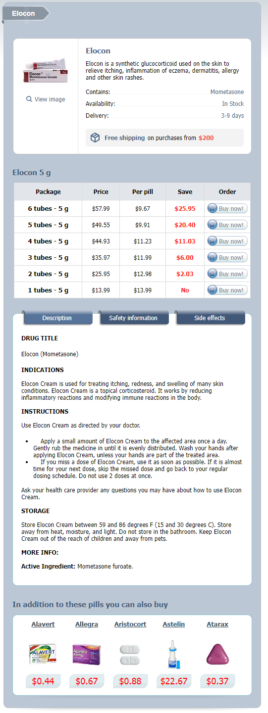
Elocon dosages: 5 g
Elocon packs: 1 tubes, 2 tubes, 3 tubes, 4 tubes, 5 tubes, 6 tubes

Buy generic elocon line
These main flexors of the knee have a secondary inner rotational affect on the tibia. The biceps femoris on the lateral facet inserts primarily into the head of the fibula. The popliteus muscle arises primarily from the lateral femoral condyle inside the capsule of the knee joint. The outer floor of the lateral condyle is sort of flat and that of medial condyle is convex. They are separated in the midline by the intercondylar eminences with its medial and lateral intercondylar tubercles. On the medial facet, the femur meets the tibia like a wheel on a flat surface, whereas on the lateral aspect, it is like a wheel on a dome. Therefore, the ligaments with the encompassing soft tissues are the one one that gives stability to knee. The menisci are attached on the periphery to this capsule particularly so medially and fewer laterally. This is as a result of laterally the passage of the popliteus tendon via the capsule produces a much less secure meniscal attachment than is present medially. The capsule is especially strengthened by the collateral ligaments and the medial and lateral hamstring muscular tissues as nicely as the popliteus muscle and the iliotibial band and these are the principal stabilizing structures posterior to the transverse axis. The major medial stabilizers of the knee are the tibial collateral ligament, the semimembranosus, the tendons of the pes anserinus and the oblique popliteal ligament of the posterior capsule. The lateral stabilizers are the iliotibial band, the fibular collateral ligament, the popliteus tendon and the biceps femoris. The anteromedial and anterolateral capsules are important in defending the anteromedial and anterolateral aspect of the knee towards subluxation and rotational excesses. The tibial collateral ligament originates on the medial epicondyle and inserts 3�4 inches below the joint line on the medial surface to the tibia deep to the pes anserinus. The fibular collateral ligament attaches to the lateral femoral epicondyle proximally and to the fibula head distally. Each meniscus is a biconcave fibrocartilaginous disc, which is thick at its attachment to the joint capsule peripherally and tapers to a thin free edge centrally. The medial meniscus is semilunar in form with a skinny anterior horn and widens posteriorly. Anteriorly and posteriorly the menisci are connected to the tibial intercondylar area by fibrous extremities or horns. The menisci get their blood provide from the superior and inferior branches of the lateral and medial geniculate arteries. The peripheral 20�30% of the medial meniscus and the peripheral 10�25% of the lateral meniscus are vascular. The remaining central portion of the menisci are avascular and depend upon diffusion of nutrients from the synovial fluid for diet. A tear sutured in the vascular portion of the meniscus (red on red) heals better surgical anaTomy and biomechanics of the knee than a tear sutured at the junction of the peripheral and central portion (red on white), or a tear sutured at the central avascular part of the meniscus (white on white). The different features attributed to the menisci are distribution of joint fluid, articular cartilage vitamin, shock absorption and deepening of the joint. The menisci defend the articular cartilage by rising the joint congruity and make contact with space and preventing focal concentrations of stress. The anteromedial band tightens with flexion, while the posterolateral band is taut in extension. It inserts on to the lateral surface of the anterior portion of the medial femoral condyle. The anterolateral band is tight in flexion and the posteromedial band tight in extension. The articular surface of the medial condyle is extended anteriorly and as a end result of the knee comes into the fully extended position, 2557 the femur internally rotates till the remaining articular surface on the medial condyle is in touch. The posterior portion of the lateral condyle rotates ahead laterally, thus, producing a screwing residence movement locking the knee in the absolutely prolonged place. Biomechanics of Knee Introduction the science of biomechanics as applied to the musculoskeletal system relates drive to motion.
Diseases
- Acute myeloblastic leukemia type 4
- Overgrowth radial ray defect arthrogryposis
- Chromosome 6, monosomy 6q2
- Hall Riggs mental retardation syndrome
- Prostatitis
- Warburg Thomsen syndrome
Buy 5g elocon mastercard
The mobility of the hip joint is comparatively preserved by this method such that the sufferers, especially Asians are able to sit cross-legged and squat comfortably. It also has couple of added advantages like instant postoperative weight bearing and permits further fine corrections during the postoperative interval, if required. Material Pelvic assist osteotomy by Ilizarov technique was performed in our institute in the interval of 1998�2004. All the patients were hospitalized and managed in accordance with a normal protocol which included preoperative analysis of affected person, preoperative planning of surgical procedure, physiotherapy plan and mobilization, distraction part of lengthening, discharge from hospital after suture removing and pin tract care, follow-up on an outpatient basis and fixator elimination. The former is measured off a supine radiograph with the affected leg maximally crossed over the alternative thigh. The latter is determined by taking a single-leg standing radiography without assist. The distinction within the adduction angle between the femoral shaft and the horizontal line of the pelvis represents the amount of help the hip abductors can at present apply along with the interference of the opposite thigh within the dropping of the pelvis. The valgus angle ought to be the single-leg stance drop angle plus 15� of over correction. Methods Preoperative Evaluation and Planning Detail case history and thorough clinical examination is carried out which would decide the management of particular case. The level of the proximal osteotomy is set based on this radiograph and should coincide with the level of the femur that crosses the ischial tuberosity; (C) Single-leg standing adduction is 40�; (D) Total valgus correction is predicated on the single-leg standing adduction plus 15� of over correction. The distal femur ought to be medially displaced; (E) Planning for the second osteotomy, the proximal axis line is the line perpendicular to the horizontal line of the pelvis, passing through the apex of the first osteotomy. The distal axis line is the mechanical axis line of the tibia prolonged proximally. The ultimate mechanical axis is perpendicular to the horizontal line of the pelvis 2548 textbook of orthoPedicS and trauma radiograph. One pelvic arch is connected to the proximal two half pins with assist of one- or two-holed rancho and another pelvic arch attached to the distal half pin. Additional tensioning of the gluteal muscular tissues was achieved by guaranteeing mild anterior angulation at the osteotomy website. The stage of the proximal osteotomy is determined from the supine cross-legged radiography. The osteotomy of the femur must be on the level of the ischial tuberosity on this view. To decide the extent of the distal osteotomy, one needs make a paper tracing of the femur and pelvis with the proximal phase of the femur maximally adducted as seen on the cross-legged view. The osteotomy should be made at the stage of the ischial tuberosity and the distal segment ought to be displaced one half-shaft thickness medially. The distal axis line is drawn as an extension of the tibial mechanical axis line, prolonged proximally. The intersection level of the proximal and distal axis strains is the level of the second osteotomy, if this degree is just too proximal, the osteotomy could be carried out extra distally if the bone is translated medially together with the varus angulation. At the proximal osteotomy, the femur should be prolonged by the amount of flexion deformity of the hip plus 5o. The extension allows the patient to lock the hip throughout standing and single-leg stance. Distal Osteotomy the distal ring positioned at the stage of the superior pole of the patella is hooked up to the femur with a transcondylar 1. This is the reference wire and is connected to the distal most ring and a hundred thirty kg tensioning carried out. The distal two femoral rings are temporarily disconnected and osteotomy is completed by rotating the distal ring externally against the proximal assemble. The two rings are then connected to one another with 4 threaded rods and conical washers. The distal ring construct is related to the arch above with the assistance of two indirect helps and threaded rods.
5g elocon buy mastercard
Sagittal T2-weighted or gradient-echo pictures create a "myelographic" effect and are helpful in evaluating compromise of the subarachnoid house. This "key-hole" appearance gets distorted in sufferers with foraminal or extraforaminal disc herniation. Similar discovering can additionally be noted in sufferers with "spondylolytic spondylolisthesis" with compromise of the exiting nerve at that degree. Degenerative spondylitic modifications are additionally manifested within the type of signal depth adjustments in endplates. The fact that discs are constitutionally avascular buildings is utilized in this technique. Epidural hematomas and abscesses appear as collections within the spinal canal, exhibiting peripheral enhancement with gadolinium on T1-weighted pictures. Gadolinium-contrast agents are also helpful in postoperative analysis of the backbone for diskitis. In extreme instances of arachnoiditis, the dural sac could appear "empty" as all the nerve roots could be adherent to the walls of the sac. On gadolinium administration, abscesses improve only on the periphery whereas related space-occupying lesion showing uniform enhancement may lead one to suspect some sort of neoplasm. Absence of abscess on these kinds of images tilts the analysis in favor of a neoplasm benign and asymptomatic. Screening of entire spine is mandatory and would additionally help establish concomitant accidents in other areas of spine. A healed fracture would exhibit normal or hyperintense (white) indicators in vertebral our bodies on T1-weighted photographs owing to normal or fatty marrow, respectively. Computed tomography scan has restricted diagnostic value in degenerative backbone pathologies. However, it offers additional insight to plan a surgical intervention in certain elements. Computed tomography scan is a obligatory preliminary screening software at most trauma centers these days. Computed tomography scan also helps detect implant-related issues such as screw loosening or breakage and most significantly a malpositioned screw. Bone Scanning (Scintigraphy) Bone scanning is an invasive radionuclide imaging scan. It can additionally be helpful in detecting osteonecrosis, in sufferers with unexplained significant back pain, in detecting stress fractures (essentially in athletes) and in figuring out the healed status of a fracture. After incorporating into bone osteoid, it emits gamma rays that get detected by gamma camera. The sooner the bone turnover, the larger deposit of 99mTc and more gamma rays emitted by the location. This is the premise of the "hot spot" indicative of a metabolically active area of bone. The best publicity occurs in bladder and is dealt with by good patient hydration before and after the scan. It is the time when osteoid incorporation is at its most and background tracer has been excreted by kidney. The half-life of 99mTc is 6 hours and by this time tracer studying turns into insignificant. These websites are usually located in rising bones on the epiphysis, metaphysis of long bones and the sacroiliac joints (a incontrovertible reality that limits its usefulness in ankylosing spondylitis). Abnormal bone turnover happens in tumors, infections, fractures and bone-soft tissue interface the place elevated metabolic actions happen. Sites with decreased bone exercise like osteonecrosis or extremely damaging tumors. A sizzling 99Tc scan with increased 67Ga exercise is highly suggestive of a bone an infection. Gallium 67 citrate (67Ga) binds with transferrin and other proteins related to irritation and infection.
Order 5g elocon with amex
Comparison of microbiological outcomes of needle puncture vs superficial swab in contaminated diabetic foot ulcer with osteomyelitis. Diabetic foot syndrome: evaluating the prevalence and incidence of foot pathology in Mexican Americans and non-Hispanic whites from a diabetes administration cohort. Outpatient administration of uncomplicated lower-extremity infections in diabetic patients. Clinical predictors of treatment failure for diabetic foot infections; information from a prospective trial. Validating the Probe-to-Bone test and other checks for diagnosing persistent osteomyelitis in the diabetic foot. Tendon Achilles lengthening for the treatment of neuropathic ulcers causes a brief lived reduction in forefoot pressure related to modifications in plantar flexor energy rather than ankle motion throughout gait. Midfoot fusion approach for neuroarthropathic feet: biomechanical analysis and rationale. Infection in diabetic osteoarthropathy: use of indium-labeled leukocytes for analysis. Diagnostic validity of three swab methods for identifying continual wound an infection. Measurement of markers of osteoclast and osteoblast exercise in sufferers with acute and continual diabetic Charcot neuroarthropathy. Treatment of diabetic foot ulcers and Charcot neuroarthropathy utilizing the patella tendon bearing brace. Bone mineral density during total contact cast immobilization for a patient with neuropathic (Charcot) arthropathy. Effects of a tendo-Achilles lengthening process on muscle function and gait characteristics in a patient with diabetes mellitus. Pattern of diabetic neuropathic arthropathy associated with the peripheral bone mineral density. Is walking barefoot a threat issue for diabetic foot illness in growing countries Serum procalcitonin and C-reactive protein concentrations to distinguish mildly infected from non-infected dibetic foot ulcer: a pilot study. The function of proinflammatory cytokines in the cause of neuropathic osteoarthropathy (acute Charcot foot) in diabetes. Infection and neuroarthropathy: the utility of C-reactive protein as a screening tool in the Charcot foot. Impact of Achilles tendon lengthening on practical limitations and perceived disability in folks with a neuropathic plantar ulcer. Performance of the probe-to-bone take a look at in a population suspected of having osteomyelitis of the foot in diabetes. The impact of zoledronic acid on the clinical resolution of Charcot neuroarthropathy. Diabetic lower extremity infection: Influence of physical, psychological, and social elements. Calcaneal bone mineral density in sufferers with Charcot neuropathic osteoarthropathy: differences in kind 1 and sort 2 diabetes. Difference in presentation of Charcot osteoarthropathy in Type 1 comparted with Type 2 Diabetes. Association between osteoprotegerin G1181C and T245G polymorphisms and diabetic Charcot neuroarthropathy: a case-control study. Six months remedy with Alendronate in acute Charcot neuroarthropathy: a randomised controlled trial. Midfoot fusion: a biomechanical comparability of plantar plating vs intramedullary screws. Prediction of end result in individuals with diabetic foot ulcers: concentrate on the variations between people with and without peripheral arterial illness. Treatment of acute Charcot foot with bisphosphonates: a scientific review of the literature. Neuropathic ulcerations plantar to the lateral column in sufferers with Charcot foot deformity; a versatile strategy to limb salvage. Superconstructs in the therapy of Charcot foot deformity; plantar plating, locked plating and axial screw fixation. Staging of Charcot neuroarthropathy along the medial column of the foot within the diabetic affected person.
Vapor (Fumitory). Elocon.
- Spasms of the gut, skin conditions, eye irritation, heart problems, bile (a fluid secreted from the liver) disorders, and other conditions.
- Dosing considerations for Fumitory.
- Are there safety concerns?
- How does Fumitory work?
- Irritable bowel syndrome (IBS). Taking fumitory does not seem to improve symptoms of irritable bowel syndrome.
Source: http://www.rxlist.com/script/main/art.asp?articlekey=96460
5g elocon with visa
Unlike the usual standing knee extension view, a fixed flexion view in Lyon-Schuss place with a hundred caudally angulated X-ray beam is a greater illustration of joint area in parallel alignment and more likely to reveal cartilage loss. For assessment of cartilage morphology, fats suppression pictures help by eliminating chemical shift artifacts. Review of the anatomical and radiological differences between fluoroscopic and nonfluoroscopic positioning of osteoarthritic knees. Ultrasound imaging has come up as a helpful adjunct modality, notably due to its non-invasive non-ionizing nature and skill to allow dynamic evaluation. It can appreciably depict patella-femoral joint and medial and lateral joint line, together with menisci and Hoffa fat pad. To summarize, radiography nonetheless is the primary line investigation, however its low sensitivity must be saved in thoughts. Recent AdvAnces in Knee Joint Recent Advances in Knee Arthroplasty From the ivory cup tibial hemiarthroplasty of 1894, by way of the development of modern knee designs in the Nineteen Seventies, to finally the declaration as probably the most successful surgical procedure of the 20th century, Total knee arthroplasty has come a long way. Simulation studies have proven improved scratch resistance and decreased polyethylene put on. New Designs Gender Specific Knee Femoral condyle is narrower in females in comparison with males. Intraoperative femoral part sizing is normally carried out with anteroposterior dimension as a reference. In a female affected person, this could lead to a too broad femoral element altering collateral ligament steadiness. New gender particular knees are actually available, though potential randomized trial has found no vital profit yet. When bearing surfaces are pressed together under load, some quantity of bonding of the surfaces takes place. Abrasive put on occurs when a tough surface mechanically cuts via a softer surface. Finally, fatigue put on (pitting and delamination) manifests when the biomaterial fails after repeated cycles of loading. Polyethylene wear not only directly impacts the bearing surface, but in addition it leads to periprosthetic osteolysis because of bioactivity of the put on and tear particles. It has been proven to be very profitable in hip arthroplasty eventualities and is now widely accepted. However, the manufacturing means of cross-linking leads to some lower in mechanical energy, fatigue resistance and fracture toughness. The significantly high-risk areas are the tibial posterior stabilized posts and the cement fixation pegs of patellar prosthesis. This may limit the potential use to solely posterior cruciate ligament retaining and deep dished designs. Clinical studies of safety at 2 and 5-year follow-up are now available, however long-term follow-up research are but to come. Patient particular instrumentation and custom chopping blocks shift the work of pc navigation from inside the operation theater to the preoperative period. The producer then fabricates single-use devices for femoral and tibial resections particular to that patient. It has following advantages and drawbacks: Advantages20 � Operating room efficiency (decreased surgical time and turnover time) � Improved implant alignment with much less number of outliers � Avoidance of systemic emboli (because of avoidance of intramedullary instrumentation) � Improved patient outcomes. Ceramic is more durable than metallic with better scratch profile and lower coefficient of friction. A solution to this drawback is proposed as a new metallic alloy, oxidized zirconium (called oxinium). Oxinium is produced by a thermally driven oxygen diffusion process, which converts the floor to a durable oxide with low friction of coefficient; essentially a ceramic. Passive methods: these perform a part of the surgical process underneath continuous and direct control of the surgeon.
Purchase elocon pills in toronto
Implant used for Correction Preferably pedicle screws be used for three-column fixation. Compression Distraction the success of the correction is dependent upon adequacy of spinal launch, and the talent and experience of the surgeon in making use of the strategies used for the correction. Most of these maneuvers are possible when one rod is in place the second rod adds to the steadiness and resistance to fatigue failure. It can be utilized in three-column osteotomy corrections, nonharmonious curves, and single lengthy curve with different sagittal contours. Uniaxial screws should plough by way of for the nut to be put after these maneuvers. True correction of vertebral rotation within the axial airplane and consequently the elimination of the thoracic and thoracolumbar prominence are troublesome to obtain with out anterior discectomy and chest wall violation with thoracoplasty. Neutral vertebra at the finish of the assemble should be locked before derotation to stop transmission of torque to the adjoining areas in order to avoid iatrogenic torsion. If the concave aspect screw fails, it can break the lateral wall and could injure the aorta. Derotation by way of Differential Rod Contouring Slight hyperkyphosis on the concave rod and hypokyphosis on to the convex rod assist in reducing the deformity in the sagittal aircraft. Coronal and Sagittal Translation Pure translation is very effective of thoracic scoliosis. It is achieved with sublaminar wire or discount screws put at the apical or periapical vertebrae on the concave facet. The rod has to be put to the proximal or distal stage and subsequently engaged to the screws to the opposite finish relying on the pliability. The pedicle screws are ideally put bilaterally and rod is engaged to the screw at on finish on the convex aspect. Compression and distraction can be used together with direct vertebral derotation in cantilever technique. Traction Halo gravity or halo pelvic traction can be used for stiffer curves preoperatively or after anterior posterior releases. Temporary Working Rods Working rods are used to scale back a segment or the whole deformity. It is most helpful in double curves and temporary stabilization in spinal osteotomy or resections. In the event of no improvement in monitoring, chill out the correction and the check the monitoring. Complications of Surgery Surgical complications can be categorised as main and minor. Major Complications Neurovascular Injury Neurovascular harm can occur with malplacement of pedicle screws or other implants. Spinal twine damage is probably the most feared and devastating complications of scoliosis surgery. There was no difference observed whether they underwent anterior or posterior procedures. Ninety % of sufferers with incomplete spinal wire damage had partial or full restoration. Injury to the spinal cord could happen on the time of surgery or in the postoperative interval. During surgery, damage can occur from an implant or an instrument, stretching during curve correction, occlusion of vessels supplying the cord or international cord ischemia secondary to hypotension. Implant breakage,15 implant migration or bony overgrowth can cause spinal wire damage in the postoperative interval. Deformity correction in kyphosis, congenital scoliosis, neurofibromatosis, skeletal dysplasia and revision surgery stand a high threat for spinal wire injury. Neurologic damage previous to treatment will increase the chance of extra postoperative neurologic harm.
Buy elocon 5g overnight delivery
From 2007 to 2010, they prospectively adopted 395 patients and followed them for a mean of 3. The incidence of wound issues, together with delayed wound therapeutic, wound dehiscence, infection, and wish for soft-tissue coverage was comparable among the groups. Proponents report improved cosmetic look and practical benefits from the single incision strategy; but, there was little data, which compares the 2 techniques. The authors did notice nine instances of overcorrection within the transarticular launch cohort. However, caution should be taken when using the transarticular lateral release as overcorrection of the lateral delicate tissue pressure may result in postoperative hallux varus. Additionally, they famous satisfactory alignment of the sesamoids and correction of the intermetatarsal and hallux valgus angles in almost all patients and one hallux varus reported. At 5 years postoperatively, the headscarf osteotomy is a dependable method for treating hallux valgus with wonderful medical results. Recent AdvAnces in Foot And Ankle Severity of hallux valgus deformity and the severity of lateral subluxation of the sesamoids have previously been debated. Additionally, postoperative incomplete discount of the sesamoids has been recognized as a potential risk issue for hallux valgus recurrence after proximal osteotomy. Seventy-six toes radiographs had been available for review at the 12 months follow-up with 72 ft (95%) having sustained sesamoid correction. In the patients with radiographs carried out at 12 months or extra, these with sustained sesamoid reductions had hallux valgus angle of 12. Complications include hallux cock-up deformity (N = 2), neuritic postoperative pain (N = 1), postoperative second metatarsalgia (N = 1), wound ulceration (N = 1), avascular necrosis of metatarsal head (N = 1), interphalangeal joint valgus (N = 1). No vital relationship was found between postoperative sesamoid position and rate of recurrence whether the topics had a primary dorsal webspace launch or not. Different procedures have been proposed, but the optimal surgical method has yet to be outlined. Among different remedy options, decompression metatarsal osteotomies have turn into extra popular in the final years. However, one of the potential drawbacks, of these procedures is the impact of the metatarsal shortening, especially in relatively brief first metatarsals. They found related results on the scientific evaluation within the three teams, with a dorsiflexion enchancment of 28� preoperatively to 37� postoperatively for the A group, 30, 8�38. All sufferers have been both very glad or glad with their outcomes whatever the group evaluated. Of notice, the authors noticed two problems: one switch metatarsalgia that underwent a Weil osteotomy in the second metatarsal, and an asymptomatic medial deviation of the second toe. Based on those outcomes, we considered that the principle explanation for first ray "insufficiency" in hallux rigidus is ache, regardless of the metatarsal length. The locked plate group demonstrated significantly much less plantar gapping and a higher mean stiffness compared to standard plates. Their findings indicate that locked plates provide further sagittal aircraft rigidity, which may allow for earlier weight bearing. In truth, the osteotomy additional destabilized the joint while a tendon transfer of the lengthy flexor tendon to the extensor hood restored nearly normal static restraints against translation, dorsiflexion and plantarflexion. In the tightrope group, general complication rate was 12%, 9% was the rate for return to theater and 2% was the rate of surgical web site infections. Interestingly, nonetheless, they discovered no correlation with the timing of surgery and the development of wound problems. They found that the charges of hardware and peroneal irritation have been comparable in each teams and concluded that there was no obvious advantage of 1 approach over the opposite in any outcome parameter despite prior reviews of hardware irritation for lateral plates and peroneal irritation with antiglide plates. Fixation of accidents to the syndesmosis in ankle fractures is a commonly researched topic. The assessment of syndesmotic accidents in the emergency room was evaluated by Jones et al. They discovered that any indirect orientation of the clamp tended to malreduce the syndesmosis, while a clamp positioned parallel to the ankle joint alongside the axis of the ankle joint achieved probably the most dependable discount. They concluded that syndesmotic screw removal could be advantageous to obtain final anatomic discount of the distal tibiofibular joint and recommend screw removal in these instances.
Order elocon visa
First of all referred pain is to be ruled out which can be from backbone and hip particularly in degenerative situations. It is necessary to know the onset (acute, continual, acute on persistent or associated with trauma) to establish the exact explanation for pathology. In inflammatory and degenerative disorders ache will be more of diffuse nature, whereas in case of traumatic injuries, ache might be positioned on the particular web site of injury. Patellofemoral arthritis will trigger ache localized to anterior facet of the knee. Pain shall be positioned at either medial or lateral joint line in case of meniscus harm. Pain which is fixed and increased at night time could also be due to severe degenerative arthritis but might counsel critical illness like tumor or infection. Pain on climbing up stairs or coming down is attribute of quadriceps or patellar mechanical problem, associated with clicking is generally because of patellofemoral malalignment. Pain on flexed knee activities like squatting and kneeling suggests patellofemoral downside. While pain on flexed weight bearing actions level to meniscus injuries, pain on strolling or working suggests degenerative situation like osteoarthritis and on twisting or turning suggests meniscus tear or chondral defect. Pain associated with tense swelling is mostly post-traumatic effusion however can be seen in acute exacerbation of rheumatoid arthritis or synovial irritation and following an infection. Aggravating elements like actions, changing positions, stairs or kneeling are to be requested together with previous trauma or surgical procedure. Relieving factors or treatment taken like rest, ice fomentation, medicine or immobilization can be noted. It must be distinguished from inhibition of motion as a end result of pain or weakness of extensor apparatus. Morning or post-inactivity stiffness could also be a standard indicator of continual inflammatory arthritis. Deformity Varus and valgus deformity of the knee is sort of common in childhood as a outcome of physiological or progress issues. In adults it might be because of arthritis, metabolic issues, malunited fractures and infection. Procurvatum and recurvatum deformity signify anterior or posterior bowing of the limb at the stage of knee, respectively. This may be as a end result of congenital dislocation of knee, poliomyelitis, congenital joint laxity and neuropathic joints. In lengthy standing inflammatory arthritis and tuberculosis a triple deformity unique to knee may be present. Swelling the onset, duration and web site of first appearance with or with out ache are to be noted. Swelling around knee may be generalized like in synovitis or hemarthrosis or localized like in bursa, ganglion, cyst or meniscus cyst. Acute onset swelling is often because of inner derangement of the knee joint or fracture. Nontraumatic swelling is usually as a result of synovitis, which may be postinfective or as a result of some arthropathy. A tense painful generalized swelling round knee could additionally be as a end result of hemarthrosis or an infection. Large swellings round knee might point to some tumorous condition like Giant cell tumor or osteosarcoma. Localized swellings such as bursa, meniscus cyst, ganglion and osteochondroma are widespread findings across the knee. Instability or Giving Way Patient may complain of "giving way" sensation of knee, which is a sudden prevalence of instability or lack of confidence in knee throughout motion. It is a mechanical dysfunction arising as a outcome of meniscus or ligamentous harm of knee leading to loss of the stabilizing impact. Traumatic or degenerative tear of ligaments produce instability or give-way in a specific path relying upon the damage to specific ligament. Giving way of knee on curb running, going downstairs or leaping could also be as a outcome of cruciate ligament tear, neuromuscular disorders, quadriceps wasting and lack of full extension of knee. Giving way of knee throughout twisting movements and strolling on uneven highway could comply with meniscus accidents.
Purchase discount elocon online
Closed reduction with out inner fixation can be tried in pure Chopart dislocations. Open discount with inside fixation is the really helpful remedy for fracture dislocations or unstable reduction after isolated dislocations. Do foot injuries significantly affect the functional end result of multiply injured patients Chopart joint fracturedislocation: preliminary open discount supplies better consequence than closed reduction. Injuries involving the Chopart joint complex are relatively uncommon and frequently missed or misdiagnosed and better degree of consciousness is needed to stop permanent incapacity. Complications of Midfoot Injuries � Compartment syndrome may develop within the foot which have to be managed by early fasciotomy decompression of all 4 compartments. Their mobility in sagittal plane helps to accommodate the foot on uneven surfaces. The distinctive anatomical features of forefoot should be considered while treating the accidents. Single metatarsal fracture may be missed initially each by affected person and the clinician. Radiological analysis is done by anteroposterior, indirect and lateral views of the foot. Sesamoid view needs to be taken to notice the harm to sesamoid bones and in addition to know the level of metatarsal heads. Fracture metatarsals are categorised primarily based on anatomical locations as metaphyseal, diaphyseal, neck and head fractures. These fractures could be studied as first, fifth and central metatarsal fractures due to some unique features involved in the remedy. Fracture of the Metatarsals the primary metatarsal bears roughly one-third of physique weight whereas lesser metatarsals bear one-sixth of physique weight each. An isolated metatarsal fracture could not displace considerably because of the intermetatarsal ligament attachments each proximally and distally. It additionally lacks the intermetatarsal attachment with the second metatarsal base dorsally. These fractures may be handled conservatively, if proved stable on confused radiograph. Isolated undisplaced first metatarsal fractures could be treated in below knee forged for 3�6 weeks. Displaced diaphyseal or intra-articular fractures can be handled with open discount and internal fixation with plate and screw. Diagnosis Thorough scientific examination ought to be done to evaluate delicate tissue standing and in addition to know associated midfoot accidents. Pain, Fifth Metatarsal Fractures these fractures represent approximately 68% of all metatarsal fractures. The base has attachment of three muscles: (1) peroneus brevis, (2) proneus tertius, and (3) abductor digiti quinti. Proximal fifth metatarsal has watershed space of blood provide at metaphyseal-diaphyseal junction. The shaft is supplied by the single nutrient artery getting into at the junction of proximal and middle third of diaphysis. The Zone I harm happens on account of a sudden inversion pressure applied to the foot. This sample of injury is usually related to lateral ligament complex injury. They are a results of repeated loading of the lateral cortex resulting in microfractures that spread toward the medial cortex. Avulsion fractures of the bottom of fifth metatarsal (Zone I) are fairly widespread in youngsters, and they should be differentiated from an apophyseal growth heart (whose lengthy axis is parallel to the shaft) or a sesamoid mendacity proximal to the insertion of peroneus brevis. The apophysis appears on the age of 8 and unites with the shaft by 12 years in girls and 15 years in boys.
Real Experiences: Customer Reviews on Elocon
Tom, 56 years: The methylprednisolone decreases edema, has an anti-inflammatory impact and protects the cell membranes from oxygen free radicals. Its highly effective ligaments be sure that the parts of the ankle mortise are held in place whilst allowing a small diploma of translational and rotational motion. Various research have shown good results with reformation of hyaline like cartilage. Medial stabilization of patella by pes anserinus transposition, however, is important in all these varieties of dislocations.
Gunock, 35 years: With the exception of the adductor pollicis, the quick thenar muscular tissues are spared with ulnar nerve involvement but involved with eighth cervical or first thoracic radiculopathy. These are most importantly the medial retinacular ligaments, next in significance the trochlear geometry and lastly the lateral retinaculum. Chondroid lesions can generally take a look at the talents of even an skilled histopathologist and this needs correlation of radiological picture. Compared to placebo, glucosamine and chondroitin sulfate18 have been discovered to scale back pain and enhance functional scores especially in reasonable to extreme instances.
Stejnar, 26 years: Demographics Our desire is to provide a reverse shoulder alternative after seventy five years age. Radiographs are obtained at 3�4 week intervals postoperatively to assess osseous healing. When the circulate of vitamins to the disc is affected, it may possibly obviously be anticipated to end in poor metabolic turnover throughout the disc and thus sooner degeneration. Mechanism of Injury Age Group � Newborn: Trauma throughout supply � Olderchild: Fall on the outstretched hand.
Malir, 62 years: Fowler also described a unilateral modification, but utilizing a 1/4" unilateral flap. Treatment of sizeable lesion with potential to fracture is curettes and packed with bone graft or bone substitutes and/or cement. Clinical Presentation the medical presentation of os odontoideum varies from negligible to gross compressive myelopathy/vascular compromise. During later stages, conservative surgical procedures like osteotomies may be helpful to restore activities of daily residing.
Kapotth, 28 years: Spastic gait disturbances and clumsiness of the fingers have been seen in 15 and 10%, respectively. Internal fixation is an option for larger secure lesions in weightbearing regions with a great subchondral bone stock, partially indifferent lesions and indifferent lesions lower than 2�3 cm2. The posterior elements start most ventrally at the base of the pedicle and extend posteriorly to the laminae, which flip toward the midline, enclosing the neural tube. In common these tips present comparable recommendation on the management of nonspecific low back ache.
9 of 10 - Review by E. Boss
Votes: 344 votes
Total customer reviews: 344
References
- Lavery HJ, Stensland KD, Niegisch G, et al: Pathological T0 following radical cystectomy with or without neoadjuvant chemotherapy: a useful surrogate, J Urol 191(4):898n906, 2014.
- Wilz DR, Gray RW, Dominguez JH, et al: Plasma 1,25-(OH)2-vitamin D concentrations and net intestinal calcium, phosphate, and magnesium absorption in humans, Am J Clin Nutr 32(10):2052n2060, 1979.
- Kaufman JJ: Ureteral injury from ureteroscopic stone manipulation, Urology 23(3):267n269, 1984.
- Brust JC. Vasculitis owing to substance abuse. Neurol Clin 1997;15(4):945-57.
- Laks H, Marello D, Drinkwater DC, et al. Prosthetic materials: the selection, use, and long term effects. In: Perloff JK, Child J, eds. Congenital Heart Disease in Adults. 2nd ed. Philadelphia: Saunders; 1998.
- Nornes H, Grip A. Hemodynamic aspects of cerebral arteriovenous malformations. J Neurosurg 1980;53:456-64.
- Igarashi T, Sekine T, Watanabe H: Molecular basis of proximal renal tubular acidosis, J Nephrol 15(Suppl 5):S135nS141, 2002.
- Yaoita H, Ogawa K, Maehara K, Maruyama Y: Attenuation of ischemia/reperfusion injury in rats by a caspase-inhibitor. Circulation 1998;97:276-281.
