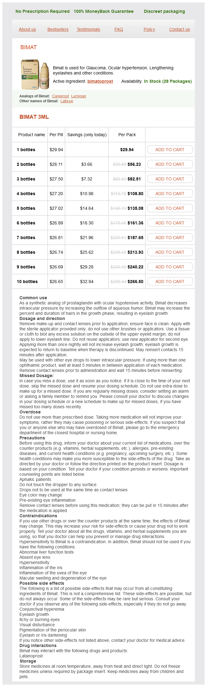
Bimat dosages: 3 ml
Bimat packs: 1 bottles, 2 bottles, 3 bottles, 4 bottles, 5 bottles, 6 bottles, 7 bottles, 8 bottles, 9 bottles, 10 bottles
Cheap bimat 3 ml buy on-line
Final discount of the direct lateral fragments of the lateral process is provisionally mounted by Kirschner wires. Isolated lateral process fractures are greatest fastened by interfragmentary mini-screw fixation. After preparation and draping, the talus is placed in two or three Bacitracin and saline baths and gently scrubbed earlier than reimplantation. Initially, two 4-mm exterior fixation half-pins are inserted bicortically into the anterior distal third of the tibia. A 4-mm half-pin is inserted bicortically into the bottom of the primary and fifth metatarsals. Finally, a 4-mm half-pin, or transfixion pin, is advanced from medial to lateral, bicortically, by way of the tuberosity of the calcaneus. An external fixation rod connects the 2 metatarsal base pins, forming a midfoot unit. It is important to leave extra rod on every finish of the midfoot unit for further tibial-rod attachment. A second, long external fixation rod is hooked up to the lateral end of the midfoot rod and related to the proximal tibial pin, controlling ankle varus and aiding dorsiflexion. Finally, external fixation rods are added, connecting the calcaneal pin to each the medial, midfoot rod and either tibial half-pin for elevated body rigidity and probably to distract the subtalar joint. Attachments: distal third of the tibia, calcaneus, base of first and fifth metatarsals. A medial malleolar osteotomy carried out distal to the shoulder of the medial ankle mortise gives poor visualization of the talar dome. Medial screw fixation of talar neck fractures, not countersunk within the talar head, generally achieves poor fixation in the metaphyseal bone. Do not use posterior-to-anterior screw fixation, with the affected person in the prone position, for a displaced talar neck fracture. The tuberosity of the tarsal navicular is a crucial landmark figuring out the plantar limit to the medial strategy. The surgeon should defend the delicate tissues posterior to the malleolus in the course of the osteotomy. The surgeon should view the Canale picture intraoperatively with C-arm fluoroscopy (flexed knee, everted foot in equinus and C-arm tube directed 15 levels caudad). Return to preinjury status is usually not achieved secondary to posttraumatic arthrosis and joint stiffness, but good functional outcomes are attained even in the most severe cases of talar neck and body fractures. Immediate postoperative remedy requires software of sterile ankle dressings and a well-padded, short-leg dressing with a posterior plaster splint. Rigid inner fixation safely permits early postoperative ankle and subtalar movement. Before hospital discharge, the patient is taught to carry out every day dressing adjustments and utility of a foot and ankle compression Ace wrap to control swelling. The affected person is to put on a detachable short-leg fracture boot and remain completely non-weight bearing for eight weeks. The fracture boot should be worn through the night for the preliminary 3 to four weeks after surgical procedure to forestall an early Achilles tendon contracture. Immediate outpatient bodily remedy after a talus harm routinely results in excessive ache and ankle swelling. However, higher and contralateral lower extremity strengthening may be useful through the preliminary, subacute, 6-week postinjury period. This may be seen as a end result of solely the medial deep deltoid blood supply to the talar physique stays intact after the injury. The Hawkins signal is an early subchondral radiolucent line indicating blood provide to the body of the talus. Fortunately, patients with isolated regional osteonecrosis of the talar dome rarely expertise late collapse. Currently, the impact of weight bearing on the progression of osteonecrosis is unknown. Protected weight bearing with a patellar tendon-bearing brace to alleviate axial load to the hindfoot and refraining from repetitive-loading sports activities are reasonable early issues to discuss with the affected person. The patient is non-weight bearing for two weeks, performing passive range of movement of the ankle and subtalar joints, isometrics of the leg, and possibly pool remedy.
Order on line bimat
The plan is modified primarily based on fracture stability, gentle tissue injury, and patient reliability. Patients with substantial (over 50%) cortical contact may start weight bearing as tolerated after delicate tissue healing has occurred. It is better to overimmobilize in questionable instances to keep away from malalignment and regain movement later with aggressive physiotherapy. Removal of symptomatic hardware (ie, nails or plate) must be delayed until fracture therapeutic and reworking are full. I prefer to take away elastic nails electively in all sufferers 6 to 9 months after harm, because the nails will turn into fully intramedullary with important continued progress, thus making late removing extraordinarily tough. Myers and coworkers10 reported a significant complication rate in high-energy tibial fractures handled with external fixation, together with delayed union, malunion, leg-length discrepancies, and pin-tract infections. Kubiak and colleagues8 reported 2 delayed unions, 2 malunions, and 3 nonunions in a series of 15 patients managed with external fixation, though these seem to have occurred in open accidents. They reported larger useful scores of their sufferers handled with elastic intramedullary nailing compared to exterior fixation. Operative methods often require additional procedures for removal of pins or outstanding nails or plates. External fixation or versatile intramedullary nailing for femoral shaft fractures in kids: a prospective, randomised examine. Ender nail fixation in lengthy bone fractures: expertise in a stage I trauma heart. Problems of operative and non-operative therapy and healing in tibial fractures. Operative treatment of tibial fractures in youngsters: are elastic secure intramedullary nails an enchancment over exterior fixation Flexible titanium nailing for the therapy of unstable pediatric tibial fracture. Chapter 17 Open Reduction and Internal Fixation of Tibial Tuberosity Fractures Ernest L. There have also been stories of related injuries such as quadriceps tendon injury, cruciate ligament tears, and meniscal injury. Patients with minimally displaced fractures could lengthen the knee, however with apparent discomfort. This is unlike Osgood-Schlatter disease, during which the onset is more persistent and there may be radiographic findings of a continual condition similar to calcification anterior to the secondary center of ossification. The second, or apophyseal stage, happens at age eight to 12 years in ladies and 9 to 14 years in boys. The third, or epiphyseal, stage is when the "tongue" of the apophysis and the epiphyseal bone are continuous. The ages for the third stage are 10 to 15 years for girls and 11 to 17 years for boys. The insertion is lateral to the midline; thus, the fracture fragment is centered lateral to the midline. This is important when considering the strategy for intra-articular visualization. Bleeding from its proximal branches because it retracts into the anterolateral compartment might lead to compartment syndrome. There is a major drive that the quadriceps mechanism is ready to generate, and this overcomes the strength of the epiphysis and the surrounding periosteum. The different mechanism of damage is sudden passive knee flexion whereas the quadriceps is contracted. In nondisplaced fractures the place patients can carry out a straight-leg increase, a long-leg solid may be used for therapy. Even in the nondisplaced fractures, percutaneous screw fixation could enable earlier immobilization and prevent 6 to eight weeks of casting. The table should enable good anterior and posterior views to be obtained with fluoroscopy.
Discount bimat 3 ml buy on line
Extensive calcification with adjustments within the shape and thickness of the cartilaginous cap ought to raise suspicion of a potential chondrosarcomatous transformation. The Taniguchii classification correlates the regional involvement of the forearm with the generalized severity of the illness. These may be particularly helpful to detail the anatomic place relative to gentle tissue structures, or when malignant transformation is suspected. The postoperative appearance of the forearm has been proven to be unrelated to the practical end result. If function is the main concern, the goal of surgery is to preserve or enhance function till reaching skeletal maturity and to not stop the deformities. Some authors5,12,15 advocate an aggressive strategy primarily based on the rationale that forearm deformities are equal to functional impairment. They feel this is the only method to forestall the development or development of deformity within the upper extremity. Symptomatic dislocation of the top of the radius is outlined as interfering with joint movement or inflicting vital ache. Procedures Exostosis excision alone is indicated when a lesion turns into symptomatic or when it alone causes limitation of forearm pronation�supination. If vital forearm deformity is present, exostosis excision is mixed with ulnar tether release with or with out radial osteotomy. Radial osteotomy is carried out in the skeletally mature or nearly skeletally mature affected person, as significant remodeling of the radius is unlikely. If the affected person has important development potential remaining, ulna-tether launch alone can result in impressive correction. The treatment for symptomatic radial head dislocation is often surgical excision once the patient is skeletally mature. In rare situations, nonetheless, exostosis excision with ulnar osteotomy could also be effective in relocating the radial head. Planning of this is essential, as the flexibility to entry the distal ulna is imperative whether the osteochondroma is located on the distal ulna or radius. If the patient has ulnar involvement solely, the incision may be placed on the subcutaneous border of the ulna between the flexor carpi ulnaris and the extensor carpi ulnaris. If the affected person has osteochondroma of each the radius and ulna, the incision has to be modified to enable publicity of both bones in addition to the distal ulna. This is usually carried out by transecting the distal ulna by way of the epiphyseal area, leaving the triangular fibrocartilage complex hooked up to the distal fragment. As stated earlier, within the skeletally immature affected person, release of the tether alone is usually sufficient. In a skeletally mature affected person, a radial osteotomy is carried out after exostoses excision and ulnar-tether launch. Layered closure is then carried out and the extremity is immobilized for 2 weeks, followed by institution of range-of-motion workout routines. In individuals close to skeletal maturity, the surgeon should carry out excision of the exostoses and ulna-tethering release related to epiphysiodesis of the distal radius to avoid any additional development of the deformity. In distraction lengthening of the forearm bones, the bone formation takes longer in comparability with the lower limb due to the dearth of weight bearing. Therefore, one of many disadvantages of this method is that the exterior fixator should be kept on for several months. To improve the bone formation and cut back the chance of fracture on the lengthening website, dynamization methods are really helpful. When the ulna is lengthened, the cordlike portion of the interosseous membrane tends to pull the radius distally. Otherwise, the cordlike portion of the interosseous membrane ought to be dissected to prevent migration of the radius. In case of exostosis excision and ulna-tethering launch, casting is carried out for 4 weeks, followed by range-of-motion exercises and splinting. If an osteotomy was performed, casting is continued until radiographic proof of healing is seen. In sufferers who require surgical procedure, we feel that ulnar-tether launch, with or with out exostoses excision, with or with out radial osteotomy, provides probably the most dependable result with the fewest issues.
Buy bimat 3 ml on line
An anterior-to-posterior drill hole is made through the epiphysis, and the anterior limb of the fascia lata is handed from anterior to posterior, exiting close to the midline posteriorly. Another drill gap that passes through the medial distal femoral epiphysis from anteromedial to posterolateral is made. The ligamentized fascia lata is pulled through the posterior capsule and into the medial femoral epiphyseal tunnel using its main suture. It is fastened in place with a biotenodesis absorbable screw (Arthrex) after tensioning in flexion. The posterior aspect of three: Anterior limb of fascia lata passed under adductor magnus tendon If the patella has a set lateral subluxation or dislocation, a modified Langenski�ld patellar reconstruction is performed earlier than the knee ligamentous reconstruction (intra-articular and extra-articular). Reverse MacIntosh (extra-articular posterior collateral ligament) process is carried out by passing the anterior limb of the fascia lata graft beneath the patellar tendon and through a window created within the medial joint capsule. The graft is then handed through a subperiosteal tunnel under the adductor magnus tendon, looped back onto itself, and secured with nonabsorbable suture. The synovium is then carefully dissected free of the undersurface of the quadriceps muscle proximally and from the patellar tendon distally. Medially, the capsule is incised proximally in a longitudinal fashion, separating the vastus medialis muscle from the vastus intermedius muscle. The quadriceps and patellar tendon are left connected to the patella and the entire extensor mechanism could be shifted medially. Initial step in the modified Langenski�ld reconstruction is to carry out a medial and lateral capsulotomy. The knee joint capsule is dissected away from the synovium medially and laterally. The synovium is also dissected free from the quadriceps tendon and the patellar tendon. Synovium is launched from the patella circumferentially, leaving the quadriceps and patellar insertions intact. The gap within the synovium is closed longitudinally with absorbable suture, leaving the patella with the quadriceps and patellar attachments extra-articular. The Grammont procedure is performed as described above, and the patellar tendon is shifted medially. Knee is positioned in full extension, and the new place for the patella is marked on the synovium. If an extra-articular posterior collateral ligament (reverse MacIntosh) procedure is carried out, the Langenski�ld reconstruction is accomplished earlier than the ligamentous reconstruction. Fascia lata graft passes by way of the advanced medial capsule and is sutured onto itself. If the popliteal angle is greater than 10 degrees, the biceps femoris tendon and medial hamstrings should be released. If the preparatory surgery has not been carried out and the Dega osteotomy is needed, it ought to be mixed with excision of the fascia lata, superknee reconstruction, or both. Alternatively, the gentle tissue releases can be performed simultaneously with the lengthening process. Placement of Femoral Fixator An arthrogram of the involved knee is obtained under fluoroscopy. In the lateral view, the femoral condyles are rotated until they superimpose each other. The center of rotation is the intersection of the posterior cortical line and the distal femoral physeal line. The external fixator rail is aligned with the femur in the sagittal view and essentially the most proximal half-pin is inserted on the degree of the bottom of the greater trochanter (this pin ought to be distal to the apophysis). The lateral view is obtained, and the posterior elements of the femoral condyles are superimposed to create the perfect lateral view. The hinge reference wire is inserted on the intersection of the posterior femoral cortical line and the distal femoral physis. The first distal half-pin is positioned on the anterior row one hole proximal to the hinge-axis pin.
Purchase bimat mastercard
Physical examination begins with a radical assessment of the pores and skin for areas of compromise, notably in clavicle fractures. Swelling from a sternoclavicular dislocation might mask initial displacement, so this area ought to be palpated for pain or crepitance. A neurologic examination to embrace the brachial plexus distribution, in addition to a vascular examination of the arm, is critical. Neurologic injury along side fracture could signify ongoing compression (ie, sternoclavicular dislocation) and will have an effect on prognosis. The "serendipity view" is useful in instances of medial clavicle fracture or dislocation. In a special patient, apical oblique radiograph taken with fluoroscopy earlier than discount of a posterior sternoclavicular dislocation. In another patient, three-dimensional contrast-enhanced computed tomography picture of left posterior sternoclavicular dislocation. Axial view of the same affected person as in D, demonstrating the harm to be a physeal fracture. The epiphysis (arrow) stays in place, whereas the metaphysis (*) is posteriorly displaced, immediately anterior to the vasculature. For proximal humeral fractures with acceptable alignment, therapy consists of sling management for consolation for several weeks, followed by a home range-of-motion program and return to actions in 6 to eight weeks. Immobilization can usually be discontinued after 3 weeks, with return to sports by 6 to eight weeks. While surgical management of distal clavicular physeal accidents has been advocated by some, the majority of patients do nicely with remedy just like clavicular shaft fractures. Older children can be handled as adults, and the surgical remedy of those accidents is mentioned elsewhere. Interposition of the biceps tendon has been famous to be the most common cause of a failed closed reduction,3 however different authors disagree. It is necessary to rule out concomitant glenoid fracture or dislocation before surgical procedure. Positioning Proximal Humerus Fractures For proximal humeral fractures, the patient is positioned in a modified beach-chair place, with the back elevated roughly 30 levels. This allows the patient to be slid slightly over the sting of the table to allow full access to the shoulder girdle. A chest pad hooked up to the table prevents the patient from being pulled off the table inadvertently when traction is utilized to the arm. Alternately, a sheet could be wrapped across the torso and secured by an assistant on the alternative facet of the table. Operative treatment of proximal humeral fractures will subsequently be discussed, as nicely as reduction of posteriorly displaced fracture-dislocations of the sternoclavicular joint. For physeal or metaphyseal fractures of the proximal humerus, operative remedy ought to be thought of for fractures with unacceptable residual displacement. Because of open progress plates, commonplace plate-fixation techniques are rarely indicated. Failure to obtain a passable closed discount could require an open reduction, and surgeons ought to be conversant in this technique as properly. Sternoclavicular Dislocation Patients with posterior sternoclavicular dislocations ought to be positioned supine with a roll between their shoulders to allow hyperextension of the shoulder girdle. It is useful to position the patient with the operative facet near the sting of the desk. Without transferring the arm, the picture can be rotated to acquire an axillary lateral view. The surgeon is making use of strain to reduce the apex anterior angulation, whereas maintaining traction with the left hand. Under the drapes, a chest pad prevents the patient from being pulled off the table with traction; a sheet wrapped around the torso and held by an assistant would accomplish the identical purpose. One of two potential incisions is made: the standard incision made within the deltopectoral interval, which is helpful for broad displacement, or a more cosmetic incision within the axilla.
Narikela (Coconut Oil). Bimat.
- How does Coconut Oil work?
- Dosing considerations for Coconut Oil.
- What is Coconut Oil?
- Are there safety concerns?
Source: http://www.rxlist.com/script/main/art.asp?articlekey=97038
Buy generic bimat
The relationship between the foramen magnum, atlas, and odontoid can be determined in lateral radiographs. The upper tip of the odontoid ought to normally be 1 cm under the anterior margin of the foramen magnum. If the effective sagittal diameter of the canal (length of the line) is lower than 19 mm, neurologic symptoms happen. This line is doubtless considered one of the finest for detecting basilar impression as a result of the osseous landmarks can normally be seen at all ages. The inferior extension of the line ought to be in contact with the posterior tip of the odontoid. The Wiesel-Rothman line is drawn connecting the anterior and posterior arches of the atlas. The McRae line connects the anterior rim of the foramen magnum to the posterior rim. The Chamberlain line is drawn from the posterior margin of the exhausting palate to the posterior margin of the foramen magnum. The McGregor line is drawn from the most caudal level of the occipital projection to the posterior edge of the onerous palate. A line is drawn connecting the anterior and posterior arches of the atlas (line 1�2). Two strains are drawn perpendicular to this line, one through the basion and the other via the posterior margin of the anterior arch of the atlas (line 3). A change within the distance (x) between these strains of greater than 1 mm in flexion and extension signifies increased abnormal translational motion. A line is drawn from the basion (B) to the posterior arch of the atlas (C) and a second line from the opisthion (O) to the anterior arch (A) of the atlas. For occipitoatlantal instability and instability with congenital or acquired (because of removal of tumor or laminectomy) defects of posterior components, we developed two techniques of occipitocervical arthrodesis at our institution. Rib graft technique Iliac graft method Many strategies of atlantoaxial fusion utilizing cables, transarticular screws, plates, and bone graft materials have been described. For isolated atlantoaxial instability, we commonly prefer the Gallie method, the Mah modified Gallie approach, and the Brooks sublaminar wire method. Somatosensory evoked potentials, transcortical motorevoked potentials, and electromyography are the preferred neurologic monitoring modalities. Flexible fiberoptic intubation with handbook in-line axial stabilization ought to be thought-about to reduce cervical motion throughout intubation maneuvers. Especially patients with congenital syndromes related to higher cervical spine instability should have periodic scientific and radiographic examinations until maturity. Patients and fogeys ought to be educated about the analysis and pure history of the dysfunction and encouraged to report any symptoms as quickly as they happen. As beforehand described, periodic statement ought to be carried out and if any development is seen, the affected person ought to be prepared for acceptable surgical stabilization when indicated. Documented vital instability at the atlanto-occipital or atlantoaxial joints is an indication for posterior arthrodesis of occiput�C2 and C1�C2 arthrodesis, respectively. In some congenital issues similar to Morquio syndrome, progression of the instability is frequent; in these cases prophylactic fusion should be thought of before neurologic symptoms happen. If the scientific signs persist or neurologic symptoms are starting to occur in the setting of serious instability, surgical therapy is indicated. Patients with progressive symptomatic seg Positioning After induction of common anesthesia within the supine position, a halo ring is applied, and the affected person is fastidiously turned to the inclined place. Lateral fluoroscopy or radiography ought to be carried out earlier than beginning the procedure to verify the alignment of the occiput and cervical spine. Approach the posterior midline incision is produced from the occiput to the meant distal fusion site (usually C2) for occipitocervical arthrodesis. Excessive lateral dissection should be avoided to stop any damage to the vertebral artery, which runs in a serpentine course in relationship to the C2 vertebra. For occipitocervical arthrodesis, the dissection is carried proximally to the occipital protuberance and distally to the extent planned to embody the fusion. For atlantoaxial arthrodesis, the dissection is began from the decrease occiput and the surgeon identifies the posterior arch of the atlas, the bifid spinous processes, and the lamina of the axis. The surgical exposure of vertebra is proscribed as much as the meant level of fusion to prevent unintentional inclusion of the adjoining degree in the fusion mass.
Discount bimat 3 ml otc
If an open strategy is used for reduction, screws positioned posterior to anterior could also be placed across the fracture web site. When application of a posterior or antiglide plate is chosen, placement of a lag screw is optionally available. Because of the biomechanical properties of this plate assemble, this lag screw is optional. If a posterior plate is utilized, the proximal screws are positioned bicortically from posterior to anterior, each proximal and distal to the fracture. The lag screw may be placed from posterior to anterior with bicortical buy achieved in each screw. Lateral displacement that permits more than a few millimeters of tibiofibular widening is considered pathologic and a sign for syndesmotic fixation. The lateral radiograph ought to be scrutinized to assess the relationship of the fibula to the articular surface of the ankle joint. In basic, on a real lateral view of the ankle, the tip of the fibula ought to be anterior to the posterior border of the diaphyseal tibia, but comparisons to the contralateral ankle can be assessed. Fixation selections vary from one or two screws, with three or four cortices drilled and 3. Although the scale and number of screws remain controversial, some parameters are agreed on. If a posterior plate is used, the syndesmosis screw will doubtless be placed exterior the plate on the lateral cortex. The Cotton take a look at is carried out following fibular fixation by pulling laterally with a hook or clamp to assess the integrity of the syndesmosis. Reduction and stabilization of the syndesmosis is achieved with a clamp positioned across the distal tibiofibular joint and a bump positioned under the leg. The distal tibiofibular anatomic relationship is assessed and the contralateral ankle is used for comparison. Supplementary Kirschner wires Multiple syndesmosis screws Posterior-placed fibula plate Locked plate Careful intraoperative radiographic assessment is essential. Laterally placed fibular hardware is associated with a higher incidence of painful hardware. Posteriorly placed fibular fixation is associated with a better incidence of peroneal tendinitis. Identification and safety of the superficial peroneal nerve within the anterior flap. Patients are then placed right into a removable practical brace that enables them to start early active-assisted and passive vary of ankle motion. At 6 weeks patients are progressed to weight bearing as tolerated based mostly on radiographic criteria. Weight bearing can be delayed for slow healing and presence of a syndesmotic screw. Patients are restricted from operating an automobile for 9 weeks following right-sided ankle fracture. They have significant improvement in perform in contrast with 6 months after surgical procedure. Younger age, male intercourse, absence of diabetes, and a decrease American Society of Anesthesia class are predictive of functional recovery at 1 yr following ankle fracture surgical procedure. It is essential to counsel patients and their families on the anticipated consequence after harm with regard to practical recovery. Looking particularly at elderly sufferers (older than 60 years), practical outcomes steadily improved over 1 12 months of follow-up, albeit at a slower fee than in the youthful patients. Our results counsel that operative fixation of unstable ankle fractures within the aged can present a reasonable functional end result at the 1-year follow-up. Ankle stress check for predicting the need for surgical fixation of isolated fibular fractures. Functional consequence of surgery for fractures of the ankle: a prospective, randomised comparison of management in a forged or a functional brace. Lower-extremity operate for driving an vehicle after operative therapy of ankle fracture. Anatomical basis of variability in accidents of the medial malleolus and the deltoid ligament.

Cheap bimat 3 ml visa
Complications included untimely consolidation in 4 sufferers, malunion of greater than 10 degrees in two sufferers, and residual limb size inequality (less than 2 cm) in two patients. There have been no stories of osteomyelitis, ring sequestra, neurologic or vascular compromise, compartment syndrome, hypertension, or hip or knee dislocations of their series. These outcomes show a significant improvement over previous reports of earlier methods of femoral lengthening by method of greater lengthening, simultaneous deformity correction, and fewer main issues. Stanitski et al24 reported tibial lengthening for 62 tibiae in 52 patients utilizing the Ilizarov approach. Twenty-eight (22%) sufferers required unplanned procedures, which included osteotomy for malunion or deformation of the regenerate and Achilles tendon for persistent equinus contracture. Distraction Period Pin tract an infection initially is treated with a brief course of oral antibiotics (7 to 10 days) and applicable pin tract care. If infection persists, consider intravenous antibiotics or removing of the wire or half-pin with curettage of the contaminated web site. Premature consolidation could additionally be because of incomplete osteotomy, sluggish distraction fee, or incorrect path of distraction. Neurologic signs could come up in the type of altered sensation or weakness of the muscle. The wire or half-pin is eliminated if direct contact or irritation of the nerve is suspected. Stretching of the nerve with fast distraction may lead to nerve damage, and it might be necessary to decrease the speed of distraction or even stop distraction briefly. Treatment consists of growing the number of bodily remedy sessions and use of dynamic splinting, particularly to prevent equinus contracture. Frame modifications may be required to appropriate the deformity and preserve a neutral mechanical axis. Paresthesia, ache with passive stretch, and ache out of proportion to the surgical process are medical indicators of compartment syndrome. Compartment pressures ought to be measured, and compartments ought to be launched as needed. The femora that gained more size (expressed as proportion of unique length) had poor healing indices. In tibia the mean tibial lengthening was 9 cm or 41% of the original tibial length. Delayed consolidation of regenerate might respond to electrical or ultrasound bone stimulator. This can be prevented by body dynamization previous to frame elimination and guarded weight bearing. Assess the regenerate bone clinically and radiographically at the time of body elimination. Consider use of a solid or brace and protected weight bearing in the setting of questionable bone regenerate. Stress fracture can happen either on the web site of half-pins, especially when the half-pin size exceeds one third the diameter of the cortex, or via the regenerate bone. Fracture is treated with reapplication of the body, casting, intramedullary rod fixation, or plate utility. Hydroxyapatite coating of external fixation pins to lower axial deformity during tibial lengthening for short stature. Femoral lengthening using the callotasis technique: examine of the complications in a collection of 70 instances in kids and adolescents. Statistical analysis of axial deformity during distraction osteogenesis of the tibia. Distraction osteogenesis of the decrease extremity with use of monolateral exterior fixation. Problems, obstacles and problems of limb lengthening by the Ilizarov approach.
Real Experiences: Customer Reviews on Bimat
Avogadro, 63 years: The implant is removed when the plafond is horizontal, no matter fibular length. An upper limb neurologic examination must be performed, including an evaluation of tone within the intrinsic muscles of the hand, because the thumb-in-palm deformity of cerebral palsy can be confused with set off thumb.
Kafa, 26 years: The surgeon must know the implications of every of those releases and restore or reconstruct them correctly on the end of the procedure. Proximal femur reconstruction is carried out for metaphyseal�diaphyseal lesions that stretch beneath the lesser trochanter, trigger extensive cortical destruction, and spare at least 3 cm of the distal femoral diaphysis.
8 of 10 - Review by F. Rozhov
Votes: 326 votes
Total customer reviews: 326
References
- Chopra A, Lavin P, Patwardhan B, Chitre D. A 32- week randomized, placebo- controlled clinical evaluation of RA- 11, an Ayurvedic drug, on osteoarthritis of the knees. J Clin Rheumatol 2004; 10(5):236-45.
- Chen GL, Yang L, Rowe TC, et al. Nonintercalative antitumor drugs interfere with the breakage-reunion reaction of mammalian DNA topoisomerase II. J Biol Chem 1984;259(21):13560-13566.
- Gilsanz V, Lozano G, Jimenez J: Renal hydatid cysts: communicating with collecting system, AJR Am J Roentgenol 135(2):357n361, 1980.
- Kelley, P., & Clifford, P. (1997). Coping with chronic pain: Assessing narrative approaches. Social Work, 42(3), 266n277.
- Cuzick J, Swanson GP, Fisher G, et al: Prognostic value of an RNA expression signature derived from cell cycle proliferation genes in patients with prostate cancer: a retrospective study, Lancet Oncol 12:245, 2011.
- Kharasch ED: Perioperative COX-2 inhibitors: Knowledge and challenges [Editorial], Anesth Analg 98:1, 2004.