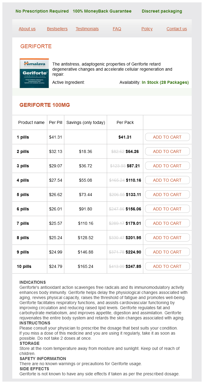
Geriforte dosages: 100 mg
Geriforte packs: 1 pills, 2 pills, 3 pills, 4 pills, 5 pills, 6 pills, 7 pills, 8 pills, 9 pills, 10 pills
Cheap geriforte master card
In many instances with recessive inheritance, two totally different mutations are present in one individual compound heterozygosity. Plexiform neurofibromas: may diffusely involve nerve, muscle, connective tissue, vascular components, and overlying pores and skin. Moderate to severe skeletal fragility; bone biopsy reveals lamellae with fish like look and excessive osteoid. Major and minor criteria of the next organ methods are evaluated in the affected person: ocular, skeletal, integumental, respiratory, and cardiovascular. Major criteria in two systems with involvement of a 3rd system are wanted to make an unequivocal diagnosis. Increased risk of malignancy at leukoplakia sites (35% of patients) � Other findings: cutaneous atrophy, hyperhidrosis of the palms and soles, telangiectasias, cracking, fissuring, bullae formation, lack of dermal ridges, hair tufts with keratotic plugs on the limbs and keratinized basal cell papillomas, alopecia, amyloidosis � Non-mucocutaneous options: � Pulmonary disease (20% of patients) � Ophthalmic manifestations: epiphoria as a result of nasolacrimal duct blockage, conjunctivitis, blepharitis, pterygium formation, ectropion, strabismus, cataracts and optic atrophy. Bone marrow failure leading to peripheral cytopenias (75% of patients develop pancytopenia, responsible for demise in 70% of patients). Gardner Syndrome � Autosomal dominant; 25% of circumstances occur as a result of spontaneous mutations. Influenza, or fungus � Osteopenia with bone fractures and scoliosis � Retention of primary enamel and other dental anomalies � Job syndrome is a subgroup with hyperextensible joints. The defect within the syndrome marked by clumped melanosomes within the fetal hair shaft, silvery hair, irregular platelets, and recurrent infections involves: A. Your affected person presents with telangiectasias, photosensitivity, acral keratoses, alopecia, and cataracts since age 5. If you think a patient has Neimann-Pick illness, you would possibly examine the skin to look for what lesion Some porphyrias can have acute attacks precipitated by various medication, infections, alcohol, dieting, and being pregnant. Sickle-shaped beanbag calcifications within the hippocampus and eyelid ("string of pearls") are related to Urbach-Wiethe syndrome (lipoid proteinosis). N-peptidase cleaves the N-terminus of collagen Type I within the extracellular area, the place tropocollagen is formed to then be included into mixed fibrils. Unlike many different icthyoses, lamellar icthyosis persists and remains extreme past childhood. Characteristic giant, squarish, "dry riverbed" scales are most outstanding within the flexures. Ataxia-telangiectasia, or Louis-Bar syndrome, is neurodegenerative and immune system dysfunction associated with infections and malignancies, particularly lymphomas and leukemias. Decreased melanosome transfer results in clumped melanosomes visible in the medulla of fetal hair shafts, useful for diagnosis. Rothmund-Thomson syndrome (poikiloderma congenitale) patients have acral verrucous keratoses that may evolve into squamous cell carcinomas. Acute attacks happen when hemoglobin or cytochromes, the end merchandise of the porphyria pathway, are depleted. Acute attacks manifest with stomach pain, peripheral neuropathy, confusion, seizure, tachycardia, and hypertension. Tuberous sclerosis 1 and a couple of result from defects in hamartin or tuberin, respectively. Epidermolysis bullosa: medical epidermiologic, and laboratory advances, and the findings of the National Epidermolysis Bullosa Registry. Epidermolysis Bullosa: scientific epidermiologic, and laboratory advances, and the findings of the National Epidermolysis Bullosa Registry. Connective Tissue, Premature Aging, and Photosensitive Disorders Andiran N, Sarikayalar F, Sara�lar M, Cag lar M: Autosomal recessive form of congenital cutis laxa: more than the medical appearance. Di Cataldo A, Haupt R, Fabietti P, Schiliro G: Is intensive follow-up for early detection of tumors effective in youngsters with Beckwith-Wiedemann syndrome Frevel T, Rabe H, Uckert F, Harms E: Giant cavernous haemangioma with Kasabach-Merritt syndrome: a case report and evaluation. Rodriguez-Revenga L, Iranzo P, Badenas C, Puig S, Carri� A, Mil� M: A novel elastin gene mutation resulting in an autosomal dominant type of cutis laxa. Kawamura A, Ochiai T, Tan-Kinoshita M, Suzuki H: BuschkeOllendorff syndrome: three generations in a Japanese household.
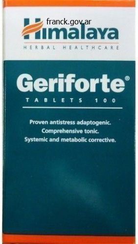
Geriforte 100 mg order overnight delivery
Sternocostal joints or chondrosternal joints between costal cartilages and the sternum. The our bodies, laminae, transverse processes and spinous processes of adjoining vertebrae are also united by a selection of ligaments. The decrease surface of the body of 1 vertebra articulates with the superior floor of the body of the next vertebra. The construction of a joint between any two vertebral bodies corresponds to that of a typical symphysis. The bony surfaces forming the joint are lined by skinny layers of hyaline cartilage. The two plates of hyaline cartilage are united to each other by a thick intervertebral disc (17. Each disc consists of an outer part called the annulus fibrosus, and an internal half the nucleus pulposus. In the younger, the nucleus pulposus is delicate and gelatinous, but this materials is steadily replaced by fibrocartilage. With advancing age, nonetheless, the annulus fibrosus turns into weak and it then becomes potential for the nucleus pulposus to burst via it. The thickness and form of intervertebral discs is different in numerous elements of the vertebral column. The discs are thinnest in the upper thoracic area of the vertebral column (which is least mobile), and thickest within the lower lumbar region. In the cervical and lumbar regions, the discs are thicker in entrance than behind (and this fact is partially answerable for the forward convexity of the vertebral column in these regions). Intervertebral discs constitute about one fifth of the size of the vertebral column. They transmit weight, act as shock absorbers, and supply resilience to the spine. A prolapsed nucleus pulposus often passes backwards and laterally and should press upon nerve roots emerging from the spinal twine at that degree. Disc prolapse occurs most incessantly in the lumbosacral area and leads to pain shooting down the back of the thigh and leg. Each vertebra has four articular processes (or zygapophyses): right and left superior, and proper and left inferior. The inferior articular sides of one vertebra articulate with the superior articular sides of the next decrease vertebra forming a sequence of zygapophyseal joints. In the cervical and thoracic region, the articular sides are flat: these are, therefore, airplane joints. Ligaments Connecting Adjacent Vertebrae Adjoining vertebrae are connected by quite a few ligaments. Anterior longitudinal ligament passing from the anterior floor of the physique of 1 vertebra to that of one other. Posterior longitudinal ligament passing from the posterior floor of the physique of one vertebra to that of another. They cross from the lower border of the lamina of one vertebra to the higher border of the lamina of the subsequent lower vertebra. They act as brakes stopping undue separation of laminae during flexion of the vertebral column. By limiting actions, they most likely shield intervertebral discs from undue compression and consequent harm. The joint between the atlas and the axis, and the joint between the atlas vertebra and the occipital bone have special options that shall be studied in the head and neck. Movements between adjoining vertebrae take place simultaneously at the three joints connecting them. However, when the actions between varied vertebrae get added collectively, the total motion turns into considerable. The vertebral column can be bent forwards (flexion), backwards (extension) and to one side (lateral flexion). On the entire, the cervical and lumbar regions are much more flexible than the thoracic region. Stability of the thoracic area of the vertebral column facilitates respiratory movements.
Syndromes
- Damage to the stomach, esophagus, liver, or small intestine. This is very rare.
- You have pain in the lower abdomen and pelvis, and other tests suggest there is fluid in the area
- Albumin level
- Convulsions
- Infant test or procedure preparation (birth to 1 year)
- Is there a lot of bleeding?
Order geriforte with a visa
Sympathetic postganglionic fibres meant for the dilator pupillae normally travel through the long ciliary nerves. Other structures within the eye that could be infected are the iris (iritis), the ciliary body (cyclitis), and a mix of each these (iridocyclitis). Corneal ulcers can also be attributable to harm or by overseas our bodies that enter the eye. Injuries to the cornea may find yourself in corneal opacities that can result in blindness. The so-called eye transplantations which are marketed a lot in the lay press are actually corneal transplants. Alterations in the curvature of the cornea can lead to an error of refraction called astigmatism (see below). Vision could be restored by removing the lens, and that is one the most typical operations accomplished on the eyeball. In myopia (near sightedness), the picture comes into focus in front of the plane of the retina. Hypermetropia is a situation by which the image comes to a focus behind the airplane of the retina. Presbyopia is a condition attributable to decreased elasticity of the lens in individuals over forty years old. Apart from diagnosis of diseases of the retina itself, such examination additionally helps in assessing the standing of patients in ailments like hypertension and diabetes. The retina consists of a nervous layer (the retina proper) and an outer layer of pigment cells. Sometimes, the nervous layer will get detached from the pigment layer (detachment of the retina). A vestibular half which supplies info to the mind relating to the place and actions of the head. The internal finish of the external acoustic meatus is closed by a skinny membranous diaphragm known as the tympanic membrane. The center ear is a small area placed deep throughout the petrous part of the temporal bone. It is also called the tympanum (from which we get the adjective tympanic applied to buildings linked with the middle ear). The cavity of the middle ear is continuous with that of the nasopharynx through a passage known as the auditory tube. Within the cavity of the middle ear, there are three small bones which are collectively known as the ossicles of the ear. The inner ear is in the form of a cavity within the petrous temporal bone having a really complicated shape. Specialised finish organs within the cochlea act as transducers that convert the mechanical vibrations into nervous impulses. These impulses journey through the cochlear part of the vestibulocochlear nerve to reach the mind. Actual notion of sound takes place within the auditory (or acoustic) areas in the cerebral cortex. Chapter 44 Orbit, Eye and Ear 955 We will now consider the different components of the ear one after the other. The cartilage of the auricle is continuous with that of the exterior acoustic meatus. The auricle has an external surface dealing with laterally, and an internal or cranial floor that lies in opposition to the side of the top. The pores and skin over the exterior surface is steady with the pores and skin lining the exterior acoustic meatus. Part of it passes forwards to turn out to be continuous with the skin over the parotid gland. The skin over the cranial surface passes backwards to turn out to be continuous with the pores and skin lining the pinnacle behind the auricle. The cartilage of the auricle is curved on it self in a sophisticated manner in order that a variety of elevations and depressions are produced.

Buy discount geriforte online
However, it is probably not attainable to see the upper a part of the rectum with a proctoscope. In passing a sigmoidoscope into the rectum the curvatures of the rectum and the presence of transverse folds inside it has to be remembered. Spread of a rectal carcinoma is often gradual but it could possibly in the end invade surrounding constructions together with a. While the decrease part of the rectum is directed downwards and forwards, the anal canal is directed downwards and backwards. The anorectal junction lies on the stage of the pelvic diaphragm (formed right here by the levator ani muscles). The rectum lies above the pelvic diaphragm within the true pelvis, whereas the anal canal lies beneath the diaphragm in the perineum. The decrease aperture of the anal canal (or anus) is within the type of an anteroposterior slit, the best and left partitions being in apposition. Posteriorly, the anal canal is separated from the coccyx by a mass of fibromuscular tissue that is called the anococcygeal ligament (or body). The perineal physique separates the anal canal from the membranous urethra and the bulb of the penis in the male (33. The decrease ends of the anal columns are united to one another by quick transverse folds of mucous membrane. The anal valves together form a transverse line that runs all round the anal canal. In distinction, the half below the line is derived from a floor depression referred to as the proctodaeum, and its lining epithelium is ectodermal. In early fetal life the two components are separated by the anal membrane which subsequently disappears. Remnants of this membrane could additionally be current in the type of small projections from the anal valves. The mucosa has a bluish appearance due to a dense venous plexus that lies between it and the muscle coat. The epithelium of the bottom part resembles that of true skin in which sebaceous and sweat glands are current. The Anal Musculature the anal canal is surrounded by a quantity of sphincters which are as follows (33. The inner anal sphincter is fashioned by thickening of the round muscle coat of the gut. When a finger is placed within the anal canal a distinct intersphincteric groove can be palpated between the decrease end of the inner sphincter and the upper margin of the subcutaneous exterior sphincter. In part the subcutaneous a half of the exterior sphincter appears like a transverse band (33. Most of fibres of the sphincter are arranged within the type of rings round the anal canal, but a few of them be a part of the perineal body anteriorly, and the anococcygeal ligament posteriorly. The superficial part of the exterior sphincter lies external to the lower part of the interior sphincter between the degrees of the pectinate line and the white line (33. The fibres of this half are attached posteriorly to the coccyx and anteriorly to the perineal body. The deep a part of the exterior sphincter lies exterior to the higher half of the inner sphincter (above the level of the pectinate line). Most of its fibres run circularly across the anal canal, but a few of them turn into steady anteriorly with the superficial transverse perinei muscular tissues, and posteriorly with the anococcygeal ligament. The following further details concerning the musculature of the anal canal are worthy of observe. The anorectal junction is closely related to the puborectalis part of the levator ani muscle (33. The fibres of the puborectalis form a sling that keeps the anorectal junction pulled forwards, thus sustaining the angle between the rectum and the anal canal. The fibres of the puborectalis mingle intimately with the upper part of the internal anal sphincter, and with the deep a part of the exterior sphincter to type a prominent ring of muscle around the anorectal junction. This ring, which can be palpated by a finger positioned in the anal canal, is known as the anorectal ring. The integrity of this ring is of great useful importance as damage to it ends in incontinence of feces.
Buy geriforte 100 mg free shipping
Dislocation and fracture of the vertebral column are very critical because of injury to the spinal twine. In demise by hanging, the dens (of the axis) dislocates backwards (by tearing via the transverse ligament of the atlas), and crushes the decrease medulla and the spinal wire. A cervical vertebra (usually the atlas) might slip forwards over the following vertebra even in the absence of harm (cervical spondylolisthesis). The other bones of the skull are firmly united to each other at joints called sutures. Its upper and posterior half contains a large cranial cavity in which the mind lies. Anteriorly, and inferiorly, the cranium types the skeleton of the face together with the walls of the orbits (in which the eyeballs lie), the cavity of the nostril, and the upper a half of the cavity of the mouth. The greater part of the roof and side walls of the cranial cavity are shaped by the best and left parietal bones. Their anterior margins join the frontal bone on the coronal suture that runs transversely across the vault. The level the place the coronal and sagittal sutures meet is recognized as the bregma, while the purpose where the sagittal suture meets the lambdoid suture is called the lambda. In the fetal skull (and for a couple of months after birth) there are gaps within the bones of the cranium in these conditions, these being stuffed by membranes. Skull Seen from behind When we view the cranium from behind we see many features seen from the top (36. Now we see more of the occipital bone, and lateral to it we see a small part of the temporal bone. Near the middle of the occipital bone we see a median projection known as the external occipital protuberance. Extending laterally from the protuberance we see a curved ridge known as the superior nuchal line. Extending downwards (and forwards) from the protuberance we see a median ridge known as the exterior occipital crest. A little above the superior nuchal traces we see the best nuchal strains (running parallel to the former). Lateral to the orbit we see a half of the temporal bone (purple) and the zygomatic bone (blue). The lateral margin of the orbit is formed by the zygomatic means of the frontal bone, above; and by the frontal strategy of the zygomatic bone, under. The medial margin of the orbit is shaped by the nasal means of the frontal bone, above; and by the frontal process of the maxilla, inferiorly. A little under the orbital margin, the anterior floor of the maxilla reveals the infraorbital foramen. Through the nasal aperture we will see some bones that lie inside the nasal cavity. Laterally we see two curved plates, the center and inferior nasal conchae projecting into the nasal cavity. The orbital opening represents the bottom of the pyramid, whereas the apex lies on the posterior finish. Posteriorly, a small part of the roof is formed by the lesser wing of the sphenoid. Anterior to the ethmoid the medial wall is formed by the lacrimal bone, and by the frontal means of the maxilla. The region of the medial wall fashioned by the lacrimal bone and by the maxilla reveals a deep lacrimal groove (for the lacrimal sac). The groove is continuous, inferiorly, with the nasolacrimal canal, the decrease finish of which opens into the nasal cavity. The superior orbital fissure is a prominent cleft that separates the posterior elements of the roof and lateral wall. It is bounded above and medially by the lesser wing of the sphenoid, and under and laterally by the larger wing. The inferior orbital fissure intervenes between the posterior components of the floor and the lateral wall of the orbit. Anteriorly, the groove ends in a canal that passes through the bony substance of the maxilla to open on its floor via the infraorbital foramen. Just in front of the temporal bone we see the greater wing of the sphenoid bone, and additional anteriorly we see the zygomatic bone.
Discount 100 mg geriforte mastercard
This is a strong band of fascia stretching throughout the ventral side of the carpus. The space between the retinaculum and the carpal bones known as the carpal tunnel. It transmits the tendons of the flexor digitorum superficialis and profundus, the tendon of the flexor pollicis longus and the median nerve (6. The retinaculum is attached medially to the pisiform bone, and to the hook of the hamate bone (6. The superficial layer is connected to the tubercle of the scaphoid, and to the tubercle of the trapezium. The deep layer is attached to the trapezium posterior to the groove for the flexor carpi radialis. This is a triangular structure consisting of thickened deep fascia that covers the central part of the palm (See later within the text). These embrace the flexor tendons, the lumbrical muscle tissue and the superficial palmar arch. Digital arteries arising from the arch, and digital branches of the median and ulnar nerves, move distally beneath cowl of the aponeurosis and enter the digits by passing beneath the free distal fringe of the aponeurosis in the intervals between the digits. Each course of once more divides into two slips that diverge to be hooked up to the 2 sides of the digit concerned. In this manner an aperture is formed between the 2 slips, and the tendons of the flexor digitorum superficialis and profundus (for the digit) move through this aperture. The lateral fringe of the aponeurosis is linked to the first metacarpal bone by the lateral palmar septum. The medial edge of the aponeurosis is linked to the fifth metacarpal bone by the medial palmar septum. The fourth and fifth digits stay in a state of flexion at the metacarpophalangeal and proximal interphalangeal joints. It causes wrinkling of the skin over the medial aspect of the palm and should thus help in offering a better grip. Fourth lumbrical from contiguous sides of tendons for ring and little fingers Contd. Insertion 107 Each muscle ends in a tendon that passes backwards on the radial side of one metacarpo-phalangeal joint and is inserted into the lateral basal angle of the extensor expansion for that digit within the following order: 1. Extension of interphalangeal joint of digit concerned Help in nice actions of fingers, as in writing or threading a needle Nerve supply Action Notes 6. Some fibres to dorsal digital expansion flexor retinaculum Abduction of thumb Median nerve (C8, T1) at metacarpophalangeal and carpometacarpal joints. Bases of 2nd and third metacarpals Transverse head: Palmar side of 3rd metacarpal bone (distal two-thirds) 1. Adjoining part of flexor retinaculum Insertion Lateral side of base of proximal phalan of thumb Action Flexion of thumb Nerve Supply Superficial head: Median nerve. Some fibres intodorsal digital growth Opposition of thumb (flexion plus medial rotation) Adducts the thumb from flexed or kidnapped position the movement is forceful in gripping Median nerve. Adjoining part of flexor retinaculum Deep department of ulnar Flexes the fifth nerve (C8, T1) metacarpal bone and rotates it laterally (makes palm hollow) 6. They flex the metacarpo-phalangeal joint and prolong the interphalangeal joints of the digit involved Dorsal Interossei 1. They flex the metacarpo-phalangeal joint and prolong the interphalangeal joints of the digit involved Contd. A palmar interosseus muscle could or will not be inserted into the base of the proximal phalanx 5. Palmar interossei take origin from, and are inserted into the first, second, fourth, and fifth digits (not the third) 109 Dorsal Interossei 1. The third digit provides origin to , and receives insertions of two muscle tissue (one on both sides, medial and lateral) three. A dorsal interosseus muscle is all the time inserted into the bottom of the proximal phalanx of the digit involved 5.
Pignut (Jojoba). Geriforte.
- Are there safety concerns?
- Acne, psoriasis, sunburn, chapped skin, hair loss, and other uses.
- How does Jojoba work?
- What is Jojoba?
- Dosing considerations for Jojoba.
Source: http://www.rxlist.com/script/main/art.asp?articlekey=96612
Order 100 mg geriforte otc
Posterior to the foramen magnum the occipital bone forms a large part of the base of the cranium. Portions of the petrous part, the squamous half and the mastoid a part of the temporal are seen on the bottom of the cranium. We shall now study the options to be seen on each of those bones when the cranium is seen from below. The alveolar process of the maxilla tasks downwards and provides attachment to the higher teeth. The posterior end of each alveolar course of types a backward projection known as the maxillary tuberosity. Within the concavity of the arch formed by the alveolar course of we see the bony palate that separates the nasal cavities (above) from the cavity of the mouth (below). The anterior a part of the palate is shaped by the palatal processes of the right and left maxillae. The part of the alveolar course of bearing the incisor teeth, and together with the adjoining part of the palate known as the pre-maxilla. Lateral to the alveolar arch we see the inferior side of the zygomatic process of the maxilla because it passes laterally to meet the zygomatic bone. The posterior borders of the horizontal plates of the palatine bones are free and kind the posterior margin of the onerous palate. A little in entrance of the posterior border we see a curved ridge known as the palatine crest. The a part of the palate shaped by the palatine bone shows the higher and lesser palatine foramina. The greater palatine foramen lies on essentially the most lateral part of the horizontal plate, just medial to the final molar tooth. The lesser palatine foramina, normally two, are current simply behind the higher palatine foramen. Just above the posterior margin of the exhausting palate there are two posterior nasal apertures. Each aperture is bounded, below, by the posterior edge of the horizontal plate of the palatine bone. The lateral wall of the aperture is shaped by one other a half of the palatine bone that known as the perpendicular plate. The perpendicular plate of the palatine bone and the medial pterygoid plate of the sphenoid bone together type the lateral wall of the area the place the nostril and pharynx meet. The sphenoid bone is giant, extending across the entire width of the bottom of the cranium and increasing additionally onto the lateral wall of the vault. When considered from under the body of the sphenoid is seen in the roof of the posterior a half of the nasal cavity and of the adjoining nasopharynx. Posteriorly, the body of the sphenoid is instantly continuous with the basilar part (or body) of the occipital bone. The pterygoid process tasks downwards from the junction of the physique of the sphenoid with the larger wing. Anteriorly, the pterygoid process is fused to the posterior aspect of the maxilla in its center half. The medial pterygoid plate is directed backwards in order that it has medial and lateral surfaces, and a free posterior border. The lower finish of the posterior border is extended downwards and laterally to type the pterygoid hamulus. At its higher end its lateral floor turns into continuous with the infratemporal surface of the greater wing (36. The anterior margin of the infratemporal floor of the sphenoid bone is separated from the maxilla by the inferior orbital fissure. Laterally, the infratemporal surface is separated from the temporal floor by the infratemporal crest. The posterior margin of the lateral part of the infratemporal surface articulates with the infratemporal surface of the squamous a part of the temporal bone.

Buy 100 mg geriforte otc
The catheter can be used for injecting a suitable distinction medium into the artery. A radiograph taken instantly after the injection shows the branches of the artery into which the dye is injected. A suitable catheter handed via the aorta can attain the opening of a coronary artery. Dye injected can outline the coronary artery and any factors of narrowing can be seen (left cardiac angiography). Catheters launched into an artery may also be used for recording pressures inside the vessel, and for obtaining samples for evaluation of blood gases. The course and relations of the femoral vein correspond to those of the femoral artery. CliniCal Correlation Canulation of Femoral Vein A canula passes into the femoral vein can go proper as much as the proper side of the heart. In the upper a half of the femoral triangle, the femoral artery and vein are enclosed in a funnel-like covering of fascia which known as as femoral sheath. The anterior wall is shaped by the fascia transversalis (which lines the inner side of the anterior belly wall). The posterior wall is shaped by the fascia iliaca (fascia overlaying the iliopsoas muscle)(10. The medial part is occupied solely by some lymph nodes and some areolar tissue and this half is identified as the femoral canal. Lymph nodes mendacity inside the canal drain the glans penis within the male and the clitoris within the feminine. The term hernia is applied to a protrusion of constructions mendacity within a cavity via an area of weak point within the wall of the cavity. For example stomach contents (like coils of intestine) can press upon an area of weak point within the belly wall. The strain progressively creates a sac like protrusion of peritoneum that passes through the stomach wall. Loops of gut (or different contents) move into the peritoneal protrusion and create a swelling. The strategy of passage of contents out of the abdominal wall is herniation and the swelling is a hernia. The peritoneal protrusion is the sac of the hernia, and the coils of intestine are the contents of the hernia. Sometimes, the opening within the stomach wall by way of which herniation takes place is slim and reduction of the hernia may not be attainable. Pressure on the loops of gut (or different contents) by the narrow opening might occlude blood supply to the coils. Such a hernia needs to be operated upon urgently to stop the coils of gut from undergoing necrosis. A femoral hernia is one by which belly contents move into the femoral canal (which represents the realm of weakness). Femoral hernia is more widespread within the feminine as a outcome of the pelvis (and in consequence, the femoral canal) is wider in this intercourse. In case of strangulation of a femoral hernia, the surgeon has to enlarge the femoral ring. Cutting of the lacunar ligament can generally result in severe bleeding attributable to an abnormal obturator artery. The obturator artery lies throughout the pelvis and is a department of the internal iliac artery. It offers off a pubic department that anastomoses with the pubic department of the inferior epigastric artery (a department of the external iliac artery). Sometimes, nonetheless, it lies medial to the ring, alongside the edge of the lacunar ligament (10. When the artery is at position y it can be broken if the femoral canal (a) is widened by incision into the medial wall. After rising from the intervertebral foramina each nerve divides into a dorsal ramus and a ventral ramus. Within the muscle, the rami from the higher 4 lumbar nerves join each other to form the lumbar plexus which is shown in 10.
Order genuine geriforte line
In some cases an examination of the upper air and food passages may be required prior to making a definitive prognosis and formulating a therapy plan. Pattern of lymph drainage Note the drainage of the infraclavicular space to the supraclavicular group of nodes. This ought to only be undertaken after in depth investigations to exclude pathology in potential major head and neck websites. This would typically embody a fine-needle aspiration of the mass for cytological assessment. Examination under general anaesthesia of the higher aerodigestive tract could additionally be required. If both aspiration cytology and a full examination under anaesthesia are adverse, an open excisional biopsy ought to be carried out, with a view to proceeding to neck dissection. Palpate the salivary and thyroid glands and listen for any overlying vascular bruits. They are commonly located anterior to the sternomastoid muscle in the anterior triangle of the neck. Dermoid cyst Thyroglossal cyst Chondroma of thryoid cartilage Midline lymph nodes Midline neck lumps Thyroglossal cyst the most common midline mass. Treatment is by excision, which ought to embody the central portion of the physique of the hyoid bone to forestall recurrences. Mumps Enlargement of the parotid glands, due to the mumps virus, is extremely widespread. All brachial arch cysts presenting in patients over 40 years of age must be thought of as a possible undiagnosed squamous cell carcinoma. Cystic hygromas Cystic hygromas are anomalies of the lymph channels and present as lateral neck swellings. Most cystic hygromas need to be eliminated owing to continued enlargement, significantly as they could encroach onto the main airways. Excision is difficult, as this benign lesion encompasses buildings such as the carotid arteries and facial nerve. In the aged they may be left untreated because the tumour is extraordinarily slow rising and the chance of metastases could be very small. Treatment is required only if the lesion is enlarging and the patient is symptomatic. Rarer major neoplasia include malignant parotid illness, rhabdomyosarcomas and neuroblastomas. Treatment is often radiotherapy in localized disease and chemotherapy with drug mixtures in systemic lymphoma. Treatment includes surgery (a total thyroidectomy) adopted by radioiodine ablation. In general, a 90% survival at 10 years may be anticipated with differentiated thyroid cancer. Common issues to have an result on the thyroid gland embrace Miscellaneous midline lumps Thyroglossal cysts, midline dermoids and a outstanding pyramidal lobe of the thyroid may all be causes of midline neck lumps in adults. Management Abnormal thyroid standing have to be managed and this will involve drug therapy. Secondary neck illness from malignancy within the upper aerodigestive tract is very common. Certain tumours of neural crest origin may current as lateral neck lumps within the grownup. The styloid course of could additionally be elongated and ossified, and subsequently palpable because it runs just anterior from the mastoid to the mandible. Normal ribs and, sometimes, an asymptomatic cervical rib may be palpated deep within the supraclavicular fossa. The prognosis is often simple, supplied the complete anatomical extent of the parotid is appreciated, together with the deep lobe which can enlarge into the oropharynx. Tuberculosis within the cervical nodes is rare in Europe but very frequent in creating international locations. If not associated with pulmonary tuberculosis, an excisional biopsy could additionally be required to affirm the analysis. However, these sufferers require long-term follow-up, as a small share will develop lymphoma in the parotid gland. Transverse strategy of axis Cervical rib Tortuous and atherosclerotic carotid artery.
Generic 100 mg geriforte amex
The margin of the acetabulum offers attachment to the capsule of the hip joint, and to the acetabular labrum. The capsule of the sacroiliac joint is attached across the margin of the auricular floor. The upper finish of the sacrotuberous ligament is hooked up to the posterior superior and posterior inferior iliac spines, and to the intervening a part of the posterior border of the ilium. The decrease end of the ligament is hooked up to the medial margin of the ischial tuberosity. The greater and lesser sciatic notches are transformed into foramina by the sacrotuberous and sacrospinous ligaments. Having emerged from the larger sciatic foramen the pudendal nerve, the nerve to the obturator internus, and the inner pudendal vessels cross behind the ischial backbone to enter the lesser sciatic foramen. The tendon of the obturator internus emerges from the pelvis by way of this foramen. The hip bone has three main centres, one each for the ilium, the ischium and the pubis. At delivery, the ilium, ischium and pubis are separated by a Y-shaped cartilage present within the region of the acetabulum. The inferior ramus of the pubis and the ramus of the ischium are at first separated by cartilage. These centres seem at concerning the age of puberty or later and fuse with the rest of the bone between 20 and 25 years of age. We have seen that the bony pelvis is made up of the 2 hip bones, the sacrum and the coccyx (9. It may be subdivided into the greater (or false) pelvis and the lesser (or true) pelvis. The walls of the larger pelvis are shaped by the broad upper elements of the 2 iliac bones (iliac fossae), and posteriorly by the bottom of the sacrum. The communication between the greater and lesser pelvis is called the superior pelvic aperture or pelvic inlet. Behind by the sacral promontory, and the ridge separating the superior and anterior surfaces of the sacrum b. The arcuate line, the pecten pubis and the pubic crest are collectively referred to as the linea terminalis. On both aspect by the pelvic surfaces of the ilium and ischium below the arcuate line c. Laterally, in that order, by the ischial tuberosity, the lesser sciatic notch, the ischial backbone and the greater sciatic notch. When the ligaments are intact the lateral margins are shaped by the sacrotuberous ligaments (that stretch from the facet of the sacrum and coccyx to the ischial tuberosity). The anteroposterior diameter is measured from the higher border of the symphysis pubis to the sacral promontory. The oblique diameter is measured from one iliopubic eminence to the alternative sacroiliac joint. The anteroposterior diameter is measured from the apex of the coccyx to the decrease border of the symphysis pubis. The oblique diameter is measured from the midpoint of the sacrotuberous ligament of 1 facet to the junction of the ischial and pubic rami on the opposite aspect. Clinical Note Sex Differences in the Pelvis It is critical in forensic medice Of all of the bones of the human skeleton sexual variations are most marked within the pelvis, and these are helpful in deciding whether a given pelvis belongs to a male or a feminine particular person. As a rule, the male pelvis is more strongly built than within the feminine, and the bones have more distinguished muscular markings. All the articular areas including the acetabulum are bigger within the male, for transmission of larger physique weight. Some points which would possibly be really useful in deciding the sex of a given pelvis are as follows. However, all the options should be taken together, nobody characteristic being decisive. The medial edges of the ischiopubic rami could additionally be markedly everted in the male for attachment of the crura of the penis. In the male, the space from the pubic symphysis to the anterior margin of the acetabulum is the same as the entire width of the acetabulum, however in the feminine distance from the pubic symphysis to the anterior margin of the acetabulum is distinctly greater than the width of the acetabulum.
Real Experiences: Customer Reviews on Geriforte
Farmon, 53 years: Functions of C5a embrace triggering mast cell release of histamines, activation of neutrophils and macrophages, and as a chemoattractant for leukocytes. The psoas sheath is opened longitudinally, and the Iliopsoas and sartorius (retracted) Rectus femoris (cut) Ilioinguinal vs. A 1-cm incision is made along the drawn line at the number of degrees from the lateral femur (0 degrees). The lesion is suspicious clinically however has a differential analysis that contains a seborrheic keratosis.
Saturas, 55 years: In distinction, the transverse tarsal joints become nonparallel (more rigid) when the calcaneus is inverted. Keeping in view the excellence between afferent and efferent fibres on one hand, and somatic and visceral constructions on the opposite, we might divide fibres in peripheral nerves into 4 broad classes as follows: a. The facial vein runs downwards and backwards just behind the facial artery, and receives tributaries similar to branches of the artery. Placed over the superficial aspect of the temporal fascia we see some subcutaneous muscles hooked up to the auricle.
Sigmor, 65 years: Superior Intercostal Artery the significance of this artery is that it gives off the posterior intercostal arteries for the first and second intercostal areas. The distal articular surfaces of the knee joint are current on the upper surfaces of the medial and lateral condyles ofthetibia. This is then called a strangulated hernia (which is an emergency requiring urgent surgery). The first thoracic ganglion is fused with the inferior cervical ganglion to kind the cervicothoracic ganglion.
8 of 10 - Review by K. Avogadro
Votes: 330 votes
Total customer reviews: 330
References
- Coutinho JM, Majoie CB, Coert BA, et al. Decompressive hemicraniectomy in cerebral sinus thrombosis: consecutive case series and review of the literature. Stroke 2009;40(6):2233-5.
- Kase CS, Williams JP, Wyatt DA, et al. Lobar intracerebral hematomas: Clinical and CT analysis of 22 cases. Neurology 1982;32:1146.
- Kim, S.C. et al. In vitro assessment of a novel dual probe ultrasonic intracorporeal lithotriptor. J Urol 2007;177:1363-1365.
- Fregene A, Ditmars D, Siddiqui A: Botulinum toxin type A: a treatment option for digital ischemia in patients with Raynaud's phenomenon, J Hand Surg Am 34:446-452, 2009.
- Yamamoto T, Hosoda Y, Takazawa K, et al: Is diabetes mellitus a major risk factor in coronary artery bypass grafting? The influence of internal thoracic artery grafting on late survival in diabetic patients, Jpn J Thorac Cardiovasc Surg 48:344, 2000.