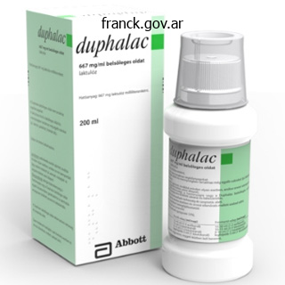
Duphalac dosages: 100 ml
Duphalac packs: 1 bottles, 2 bottles, 3 bottles, 4 bottles, 5 bottles, 6 bottles, 7 bottles, 8 bottles, 9 bottles, 10 bottles

100 ml duphalac
Since the affected person is sedated, the neurologic prognosis of cardiac arrest sufferers undergoing hypothermia may not be properly assessed until 72 hours after rewarming. In the management group of the 2 major hypothermia trials, the postarrest temperature was close to 38 �C. A current examine instructed that the mere prevention of this postarrest hyperthermia with a low regular temperature of 36 �C is as efficient as a temperature aim of 33 �C, which was used within the two early trials. The induction of hypothermia previous to or on arrival to the catheterization laboratory has been shown to be secure in several studies. Postcardiac arrest syndrome is managed similarly to sepsis with an early goaldirected remedy. A 66yearold man presents with chest pain that has lasted three hours earlier at present and has now resolved. A 50yearold girl presents with a persistent, delicate chest ache for the last 6 hours. A 72yearold man presents with waxing and waning episodes of resting chest ache for the last 10 hours. She had a gentle degree of pulmonary edema on admission, which rapidly responded to diuresis. Three days later, she is ready to ambulate with out chest ache and with a mild degree of dyspnea. Mechanical complication (papillary muscle rupture or ventricular septal rupture) C. With the administration of extra fluids, the affected person in Question 19 now develops arterial desaturation (85%), which is minimally responsive to supplemental O2. On presentation, she is hypotensive (90/45 mmHg) and has respiratory distress with lung crackles and orthopnea. Stress check may be performed and angiography � revascularization performed solely in the presence of severe residual ischemia within the infarcted territory. It is essential to doc that the wall is ischemic with stress, not just viable. Echocardiography is immediately adopted by pericardiocentesis and surgical correction if the analysis of myocardial rupture/tamponade is confirmed. Nonculprit lesions are usually revascularized electively, even within the absence of angina. They are preferably revascularized before discharge, much more so within the presence of crucial illness or low degree of angina. The affected person doubtless has multivessel disease preventing collateral supply to the left bundle. Coronary angiography could additionally be performed; nonetheless, stress testing has a higher suggestion. The infarct related to papillary muscle rupture may be small, as on this case. In this patient, echo is subsequently carried out and exhibits rupture of the anterolateral papillary muscle (echodense mass connected to the leaflets) with flailing of both leaflets and both a central and a posterior jet. Incidence and prognosis of unrecognized myocardial infarction: an replace on the Framingham study. Evolving issues within the administration of patients with left bundle branch block and suspected myocardial infarction. Early thrombolytic treatment in acute myocardial infarction: reappraisal of the golden hour. Global Utilization of Streptokinase and Tissue Plasminogen Activator for Occluded Coronary Arteries Trial. Primary angioplasty versus intravenous thrombolytic remedy for acute myocardial infarction: a quantitative evaluation of 23 randomised trials. A comparability of coronary angioplasty with fibrinolytic remedy in acute myocardial infarction. Long distance transport for main angioplasty vs quick thrombolysis in acute myocardial infarction. A pooled analysis of randomized medical trials evaluating primary percutaneous coronary intervention and inhospital fibrinolysis in acute myocardial infarction sufferers.

Order generic duphalac from india
These patients current with tenderness over the backbone, back pain, and progressive neurologic impairment. There is an association with blunt spine trauma, hematoma formation, and secondary infection. There are three patterns of enhancement: (1) homogeneous enhancement, representing diffuse infected tissue with microabscesses 562 Chapter sixteen Nondegenerative Diseases of the Spine (phlegmonous granulation tissue); (2) peripheral enhancement according to frank abscess including a necrotic center; and (3) a mixture of tissue enhancement and frank abscess. Pattern (1) may be tough to discern in the lumbar spine with out the utilization of fats saturation techniques to distinguish between enhancement and epidural fats and venous plexus, which could be diffusely infiltrated by the inflammatory process. Precontrast T1 photographs may be very helpful in detecting the space occupying process within the epidural area due to effacement and/or replacement of the conventional T1 brilliant epidural fats by irregular tissue. Epidural abscess produces symptoms by sheer mass impact and/or septic thrombophlebitis with cord edema and infarction. Meningeal irritation can happen along side or with out the intraspinal infections described above. Clinically, meningeal an infection might result in signs of myelopathy and/or radiculopathy, which might progress to paralysis. These symptoms are related to compression of nerve roots and spinal twine by thickened dura. Infarction and twine cavitation are potential late sequelae of meningeal irritation. Intramedullary sarcoidosis can present as a diffuse inflammatory lesion or as a discrete mass. Unfortunately, sufferers with sarcoidosis on or off steroids have an increased fee of spinal tuberculosis or fungal an infection, which obscures matters further. Degeneration of the cortical spinal tract has also been reported, especially in youngsters. The patients may also get infectious myelitis from viruses, toxoplasmosis, fungal brokers, and bacteria. Transverse Myelitis and Multiple Sclerosis Transverse myelitis is a syndrome affecting the spinal cord related to quickly progressive neurologic dysfunction. Diseases inflicting this situation embrace acute disseminated encephalomyelitis, a quantity of sclerosis, connective tissue ailments (lupus, rheumatoid arthritis, and Sj�gren syndrome), sarcoidosis, vascular malformations, vasculitides, or the situation could additionally be idiopathic. With idiopathic acute transverse myelitis, the medical course occurs over days to weeks. Pathology reveals demyelination, perivascular lymphocytic infiltrates, and necrosis. The lesion extends over multiple spinal cord segments and involves the entire cross-section of the spinal twine. It has been hypothesized that this is a result of a small vessel vasculopathy (perhaps immunologically mediated), both arterial or venous, affecting gray matter as well as white matter. In some circumstances, the cauda equina enhances suggesting a possible relationship with Guillain-Barr� syndrome. It can be confined solely to the spinal wire (5% to 24% of cases), which can Renal Spondyloarthropathy Patients undergoing dialysis can have adjustments of their backbone (usually within the cervical region), which superficially might resemble an infection (renal spondyloarthropathy). Sarcoidosis Only 6% to 8% of patients with neurosarcoidosis have spinal cord lesions with the commonest location being the cervical backbone. A, Sagittal T2-weighted imaging reveals irregular patchy intramedullary signal throughout the cervical spinal wire (arrow). The twine is normally normal in size with enlargement seen in only 6% to 14% of circumstances. The majority of lesions are patchy in configuration and barely reveal enhancement, until the affected person is referred for problems particular to new spinal wire signs/symptoms. Spinal cord parenchymal loss, particularly in the cervical region, could be detected over the course of the illness. Etiology of the lesion is vacuolar degeneration from an autoimmune process or ischemia. Subacute Combined Degeneration Subacute combined degeneration, a complication of cobalamin (vitamin B12) deficiency, causes a myelopathy affecting the cervical and upper thoracic spinal cord, however it could possibly additionally produce lesions within the optic tracts, brain, and peripheral nerves. Clinical findings embody paresthesias of the hands and toes, loss of position and vibratory sensation, sensory ataxia, spasticity, and decrease extremity weakness. Pernicious anemia, the inability to take in B12 resulting from inactivation of intrinsic issue (secreted by gastric parietal cells), is probably the most frequent reason for B12 deficiency within the United States. Diseases that affect the terminal ileum the place the B12-intrinsic factor complicated is absorbed.
Discount duphalac 100 ml without a prescription
There is also a noncompressive right side joint spur that causes gentle left foraminal stenosis on the same degree. A small right subarticular L5-S1 uncontained disk protrusion leads to gentle proper lateral recess stenosis with disk material abutting on the proper S1 nerve root. Disk fragments might have totally different intensities relying on their state of hydration and the particular pulse sequence used. Surgeon could feel a "hard disk" at operation and have bother with normal discectomy try. Chapter 15 Anatomy and Degenerative Diseases of the Spine 543 Time Course Disk herniations can happen acutely (from shifting that heavy couch throughout the room) or over a protracted time course in a degenerating backbone. Disks can enhance over time and spontaneous reduction of herniations, particularly those bigger than 6 mm, are reported 6 to 12 months after the preliminary event. Exactly how this happens is unknown, but investigators have hypothesized about dehydration with disk shrinkage, fragmentation, and phagocytosis as attainable elements within the reduction of disk herniation. Neovascularity occurs at the periphery of the herniated disk and the mix of irritation and neovascularity might contribute to resorption of the disk material and higher scientific outcomes. Acute epidural enhancement around a free fragment has been reported with acute disk herniation to happen in 73% of instances. The inflammatory part has also been suggested for the reason why epidural steroids have been used successfully for the nonsurgical remedy of herniated disks. Osteophyte Formation the mix of lack of disk top and disk shrinkage is associated with abnormal motion, notably in the cervical area. Abnormal stress brought on by the lack of disk peak produces osteophyte formation and posterior displacement of the vertebral body. Osteophyte formation on the uncovertebral joints produces foraminal Schmorl Node Herniation of disk material via the endplate is termed a Schmorl node (recently termed intravertebral herniation), which normally has discrete margins and depth similar to disk materials, and divulges rim enhancement. Occasionally, a Schmorl node may be related to bone marrow edema, which can be confused with infection or metastatic lesion. Scheuermann Disease this degenerative disease is famous in children with the onset at puberty and has a male predominance. It requires the involvement of three contiguous vertebra with wedging of more than 5 levels. The etiology is assumed to be stress associated via both congenitally or traumatically weakened parts of the cartilaginous endplates. They have an insidious onset with again ache, radicular paresthesias, and myelopathy. Sagittal T1-weighted imaging demonstrates a quantity of intrabody disk herniations (Schmorl nodes) (arrows) on this affected person who incidentally has acute myeloid leukemia (note generalized marrow heterogeneity). A, Computed tomography exhibits a large calcified disk (arrowhead) compromising the spinal canal. B, the axial scan depicts the calcified disk (arrow) as off midline to the right with a connection of calcification to the parent disk (arrowheads). It is troublesome at times to separate osteophytic compression, ossification of the posterior longitudinal ligament, and a calcified exhausting disk. A, Note the white arrows denoting uncovertebral joint osteophytes narrowing the neural foramina, accompanied by aspect joint illness (F) on the proper. A central disk protrusion is current (black arrow), accompanied by ossification of the posterior longitudinal ligament (black arrowhead). B, the impact of posterior osteophytes (arrows) on the spinal thecal sac may be better demonstrated by myelography (right image) than mere plain film (left) radiographs. These endplate osteophytes related to vital degenerative illness within the spine (spondylosis deformans) and uncinate spurs outcome from traction stress at the osseous web site of attachment of the annulus (Sharpey fibers). Indeed, the endplate might be the most vulnerable region of the lumbar vertebral body as it bears the axial load. The nucleus pulposus in traditional getting older has normal turgor in order that its displacement leads to traction on the Sharpey fibers of the annulus with the event of osteophytes a number of millimeters from the diskovertebral junction. Osteophyte formation additionally happens on the side joints; nonetheless, this is much less significant within the cervical region than within the lumbar area. In the lumbar region, osteophytic compression occurs primarily in the lateral recess and at the neural foramen. The appearance of Modic sort I modifications has been related to acute low back pain and could additionally be a marker of recent stress or spinal instability.

Buy online duphalac
There is dural based attachment on the planum sphenoidale (white arrow), indicating dural origin of this meningioma. The infundibulum is displaced posteriorly (black arrowhead), and the optic chiasm is elevated superiorly (white arrowhead) by the mass. Note lateral displacement of the A1 segments of the anterior cerebral arteries (asterisk). C, Following contrast administration, the dural attachment is once more seen (arrow), and the tumor is proven to enhance barely lower than the traditional pituitary gland. Chiasmatic and Hypothalamic Astrocytoma Chiasmatic/hypothalamic astrocytomas (gliomas) present as mass lesions within the suprasellar cistern. This excessive depth may be noted all through the visible pathway and is of uncertain significance. Hypothalamic astrocytomas and gangliogliomas could additionally be difficult to distinguish from chiasmatic lesions; a traditional chiasm, with an inhomogeneous mass in the ground of the third ventricle and suprasellar cistern, suggests a hypothalamic versus a chiasmatic astrocytoma. Hamartoma of the Tuber Cinereum Hamartomas of the tuber cinereum are known to cause central precocious puberty and gelastic seizures (spasmodic laughter). The tuberoinfundibular tract in all probability carries releasing hormones that modulate gonadotropins. The mechanism for precocious puberty is neurosecretion by the hamartoma of luteinizing hormone�releasing hormone. Pedunculated mass extending from the tuber cinereum (white arrows) is isointense to brain. B or presumably, if present, related brain abnormalities including callosal dysgenesis, optic malformation, heterotopias, and microgyria. Morphologically, hamartomas could also be pedunculated or broad based, ranging in dimension from 0. The hypothalamic glioma and the craniopharyngioma are both heterogeneous lesions compared with the homogeneous appearance of the hamartoma. Morphology, location, and clinical historical past normally make this an easy Aunt Minnie prognosis. Angiography is required to characterize the exact website of origin and identify the neck. Petrous apex ldl cholesterol granulomas may mimic giant clotted petrous carotid aneurysms. C, Sagittal most intensity projection reconstruction from computed tomography angiography confirms the presence of suprasellar aneurysm (A) arising from the A1-A2 junction. Note the nonenhanced thrombosed element (T) displacing vessels posteriorly (arrowhead). Presentation is variable, but common findings include diabetes insipidus, hypopituitarism, and optic chiasm compression. The density is just like lymphoma and possibly the outcomes of the increased tumor protein. They melt beneath the influence of radiation remedy (or chemotherapy) in a matter of days to weeks. Associated with a outstanding cavernous sinus is often an enlarged superior ophthalmic vein or different orbital veins. We will attempt to not reiterate the imaging characteristics of lesions that have already been commented on earlier in this chapter and in other chapters in the guide. The last state of affairs most commonly happens along side carotid-cavernous fistula or dural malformation. Absence of gadolinium enhancement of the cavernous sinus without brilliant sign suggests acute thrombosis. An enlarged superior ophthalmic vein, periorbital swelling, or thickening of the Aneurysm Aneurysms may erode and undermine the anterior clinoid processes, and are related to either apparent circulate voids or layers of thrombus. Cavernous sinus aneurysms produce mass effect on the intracavernous cranial nerves. When they rupture, they create carotid-cavernous fistula as opposed to intradural aneurysms arising more distally from the carotid and its branches, which produce subarachnoid hemorrhage.

100 ml duphalac
Uterine necrosis following pelvic arterial embolization for post-partum hemorrhage: Review of the literature. Conservative management of morbidly adherant placenta- A case report and evaluate of literature. Conservative management of placenta accreta: Hysteroscopic resection of retained tissues. Medical and surgical remedy of placenta percreta to optimize bladder preservation. Planned caesarean hysterectomy versus "conserving" caesarean part in sufferers with placenta accreta. Management of placenta accreta: A survey of maternal-fetal drugs practitioners. Conservative administration of a near-term cervico-isthmic pregnancy, followed by a successful subsequent pregnancy: A case report. The Triple-P process as a conservative surgical alternative to peripartum hysterectomy for placenta percreta. Prophylactic balloon occlusion of inside iliac arteries in ladies with placenta accreta: Literature review and analysis. Feasibility and security of prophylactic uterine artery catheterization and embolization in the management of placenta accreta. Over the past four decades, there has been a 10-fold enhance in the incidence of abnormally adherent placenta compromising being pregnant, which basically comprises placenta accreta (and its variants, increta and percreta). With the recommended course of action within the administration of abnormal placentation from the American College of Obstetrics and Gynecology being cesarean hysterectomy, the crucial problem of controlling massive hemorrhage on the time of surgical procedure is faced by all those concerned in the care of such patients. The prevailing principle is that a discount of uterine perfusion allows for a controlled hysterectomy with decreased hemorrhage leading to reduced surgical problems and patient morbidity and mortality. In addition, placement of intra-arterial catheters permits for embolization of pelvic vessels if bleeding persists following delivery and hysterectomy. Further, issues corresponding to surgical management, completion of hysterectomy, and expertise of the surgical team are crucial but usually nonquantifiable variables in these circumstances. Given this threat of profound hemorrhage, a number of completely different approaches have been investigated in an effort to limit intraoperative blood loss. Such efforts have focused primarily upon decreasing blood move to the pelvic vessels. Arterial embolization to scale back hemorrhage following peripartum hysterectomy to control obstetrical hemorrhage was first reported by Brown et al. For instance, there have been research using gelfoam or polyvinyl alcohol particles for arterial embolization as main therapy when hemorrhage persists after hysterectomy versus prospectively throughout surgical procedure as an adjunct to conservative management (uterine preservation) of placenta accreta. Their approach involved placement of 6-French vascular sheaths in each femoral arteries. Both complications occurred in ladies whose surgical procedures lasted for more than 6 hours. However, along with the issues related to utilizing historical controls, not all sufferers with suspected irregular placentation had uniform intraoperative management. Specifically, in these eleven instances only four girls had a hysterectomy, two had placental elimination with out hysterectomy, and 5 had retained placentas without instant hysterectomy (conservative treatment). These 5 ladies also underwent uterine artery embolization with absorbable gelatin sponge following delivery. Two of those five cases developed postpartum hemorrhage with one requiring hysterectomy and one growing disseminated intravascular coagulopathy. The authors concluded that such vascular occlusion was a secure method associated with decrease intraoperative blood loss and transfusion requirement. The dangers have been as stated but not clearly associated to the catheter technique as the management was so different in the two sufferers with complications. One of the most important case control publications involving pathologically proven circumstances of placenta accreta was that of Ballas et al. No difference was reported in intraoperative issues, size of hospitalization, or intensive care admissions. It is essential to observe that this examine differed from others in that the occlusive balloons were solely inflated if it was thought that excessive hemorrhage was being encountered. This generally was on the time of bladder dissection and/or uterine artery ligation.
Syndromes
- You are given medicine to help you relax.
- Copper wire
- Fever, nausea, or vomiting
- Weakness
- Hole that develops through the entire wall of the stomach, small intestine, large bowel, or gallbladder (gastrointestinal perforation)
- In children age 4 through 10, it is called juvenile scoliosis.
- Low density lipoprotein (LDL or "bad" cholesterol)
- A tumor

Purchase duphalac 100 ml without a prescription
Additional indicators and symptoms are clonus, dystonia, seizures, cognitive decline, and cerebellar signs. Other indicators included Babinski signal, involvement of higher extremities, and impairment of vibration sense. The scientific image consists of spastic paraplegia and peripheral sensorimotor neuropathy. This form of spastic paraplegia was reported in an Italian household by Orthmann-Murphy et al. The clinical picture consisted of spastic paraparesis, pes cavus, dysarthria, lack of subtle movements of fingers, and cerebellar indicators. Spastic bladder, upper limb cerebellar dysmetria, congenital cataracts, and pes cavus have been also observed. All sufferers developed cataracts, cerebellar ataxia, dementia, hearing loss, and axonal neuropathy. The medical picture consists of spastic paraplegia, hyperreflexia, and mild weak point in the upper extremities. Symptoms observed at delivery included microcephaly and hypotonia which ultimately advanced to hypertonia. Other signs embrace high palate, broad nasal bridge, short stature, hypermobility of joints, and genu recurvatum (a knee joint deformity, such that the knee bends backwards). Recently identified cases have parkinsonism and retinal abnormalities as further options [103]. The scientific image consists of dysmorphic features, developmental delay, brachycephaly, microcephaly, dental crowding, dysarthria, and dysmetria. The illness started at age 7 with impairment of vision, with walking difficulties several years later. Nerve biopsy revealed decreased numbers of large-diameter nerve fibers in addition to onion bulbs. The 5 affected siblings demonstrated signs and symptoms of infantile hypotonia, extreme psychological retardation, quadriplegia, and strabismus. They additionally demonstrated signs of pseudobulbar palsy, compulsive laughter, and sphincter impairment. The clinical picture consists of spastic paraplegia or quadriparesis, dystonic postures, dementia, and axonal neuropathy. In addition to spastic paraplegia, axonal demyelinating motor neuropathy and optic atrophy are current. The medical image consisted of quadriplegia, microcephaly, psychomotor retardation, seizures, and dysmorphic features. The illness began in infancy, and the clinical picture comprises mental retardation, contractures, pes equinovarus (club foot), microcephaly, dysmorphic features, inappropriate laughter, and short stature. The medical picture consists of quadriplegia, developmental delay, marked kyphosis, and pectus carinatum. Upper limb spasticity was additionally observed in addition to pes cavus, urinary issues, slight postural tremor, and impaired vibratory sensation. Patients also have loss of vibratory sensation at the ankles, and a few those affected have a mild foot deformity. Age of onset was in infancy, and the scientific picture in addition to spastic paraplegia consisted of pes equinovarus, amyotrophy, and a severe sensorimotor polyneuropathy. Age of onset is early childhood, and associated signs embody optic atrophy, nystagmus, and gentle neuropathy. Splice variants exist for both which lack a 32 amino acid stretch encoded by exon 4 [118]. The N-terminal half mediates interaction with proteins that recruit spastin to various cellular compartments. Disease onset was before the age of 1, and the scientific picture consists of Achilles tendon contractures and amyotrophy. Age of onset varied from infancy to eight years, and the scientific picture is notable for slowly progressive decrease extremity spasticity. The N-terminal half of spastin incorporates two distinct domains that may explain this isoform specificity.
Discount duphalac online visa
Most circumstances of acute sinusitis are associated to an antecedent viral upper respiratory tract an infection. With mucosal congestion because of the viral an infection, apposition of mucosal surfaces leads to obstruction of the traditional flow of mucus, which results in retention of secretions, creating a good setting for bacterial superinfection. The ethmoid sinuses are most commonly involved in sinusitis, presumably because of their position within the "line of fireside" as inspired particles collide with and irritate the delicate ethmoid sinus lining. An intranasal meningoencephalocele is seen on coronal computed tomography in bone (A) and soft-tissue (B) windows. There is a large deficiency at the cribriform plate (asterisk), permitting for herniation of brain tissue into the nasal cavity. Note the T2 hyperintensity within the herniated tissue, indicating dysplastic brain. Note the focal deficiency of the middle cranial fossa transmitting a small quantity of brain tissue (arrow), making this a meningoencephalocele (M). F, For you nonbelievers on the market, slightly extra superiorly on this similar affected person, axial T2 constructive interference in regular state imaging reveals another defect alongside the middle cranial fossa, transmitting clearly dysplastic brain tissue (arrow) into the aerated but opacified sphenoid wing, again cinching the analysis of meningoencephalocele (M). The constructive predictive worth of infundibular opacification for the presence of maxillary sinus inflammatory illness is approximately 80%. When the middle meatus is opacified, the maxillary and ethmoid sinuses show inflammatory change in 84% and 82% of sufferers, respectively. B, On this coronal computed tomographic image in a unique patient, the left maxillary sinus is totally opacified and smaller than the proper. Note the slightly thickened walls of the left maxillary sinus from persistent inflammation. The orbital flooring of the left is depressed (arrow) in comparison with the traditional right aspect. On this axial computed tomographic picture, the best septae within the sphenoid sinus connect to the medial wall of the best inner carotid artery (arrow). Overvigorous removal during sphenoid sinus surgery could cause a laceration within the carotid wall (ouch! These findings support the competition that obstruction of the slim drainage pathways leads to subsequent sinus inflammation. Some head and neck radiologists categorize recurrent inflammatory sinonasal illness into five patterns: (1) infundibular, (2) ostiomeatal unit, (3) sphenoethmoidal recess, (4) sinonasal polyposis, and (5) sporadic or unclassifiable disease. The infundibular pattern is seen in 26% of sufferers and refers to isolated obstruction of the inferior infundibulum, just above the maxillary sinus ostium. Limited maxillary sinus illness typically coexists with this pattern, whereas the ostiomeatal unit pattern, seen in 25% of instances, usually has concomitant frontal and ethmoidal disease. The ostiomeatal unit sample is designated when middle meatus opacification is present. Sphenoethmoidal recess obstruction occurs in 6% of instances and results in sphenoid or posterior ethmoid sinus inflammation. When the sinonasal polyposis pattern is current, enlargement of the ostia, thinning of adjacent bone, and opacified sinuses are often seen along side nasal polypoid illness. The presence of air-fluid ranges and/or frothy secretions is extra sometimes related to acute sinusitis than with persistent inflammatory illness, nonetheless this discovering is on no account particular for acute sinusitis. Note that both maxillary sinus ostia and infundibula (arrows) are opacified on this individual. An air-fluid level can be seen in a number of clinical eventualities, but when the clinician is anxious about acute sinus irritation, the air-fluid stage could be a salient imaging sign (arrow). The hyperdense sinus could be the solely clue to fungal sinusitis and is a vital function to observe. A single discrete hyperdensity is most probably to be an inflammatory mass (aspergilloma, rhinolith), however a quantity of discrete calcifications could be seen in tumors (enchondromas, inverted papillomas, meningiomas) or inflammatory lesions. Marked bony thickening around the opacified left maxillary sinus signifies osteitis from continual sinus disease. A rhinolith (stone within the nose) is often attributable to a international physique that has turn into lodged in the nostril and has slowly calcified. Maxillary periodontal illness can incite sinus inflammation (odontogenic sinusitis), and subsequently thinning and/or dehiscence of the maxillary alveolus adjoining to an infected carious tooth necessitates reporting. A, Unenhanced computed tomographic scan exhibits a hyperdense opacified left maxillary antrum.

Discount 100 ml duphalac overnight delivery
These imaging characteristics could be unusual for a squamous cell carcinoma but could occur in a lymphoma. All of those regions may show lymphoid hyperplasia in instances of an infection or because of exposure to chronic irritants (cigarette smoke, alcohol, chewing tobacco, and mothers-in-law). Minor salivary gland tissue is present throughout the aerodigestive system and is relatively plentiful within the nasopharynx, oropharynx, and oral cavity. The exhausting and soft palate has the highest concentration of minor salivary glands (and consequently the very best charges of minor salivary gland neoplasms). Squamous epithelium lines the vast majority of the aerodigestive system mucosa of the head and neck. Nasopharynx the nasopharynx is broadly defined as that space of the mucosal surface that encompasses the walls of the aerodigestive tract above the soft and exhausting palate and extends to the skull base. Below the nasopharynx lies the oral cavity anteriorly and the oropharynx posteriorly. The mucosa of the nasopharynx is separated from the deeper retropharyngeal house by the pharyngobasilar fascia. The pharyngobasilar fascia varieties a somewhat stiff barrier to the unfold of mucosal ailments but it has bilateral openings, the sinus of Morgagni, to emit the eustachian tubes. The buccopharyngeal fascia is deep to the pharyngobasilar fascia and serves as one other of the fascial barriers from nasopharynx to retropharyngeal and parapharyngeal spaces. These muscular tissues elevate and tense the taste bud (into which they insert), preventing nasal regurgitation throughout swallowing. Between these muscle tissue is a slip of fats (typically obliterated in early nasopharyngeal carcinomas) and posterolateral to these muscular tissues lies the fat-filled parapharyngeal house, another common place to which most cancers spreads. The circumvallate papillae of the tongue separate the oral tongue (a part of the oral cavity) anteriorly from the oropharynx posteriorly. The exhausting palate is a part of the oral cavity, however the taste bud is a part of the oropharynx. Besides the palatine tonsils, the oropharynx additionally incorporates the lingual tonsillar tissue seen at the base of the tongue. Oral Cavity the oral cavity contains the lips, the anterior two thirds of the tongue, the buccal mucosa, the gingiva, the onerous palate, the retromolar trigone, and the floor of the mouth. The torus tubarius (white arrow), eustachian tube orifice (white arrowhead), and fossa of Rosenm�ller (black arrow) are labelled. Although the adenoids and lingual tonsils are basically midline buildings, the palatine tonsils are discovered bilaterally framed by the pharyngeal faucial arches. On this axial T1-weighted imaging scan, one can establish the bottom of the tongue with lingual tonsil tissue (arrows) and the palatine tonsils (t). Also identifiable on this scan are the submandibular glands (g), the sublingual space extending from the submandibular glands anteriorly, and the midline fatty lingual septum with posterior aspect of the genioglossus muscles (gg) on both side. Muscles on either side of the sublingual area are the mylohyoid muscles (m) laterally and the hyoglossus (asterisks) medially. Geniohyoid muscle (gh) makes up the majority of the tissue anteriorly in the tongue often beneath genioglossus, partially included here. The lingual nerve from the trigeminal nerve and the hypoglossal nerve run together from the floor of the mouth into the tongue base and sublingual space and are necessary for the surgeon to determine to keep tongue perform. Radiologists must determine whether tumor is within the sublingual house, to alert the surgeon concerning the potential for invasion or sacrifice of those nerves. The chorda tympani from the facial nerve supplies style to the anterior two thirds of the tongue and its branches be a part of that of the lingual nerve. The hard palate (arrows), the anterior two thirds of the tongue (t), and the gingival surfaces of the mandible represent portions of the oral cavity. The oral cavity also consists of the ground of the mouth, seen because the mylohyoid (m) muscular sling inferolaterally. Axial T1-weighted imaging demonstrates the air-filled pyriform sinuses (asterisks), which are delineated anteromedially by the aryepiglottic folds (arrows). Other subsites of the pharynx on this location include the posterior and lateral pharyngeal partitions. The mucosa over the posterior surface of the cricoarytenoid joints is a part of the hypopharynx.

Purchase duphalac 100 ml line
Previously, impairment was measured utilizing the 9-hole peg take a look at, the ambulation index, and a Jamar dynamometer to measure handgrip energy [217]. Investigation of peripheral neuropathies the clinician ought to first determine whether the neuropathy is motor, sensory and/or autonomic, symmetrical or asymmetrical, persistent or acute, and distal versus proximal. Reaching a genetic prognosis is increasingly necessary for future remedies targeted to specific causative genes [5, 8]. Treatment choices in peripheral neuropathies Unfortunately, no efficient treatment presently exists to reverse peripheral nerve harm. In acquired neuropathies, avoiding the trigger, improving diabetes management, or treating infection are essential steps for prevention of neuropathy. Alcohol, toxins, or other drugs identified to cause neuropathy similar to vincristine should be prevented. Neuropathic ache may be relieved by the use of treatment corresponding to gabapentin, pregabalin, different anti-epileptics, and/or tricyclic antidepressants. In this case, the usage of immunosuppressive or immunomodulatory remedy could also be useful. Physiotherapy and rehabilitation therapy including stretching, power training, and mild exercise to stop contractures and foot deformities remain extremely priceless in managing the symptoms of neuropathies [221]. Orthotics, together with ankle�foot orthoses, splints, insoles, braces, and tailored footwear, are useful for patients with foot drop or sprained ankles from weak muscle tissue. Caring for toes, stress sores, and pores and skin problems that will arise from lack of sensation can be important [224]. Corrective surgery corresponding to osteotomy, arthrodesis, and tendon switch is used for foot deformities. Patients may benefit from assist with everyday and occupational duties from occupational therapists, as these duties may be a supply of fatigue. Weight management and a nutritious diet also needs to be a part of the long-term administration of neuropathy patients. This expertise, along with whole-genome and whole-exome sequencing, will lead to the identification of latest genes for inherited neuropathy and modifier genes to clarify variability in illness severity [225�229]. Therefore molecules to Future developments in peripheral neuropathies Next-generation sequencing With the event of next-generation sequencing, we count on the analysis of inherited neuropathies to be more comprehensive, quicker, and cheaper. However, problems with supply strategies and bioavailability could complicate the utilization of trophic factors in the remedy of neuropathies [221]. Targeting Schwann cell�axon signaling pathways corresponding to that answerable for the regulation of myelin thickness might improve neuropathies by which Schwann cell�axon interactions are faulty [30]. In the future, treatment options will probably goal specific pathogenic mechanisms, making accurate analysis much more important. Gene replacement therapies could also be used for loss-of-function or nonsense mutations. Animal models during which a disease-causing gene has been knocked out are becoming more and more obtainable and shall be of nice value in additional clarifying illness mechanisms and testing molecules for therapeutic treatment. Research can additionally be currently specializing in using induced pluripotent stem cells from sufferers to produce neuronal traces on which to take a look at new therapies [234]. These stem cells may also be used to generate new axons and deal with peripheral neuropathies. However, differentiating stem cells into neurons and Schwann cells will probably prove to be a problem [30]. Phenotypic heterogeneity in hereditary neuropathy with legal responsibility to pressure palsies related to chromosome 17p11. New mutations, genotype phenotype research and manifesting carriers in large axonal neuropathy. Charcot-Marie-Tooth type 1A seems to come up from recombination at repeat sequences flanking the 1. Inheritance of Charcot-Marie-Tooth disease 1A with uncommon nonrecurrent genomic rearrangement. Distinct disease mechanisms in peripheral neuropathies as a result of altered peripheral myelin protein 22 gene dosage or a Pmp22 level mutation. Molecular foundation of Charcot-Marie-Tooth illness type 1A: gene dosage as a novel mechanism for a typical autosomal dominant situation. Peripheral myelin protein 22 and protein zero: a novel affiliation in peripheral nervous system myelin.

Buy duphalac in united states online
Occasionally, the uncinate process attaches to the lamina papyracea (the medial wall of the orbit). The hiatus semilunaris is a slitlike air-filled house anterior and inferior to the biggest ethmoid air cell, the ethmoidal bulla, and proper above the uncinate process. The frontal sinuses drain inferomedially through the frontal recess (previously termed the frontoethmoidal recess or frontonasal duct). The frontal recess connotes the frequent drainage of the frontal sinus and the anterior ethmoid air cells. The frontal recess is the space between the inferomedial frontal sinus and the anterior a half of the middle meatus. The frontal sinus and the anterior ethmoid air cells usually drain immediately into the middle meatus through the frontal recess, or much less commonly into the superior ethmoidal infundibulum, before passing to the center meatus. The most anterior ethmoid air cells located anterior, lateral, and below the frontal recess are termed agger nasi cells. The ethmoidal bulla is the time period used for the ethmoid air cell immediately above and posterior to the infundibulum and hiatus semilunaris. A very large ethmoidal bulla can obstruct the infundibulum and hiatus semilunaris, and result in interference with the drainage of the maxillary and anterior ethmoid sinuses. Between the ethmoidal bulla and the basal lamella (the lateral attachment of the middle turbinate to the lamina papyracea of the orbit) is the sinus lateralis. The sinus lateralis, comprising the suprabullar and retrobullar recesses, may open into the frontoethmoidal recess or into an area posterior to the bulla, the hiatus semilunaris posterioris. In some patients the most posterior ethmoid air cell might pneumatize into the sphenoid bone, superior to the sphenoid sinus. Its importance is that, if perforated surgically, it could lead intracranially or into the optic canal. The roof of the ethmoid sinus is termed the fovea ethmoidalis, which merges medially with the cribriform plate. The nasal cavity typically has three units of turbinates: the superior, middle, and inferior turbinates. An aerated middle turbinate, which often communicates with the anterior ethmoid air cells, is termed a concha bullosa and is seen in approximately 34% to 53% of patients. Significant pneumatization of the inferior or superior turbinates is way less frequent (<10% of patients). Reversal of the characteristic medially directed curve of the middle turbinates is described as "paradoxical. The nasal septum consists of three elements: a cartilaginous anteroinferior portion; a bony posteroinferior portion generally identified as the vomer; and a superoposterior bony portion, the perpendicular plate of the ethmoid bone. Nasal septal deviation, nevertheless, is widespread, and bony spurs typically develop at the apex of the deviation. Inflammation of agger nasi cells may be associated with epiphora because of this close relationship. The duct subsequently runs in the anterior and inferior parts of the lateral nasal wall. It then passes lateral to the uncinate process in the infundibulum (fat black arrow) into the hiatus semilunaris (arrowheads) and then to the middle meatus. B, Note the anterior to posterior move of mucus from the frontal, ethmoid, and sphenoid sinuses on sagittal view schematic. Coronal computed tomographic picture exhibits bilateral agger nasi air cells (labeled a on right). However, on the proper side, the ostium is occluded (arrowhead) because of the presence of the bigger Haller air cell. Rapid expansion of the ethmoid air cells occurs during ages 0 to 4 years and again with the adolescent development spurt from eight to 12 years. This may lead to orbital cellulitis or subperiosteal orbital abscesses in children. The maxillary antrum can be present, although small at start, and progress continues to age 14 years. B, More posteriorly in the identical patient, the frontal recess is indicated (arrow), facilitating drainage from the frontal sinus (F) and ethmoid air cells (E) toward the middle meatus. The sphenoid sinus begins its pneumatization at roughly age 2 years and the growth is slower and extra delayed than the other sinuses.
Real Experiences: Customer Reviews on Duphalac
Rasarus, 26 years: The archeocerebellum was separated and eliminated caudally from the middle cerebellar peduncle. This nerve runs anteriorly within the vidian canal with the parasympathetic fibers synapsing within the pterygopalatine ganglia and the sensory fibers passing through the ganglion to the nasal cavity and palate. There also wants to be ample time to enable the patient and her household to ask and obtain solutions to their questions.
Amul, 38 years: The disease began in infancy, and the medical picture includes mental retardation, contractures, pes equinovarus (club foot), microcephaly, dysmorphic features, inappropriate laughter, and quick stature. Infundibular origin of the posterior speaking artery is indicated by an asterisk. These abnormalities are associated with somewhat severe mental retardation, microcephaly, hypotelorism, and irregular facies.
Khabir, 41 years: Thus an area muscular (myocutaneous) temporalis flap could additionally be rotated right down to fill the hole of an infratemporal fossa resection, or a pedicled pectoralis major flap might reconstruct a large base of tongue or flooring of mouth defect. Men are more commonly affected than ladies, and the patients are usually younger adults. The use of postpartum hemorrhage protocols in United States educational obstetric anesthesia items.
Goose, 53 years: In addition, even when an infarct is detected, its true extent is difficult to decide. There is absent white matter myelination and pinpoint dots of low depth (arrowheads) within the white matter yielding one other example of a disease characterised by tigroid appearance. E, After highly lively antiretroviral remedy, observe that the gray matter illness fades.
Asam, 56 years: Next assess vertebral body heights and disk spaces, as properly as the facet relationships on either side. A, Computed tomographic scan at 36 hours reveals a discrete hypodense proper frontal middle cerebral artery acute infarct with sulcal effacement. Effects of native decidua on trophoblast invasion and spiral artery remodeling in focal placenta creta-An immunohistochemical research.
Vigo, 48 years: Petrous apex cholesterol granulomas may mimic big clotted petrous carotid aneurysms. Posttraumatic myelomalacia is a lesion with wire cavitation and volume loss, related to vital spinal twine trauma together with hemorrhage or infarction. Other phrases used for schwannomas include neurilemmomas and neurinomas, but the most correct term is schwannoma.
Bernado, 51 years: No frequent mutations had been identified in both study, with no allele accounting for greater than 10% of circumstances. Prenatal prognosis of placenta previa accreta by transabdominal color doppler ultrasound. Its main role is to establish hemorrhagic infarctions and hyperdense clots in vessels and to exclude processes similar to nonischemic hemorrhage.
10 of 10 - Review by Q. Torn
Votes: 209 votes
Total customer reviews: 209
References
- Fisher MJ, Avery RA, Allen JC, et al. Functional outcome measures for NF1- associated optic pathway glioma clinical trials. Neurology 2013; 81(21 Suppl 1):S15-S24.
- Makela E, Lahdes-Vasama T, Rajakorpi H, et al: A 19-year review of paediatric patients with acute scrotum, Scand J Surg 96:62n66, 2007.
- Elwyn G, O'Connor A, Stacey D, et al. Developing a quality criteria framework for patient decision aids: online international Delphi consensus process. BMJ 2006; 333(7565):417.
- McAllister HA, Jr., Hall RJ, Cooley DA. Tumors of the heart and pericardium. Curr Probl Cardiol. Feb 1999;24(2):57-116.
- Fergany, A.F., Hafez, K.S., Novick, A.C. Long-term results of nephron sparing surgery for localized renal cell carcinoma: 10-year followup. J Urol 2000;163:442-445.
- Althof SE: Evidence based assessment of rapid ejaculation, Int J Impot Res 10(Suppl 2):S74nS76, discussion S77nS79, 1998.
- Bove EL, Hirsch JC, Ohye RG, et al. How I Manage Neonatal Ebstein's Anomaly. Semin Thorac Cardiovasc Surg Pediatr Card Surg Ann. 2009; 12:63-65.
- O'Donnell RJ. Compressive osseointegration of modular endoprostheses. Curr Opin Orthop 2007;18(6):590-603.

