
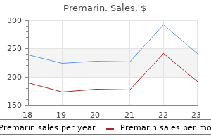
Premarin dosages: 0.625 mg
Premarin packs: 14 pills, 28 pills, 56 pills, 84 pills, 112 pills

Generic premarin 0.625 mg buy on-line
Abscesses could appear as a poorly marginated gentle tissue mass within the expanded anterior cervical area with single or multiloculated low-density middle, with or with out gasoline collections, and normally thick abscess wall. It could result from extension of cellulitis or abscess from adjoining areas or after direct penetrating trauma. Malignant neoplasms Contiguous tumor extension Obliteration of the fat of the anterior cervical area due to an infiltrating mass with its middle within the adjoining infrahyoidal cervical spaces. These malignant tumors get away of their area of origin and invade the anterior cervical space. Also seen are related cellulitis with obliteration of adjacent fat planes, thickening of the left sternocleidomastoid muscle and platysma, and infiltration of the subcutaneous fat. Radial tears of the anulus fibrosus are sometimes clinically significant and are related to disk herniations. The term disk herniation usually refers to extension of the nucleus pulposus via an annular tear past the margins of the adjoining vertebral physique finish plates. Posterior and posterolateral herniations could cause compression of the thecal sac and contents, in addition to compression of epidural nerve roots in the lateral recesses or inside the intervertebral foramina. Lateral and anterior disk herniations are much less common however could cause hematomas in adjacent structures. Disk herniations that happen superiorly or inferiorly end in focal depressions of the vertebral cortical end plates. The distal end of the conus medullaris is normally situated on the T12�L1 degree in adults. Lesions inside the thecal sac are categorized as intradural intra- or extramedullary. The upper two cervical vertebrae have completely different configurations than the opposite vertebrae. The atlas (C1) has a horizontal ringlike configuration with lateral masses that articulate with the occipital condyles superiorly and the superior aspects of C2 inferiorly. The dorsal margin of the higher dens is secured in place in relation to the anterior arch of C1 by the transverse ligament. Various anomalies occur in this region, such as atlanto-occipital assimilation, segmentation. The lower 5 cervical vertebral bodies have rectangular shapes with progressive enlargement inferiorly. Superior and lateral projections from the C3 to C7 cervical vertebral bodies type the uncovertebral joints. The transverse processes are situated anterolateral to the vertebral bodies and contain the foramina transversaria within which the vertebral arteries and veins are situated. The posterior parts include paired pedicles, articular pillars, laminae, and spinous processes. The 12 thoracic vertebral bodies and 5 lumbar vertebral bodies progressively enhance in size caudally. The posterior parts include the pedicles, transverse processes, laminae, and spinous processes. The transverse processes of the thoracic vertebrae also have articulation websites for ribs. Anterior and posterior longitudinal ligaments join the cervical vertebrae, and interspinous ligaments and ligamenta flava present stability for the posterior parts. The medullary compartments of the vertebrae are comprised of bone marrow and trabecular bone. Pathologic processes corresponding to tumor, irritation, and an infection end in bone lysis with or without extension of the intramedullary lesions via destroyed cortical bone. The two major parts of normal disks, the nucleus pulposus and anulus fibrosus, are seen as structures with intermediate attenuation. The mixture of assorted elements, corresponding to decreased turgor of the nucleus pulposus and lack of elasticity of the anulus with or with out tears, ends in degenerative changes in the Computed Tomography of Spinal Abnormalities territories of the spinal cord (cervicothoracic: cervical and higher three thoracic ranges, midthoracic: T4 stage to C7 degree, and thoracolumbar: T8 stage to lumbosacral plexus). The cervicothoracic vascular distribution is supplied by radicular branches arising from the vertebral arteries and costocervical trunk. The midthoracic territory is commonly provided by a radicular branch at the C7 stage.
Order premarin 0.625 mg with visa
Localized perforation (arrow) has led to abscess formation on this patient with diverticulitis. Terminal ileum with wall thickening (arrowhead) and associated fibrofatty mesenteric proliferation (arrows). Small bowel graft versus host disease with wall thickening (arrows) following bone marrow transplant for lymphoma. Rectosigmoid wall thickening (arrows) because of chronic radiation enteritis a number of years following treatment of a gynecological malignancy. Patient who had previously obtained antibiotics returned 2 weeks after appendectomy with belly ache. Note the everyday sample and look of the colonic wall (arrows) on this situation. Patient with belly pain and hematochezia has a thickened descending colon wall (arrow). Typical pattern of pneumatosis and wall thickening (arrows) within the setting of ischemic/infarcted bowel. Note the pneumatosis encircling luminal contents when the bowel is seen in transverse section. Comments High-attenuation adnexal masses with fluid�fluid ranges on nonenhanced scans could also be an related finding. Identifiable causes are more common in adults and embrace polypoid neoplasms and Meckel diverticula. Congenital lesions Duplication cysts may communicate with the bowel or be associated with rectogenitourinary fistulas, duplications of genital constructions, or skeletal abnormalities. In middle-aged or older sufferers with appendicitis or mucocele, rule out underlying cecal malignancy (see additionally. Differential diagnosis includes ischemia/infarction and less ominous etiologies, similar to scleroderma. Intramural air could additionally be difficult to recognize on standard gentle tissue windows and ranges. Note the ribbon look (arrowheads) of the intussuscepted small bowel mesenteric fat. Localized proper decrease quadrant mesenteric air and fluid (arrows) following appendiceal perforation. Isolated cystic lots 5 cm in women of childbearing age are most often benign practical cysts, but when 2. Includes adenomas, fibromas, serous or mucinous cystadenomas, and dermoids/teratomas. Meigs syndrome is the affiliation of ovarian fibroma, hydrothorax (usually right-sided), and ascites. Other fat-containing adnexal masses, corresponding to lipomas and lipoleiomyomas, are rare. May seem cystic or solid, depending on the amount of related internal debris/hemorrhage. Polycystic ovaries (Stein�Leventhal syndrome) often manifest as bilateral ovarian enlargement, however the cysts are small (usually 1. Functional cysts Often a consideration in girls of childbearing age (see comments on carcinoma). Caution should be exercised in the setting of household history of ovarian carcinoma and for the bigger cystic lesions. May secondarily involve abdominal wall, bowel, or ureters, inflicting hydronephrosis. Typical look of a cystic and solid carcinoma (arrows) involving each adnexa. Axial (a) and coronal (b) views of a heterogeneous advanced mass comprised of fat, delicate tissue, and calcific attenuation.
Premarin 0.625 mg order on-line
The narrowing may result from disk herniations, posterior vertebral body osteophytes, hypertrophy of the ligamentum flavum and facet joints, synovial cysts, excessive epidural fat, epidural 411 neoplasms, abscesses, hematomas, spinal fractures, spondylolisthesis, or spondylolysis. The anterior column consists of the anterior half of the vertebral physique and disk, along with the anterior longitudinal ligament. The center column consists of the posterior half of the vertebral physique and disk, along with the posterior longitudinal ligament. The posterior column consists of the posterior bony vertebral arch, inter- and supraspinous ligament complicated, ligamentum flavum, and aspect joints and capsule. Compression fractures involving only the anterior column with disruption of the anterior end plate (often between T6�T8 and T12�L3) with an intact spinal canal are thought of steady. Seat belt fractures (Chance fractures) are flexion-compression fractures involving the anterior, middle, and posterior columns and are typically unstable. Fracture-dislocation fractures represent unstable fractures involving all three columns from mixtures of compression, rigidity, and rotation/shear forces. Syringohydromyelia in 20% to 40%; hydrocephalus in 25%; basilar impression in 25%. Posterior protrusion of spinal contents and unfolded neural tube (neural placode) via defects in the bony dorsal parts of the involved vertebrae or sacral parts. The neural placode is usually situated on the decrease lumbar-sacral region with resultant tethering of the spinal cord. If the neural placode extends above the adjacent skin floor, the anomaly is labeled a myelomeningocele; with or without syringohydromyelia. Anomalous growth of the lower spinal wire, vertebral column, sacrum, and meninges, with or with out affiliation with anomalies of genitourinary tract (epispadias, caudal regression syndrome, and anomalies of the genitourinary system and hindgut). With lipomyelomeningocele, the dorsal lipoma that extends in to the spinal canal is asymmetric, resulting in rotation of the placode and meningocele. Intradural lipoma Focal dorsal dysraphic spinal cord hooked up to a lipoma with low attenuation that always extends from the central canal of the spinal twine to the pial surface; intact dorsal dural margins and posterior vertebral elements. Division of spinal twine in to two hemicords normally from T9 to S1, with or without fibrous or bony septum partially or completely separating the two hemicords. Often related to tethering of the conus medullaris, osseous anomalies (spina bifida with laminar fusion, butterfly vertebrae, hemivertebrae, and block vertebrae). Diastematomyelia Developmental anomalies associated to irregular splitting of the embryonic notochord with irregular adhesions between the ectoderm and endoderm. Can present in children with clubfeet or adults and youngsters with neurogenic bladder, decrease extremity weakness, and continual ache; with or with out association with nevi, lipomas. Comments Independent bony structure positioned superior to the C2 body at site of normally expected dens, often associated with hypertrophy of the anterior arch of C1, with or with out cruciate ligament incompetence/ instability (with or without zone of excessive sign on T2-weighted pictures in spinal cord). Os odontoideum related to Klippel� Feil anomaly, spondyloepiphyseal dysplasia, Down syndrome, and Morquio syndrome. Etiology suggested to be regular variant or childhood injury (before age 5�7 y) with fracture/separation of the cartilaginous plate between the dens and body of axis. Sagittal image reveals extension of the cerebellar tonsils beneath the foramen magnum to the level of the posterior arch of C1, in addition to a normal-shaped fourth ventricle. Sagittal image shows a small posterior cranial fossa, inferior extension of the cerebellum through a widened foramen magnum, and an abnormal-shaped fourth ventricle. Sagittal (a) and coronal (b) pictures present a corticated bony structure positioned superior to the C2 physique on the web site of a normally anticipated dens (arrows). Comments Congenital nonunion of the higher margin of the dens with a terminal ossicle positioned superior to the transverse ligament. Developmental variation with potential predisposition to spinal wire damage from traumatic accidents or disk herniations, in addition to early symptomatic spinal stenosis from degenerative adjustments. Achondroplasia represents a congenital type of osteochondrodysplasia that leads to short-limbed dwarfism (decreased price of endochondral bone formation). Anomalies on the craniovertebral junction: Small foramen magnum, basioccipital hypoplasia, odontoid hypoplasia, basilar invagination, hypertrophy of posterior arch of C1, platybasia, and atlanto-occipital dislocation. Represents congenital fusion of two or more adjacent vertebrae ensuing from failure of segmentation of somites (third to eighth weeks of gestation). Can be associated with Chiari I malformations, syringohydromyelia, diastematomyelia, anterior meningocele, and neurenteric cyst. Disordered embryogenesis during which the paramedian centers of chondrification fail to merge, resulting in failure of formation of the ossification heart on one side of the vertebral body; with scoliosis. Disordered embryogenesis at multiple degree with asymmetric malsegmentation, with scoliosis.

Premarin 0.625 mg buy amex
Fujita S, Shimoda T, Yoshimura K, Akasu T, Moriya Y: Prospective analysis of prognostic elements in sufferers with colorectal cancer undergoing curative resection. An anatomic feature of the perirenal adipose tissue is its abundance posterior and lateral to the decrease pole of the kidney. Cross-sectional imaging by demonstrating specific characteristics of the pathology shifts the significance of renal relationships to the understanding of the mechanisms of disease unfold � especially subperitoneal spread and direct invasion to contiguous organs. The anterior floor of the proper kidney mostly lies in the renal impression of the best lobe of the liver. On the left, the majority of the anterior lateral surface is adjacent to the spleen and a small space is expounded to the splenic flexure of the colon. Vascular Anatomy the primary renal arteries arise laterally from the aorta beneath the superior mesenteric artery. The left renal artery courses in the perirenal area posterior to the left renal vein and the anterior pararenal house in the area of the splenic vessels, physique of pancreas, and inferior mesenteric vein. A small proportion of renal neoplasms come up from the urothelium of the renal collecting system, typically transitional cell carcinomas. These plexuses drain in to lymphatic trunks, which run from the renal hilum alongside the renal vein to the paraaortic nodes, which then drain in to the cisterna chyli and predominantly the left supraclavicular nodes through the thoracic duct. The lymphatic drainage for the proximal ureters is to the paraaortic nodes in the region of the renal vessels and gonadal artery. The middle ureteral lymphatics drain to the frequent iliac nodes and the lower Spread of Disease enhancement, whereas sarcoma and medullary cell carcinoma are suggested by their permeative look and wide zone of transition. These embrace subperitoneal unfold within fascial planes, lymphatic and hematogenous unfold and extension inside veins, in addition to direct unfold across fascial planes. This permits for extent of 315 tumor (T) to be distinguished from lymph node metastases (N) and distant metastases (M) (Table 13�1). Direct extension throughout the perirenal area but confined by the renal fascia constitutes further development of disease (T3). The tumor could prolong to the ipsilateral adrenal gland, most frequently occurring from higher renal pole lesions. Patterns of Spread of Renal, Upper Urothelial, and Adrenal Pathology tumor enhancement. Tumor thrombus in the renal vein and inferior vena cava beneath the respiratory diaphragm versus supradiaphragmatic tumor thrombus within the inferior vena cava. Ten to fifteen p.c of patients have regional nodal involvement without distant unfold. Lymphatic spread might continue above or under the level of the renal hilum, with subsequent spread to the b. The lymphoma encases the renal vasculature, spreading along the scaffold of the vessels to invade the perirenal area and the kidneys. The tumor is poorly marginated, aggressive, and infrequently presents with associated adenopathy. Most grownup circumstances of perirenal abscess are secondary to direct extension from an ascending urinary tract infection, resulting in a renal cortical abscess that perforates the renal capsule. Other causes are direct extension from surrounding extraperitoneal structures, such as colonic diverticulitis, perforated colon most cancers, retrocecal extraperitoneal appendicitis, contaminated pancreatitis, and pelvic infections. Remote websites of main infection embrace furunculosis, respiratory infections, and wound infections; Staphylococcus aureus is the commonest offending organism. Complications of a perinephric abscess embody sepsis and direct spread across fascial planes. Direct extension of a perirenal abscess posteriorly is to the posterior pararenal area above the renal hilus and to the posterior pararenal area or on to the psoas compartment below the renal hilus. The majority of the remaining arise in the renal pelvis with less than 2% originating in the ureters and urethra. Delayed imaging is determined by contrast filling the lumen outlining a filling defect or stricture. These are direct invasion of the renal parenchyma and subperitoneal unfold by tubular extension along the urothelium, lymphatic unfold, and hematogenous spread. There is a bimodal age occurrence, with a peak incidence underneath 5 years and a second peak within the fourth decade. The tumor in children is related to Beckwidth�Wiedemann syndrome and hemihypertrophy. The functioning tumors are distinguished by hormonal secretion and present as Cushing syndrome, adrenogenital syndrome, precocious puberty, or Conn syndrome.
Buy premarin 0.625 mg without a prescription
Osseous manifestations range from a chest wall soft tissue mass (arrows) secondary to a totally destroyed rib (a), to a solitary osteolytic humeral head lesion (b), to a combined osteolytic and osteoblastic lesion in the femoral neck (c), and diffuse osteoblastic manifestations (d). A coarse trabecular pattern that may mimic a hemangioma is seen on this vertebral physique. Relatively well-defined radiolucent areas with endosteal scalloping with or without slight bone growth and ranging degrees of periosteal new bone formation and sclerosis are typical within the long bones. More aggressive lesions with cortical violation and interrupted laminated periosteal response could mimic acute osteomyelitis or Ewing sarcoma. Well-defined lytic lesions with or with out sclerotic borders may be discovered within the skull and pelvis. Larger osteolytic areas within the skull typically depict beveled edges brought on by the uneven destruction of the inside and outer tables. In the mandible, radiolucent lesions in regards to the teeth may result in the "floating tooth" appearance. In the backbone, a collapsed vertebral body (vertebra plana) with intact intervertebral spaces or, less frequently, a lytic and sometimes slightly expansile lesion involving the vertebral body and/or the posterior elements could also be found. Osteolytic lesions of variable measurement with endosteal scalloping simulating a number of myeloma preferentially positioned within the proximal humerus or proximal femur is the commonest presentation. Subchondral amyloid deposition might result in avascular necrosis brought on by perivascular amyloid deposition with subsequent vascular occlusion. Subchondral cyst formation and erosions within the hand and wrist (especially carpal bones) associated with periarticular or diffuse osteoporosis may simulate rheumatoid arthritis, though extensive nodular gentle tissue masses, well-defined cystic lesions with or with out surrounding sclerosis and preservation of the joint house are extra characteristic for amyloidosis. Single or multiple, occasionally expansile, well- to poorly defined osteolytic lesions of the axial and appendicular skeleton. Brown tumors could endure necrosis and liquefaction, producing cysts, or with correct treatment (removal of the parathyroid adenoma) become more and more sclerotic. Central (intraosseous) or eccentric (subperiosteal), well-demarcated osteolytic lesion, often related to cortical violation, a solid or interrupted periosteal reaction, and a big gentle tissue mass is a standard presentation. Minimal to massive calcification throughout the lesion is occasionally also encountered. One or more cystic lesions usually with partial calcification could additionally be found in the subchondral and deeper osseous areas simulating enchondromas. Eosinophilic granuloma is both the commonest and most benign variant, representing about 70% of instances. Spontaneous therapeutic of a solitary lesion happens, typically progressing from the periphery toward its center, and ultimately leading to its disappearance or transformation in to a sclerotic focus. Hand�Sch�ller�Christian disease is characterized by the triad of exophthalmos, diabetes insipidus, and enormous lytic skull lesions ("geographic cranium"). Letterer�Siwe illness is the acute disseminated variant in youngsters younger than 2. Bone lesions are much less frequent however could include a quantity of widespread lytic lesions within the skull ("raindrop" pattern). Hepatosplenomegaly, lymphadenopathy, and nonitching eczematous pores and skin lesions are commonly associated. Secondary amyloidosis is associated with chronic renal disease, rheumatoid arthritis, lupus erythematosus, ulcerative colitis, persistent suppurative illness, and lymphoproliferative problems. Other manifestations of hyperparathyroidism are normally also apparent and embrace osteopenia, subperiosteal, endosteal, and subchondral bone resorption, intracortical tunneling, chondrocalcinosis, and paraarticular calcifications and vascular calcifications. Lesions are late sequelae of intramedullary or periosteal hemorrhage/hematoma occurring in 2% of hemophiliacs. Hemophilic arthropathy, including dense joint effusions and joint contractures, avascular necrosis, particularly of the femoral head and talus, spontaneous fractures, and soft tissue hematomas, may be evident. Intraosseous urate deposition with subsequent calcifications normally originates from the adjoining joint, penetrates the cartilaginous floor, and extends in to the spongiosa. An eccentric lesion is seen within the midshaft of the femur with cortical destruction, soft tissue extension, and periosteal response. Diffuse lytic and sclerotic involvement of the vault and base of the skull is seen. Vertebral manifestations include a poorly defined osteolytic lesion (a), a well-demarcated osteolytic lesion with a sclerotic border (b), and a vertebra plana (c). Expansile lesion with central calcification is seen within the left ilium of a affected person with von Willebrand illness. In the diametaphyses, they embody serpiginous peripheral rim of calcification or sclerosis surrounding an oblong space of bone rarefaction.
Generic premarin 0.625 mg buy line
Stanford classification of dissections: Type A: Entry of dissection in ascending aorta (70%) Type B: Entry of distal to left subclavian artery (30%) Intimo-intimo intussusception is observed in case of complete circumferential dissection. Typical complications embrace occlusion of aortic side branches and continuation in to iliac arteries. Fusiform aneurysm of the ascending aorta (a) without involvement of the aortic arch or supra-aortic vessels (b). Soft tissue stranding of perianeurysmal fats and local lung atelectasis are extremely suspicious of an imminent aortic rupture. A contrast-filled double channel spirals down the whole aorta and likewise entails supra-aortic vessels (Stanford type A) (b). The false lumen is the smaller lumen, recognizable by the beaklike angulation with the aortic wall and presence of intraluminal cobwebs (c). Stanford type B aortic dissection (a) with a partly thrombosed larger false lumen (b). Protocols for aortic imaging should at all times start with noncontrast helical pictures of the chest. On postcontrast scan, hypoattenuating as in contrast with hyperattenuating aortic lumen ("flip-flop" pattern) is common. Widespread easy wall thickening and narrowing of the aorta and adjacent main vessels. Diagnostic pearls: Concentric smooth wall thickening and native aortic and arterial stenosis. Dystrophic wall calcifications might happen, also homogeneous involvement of huge supra-aortic arteries. Penetrating atherosclerotic ulcer induced by rupture of an intimal atherosclerotic plaque, allowing blood to gain entry to the aortic media. Disease of unknown etiology, which is typically noticed in Asian countries/populations. Histologically, the irritation starts with a mononuclear infiltration of the adventitia, adopted by granulomatous adjustments within the media and subsequent fibrosis and thickening of the intima and media. Type I impacts predominantly young adults (20�30 y) with none gender preponderance. Collateral pathways embody the posterior system (azygos-hemiazygos vein/paravertebral veins), the anterolateral system (internal mammary veins/ anterior thoracic veins), and the superior system (anterior jugular venous system/external jugular vein/ transverse arch). Diagnostic pearls: Diagnosed by visualization of underlying cause and the exact degree of the occlusion, in addition to display of collateral pathways. Tortuous intercostal arteries function collaterals and thus result in marked inferior rib notching. Two aortic arches form an entire vascular ring that can compress the trachea and/or esophagus. Diagnostic pearls: the arches might compress either side of the trachea, normally extra on the proper facet. The two arches be part of posteriorly to kind the descending aorta which is normally on the left aspect (but may be right-sided or within the midline). The proper subclavian and customary carotid arteries arise from the best arch and the left from the left arch. The proper subclavian artery arises as the final department of the aortic arch and crosses the mediastinum from left to right behind the trachea and the esophagus. Diagnostic pearls: Tubular opacity behind the esophagus in contiguity with the aberrant proper subclavian artery, which is seen on the proper posterior aspect of the trachea. A rare congenital abnormality however one of the two commonest types of vascular ring, a class of congenital anomalies of the aortic arch system by which the trachea and esophagus are utterly encircled by related segments of the aortic arch and its branches. Although the double aortic arch has varied forms, the common defining feature is that each the left and right aortic arches are present. Seventy-five p.c happen within the left descending aorta, and the smaller arch is normally (80%) anterior. Double aortic arch Left aortic arch/aberrant proper subclavian artery (continues on page 658) Mediastinal Vessels 657 a b c. Penetrating atherosclerotic ulcer of the aorta: hypodense atheroma with central ulceration on the flow of the aortic arch (c).
Diseases
- Myoclonic epilepsy with ragged red fibres (MERRF syndrome)
- Autoimmune hepatitis
- Coloboma of macula
- Primary hyperoxaluria
- Silicosis
- Hip dysplasia (human)
Premarin 0.625 mg purchase with mastercard
The respiratory bronchi (central or proximal portions of the acinus) are destroyed. Paraseptal emphysema; only alveolar ducts and sacs (peripheral portion of the acinus) are destroyed (C). Panlobular (panacinar) emphysema; notice that the acinus and secondary lobule are destroyed in full (D). It may represent an early type of bullous lung disease that may progress to bullous emphysema. Paraseptal emphysema is proscribed in extent and usually not related to medical illness, with the exception of a spontaneous pneumothorax. Irregular (paracicatricial or scar) emphysema is always related to localized. Clinical abnormalities on this form of emphysema are primarily related to the underlying lung illness. Bullae are frequently associated with emphysema however may be found as a localized process in in any other case regular lungs (primary bullous disease). A bulla is defined as an air-filled thinwalled ("hairline") intrapulmonary cavity 1 cm in diameter. The pulmonary interstitium is the supporting construction of the lung and may be divided in to two compartments: (1) the central or axial interstitial house, consisting of the connective tissue surrounding main airways and pulmonary vessels, and (2) the peripheral interstitial house, together with the connective tissue of interlobular septa, as nicely as around the centrilobular arterioles and bronchioles. From an anatomical point of view, any distinction between the central and peripheral interstitium is bigoted. A key finding of interstitial lung disease is thickening of interlobular septa (reticular thickening), primarily seen in the peripheral. Depending on the underlying illness, other typical findings embody nodular and nonnodular thickening of interlobular septa, centrilobular nodules, and honeycombing. Nodular thickening of interlobular septa and peribronchial noduli are commonly related to interstitial ailments affecting the lymphatics, similar to metastatic unfold. A bronchocentric pattern affects the acinus, together with all constructions distal to the end-terminal bronchiole. It is initiated through inhalation of particles and subsequent mural infection/ irritation. The peripheral V Thorax 596 16 Lungs a coexistence of pulmonary fibrosis and obstructive airway disease with cystic areas various from 1 to 10 cm in diameter. Typically noticed in sufferers with asbestosis, but in addition those with pulmonary fibrosis and lymphangitic carcinomatosis, are thin subpleural traces, 2 to 10 cm long, paralleling the chest wall (curvilinear subpleural lines), as properly as nontapering bands of fibrous tissue radiating from the lung periphery. Irregular and serrated thickening of bronchi and vessels suggests fibrosis, whereas a clean thickening of these constructions favors edema and infiltrates. Besides these edematous and infectious processes, central interstitial thickening is associated with lymphangitic carcinomatosis, lymphoma, and sarcoidosis. In air-space (alveolar) illness, the air in peripheral airways is replaced by fluid, cells, or strong substances, leading to an increased regional lung density. The following situations could additionally be underlying causes: (1) low osmotic blood stress. With development of the disease, these nodules coalesce and type bigger areas of consolidation, obscuring pulmonary vessels and inflicting characteristic air bronchograms. However, air bronchograms are additionally encountered in atelectasis and, hardly ever, in intensive interstitial illness, corresponding to sarcoidosis. Diffuse air-space illness tends to contain central parts of the lungs, whereas diffuse interstitial processes are predominantly observed in the lung periphery. Relatively high attenuation values are found in acute pulmonary hemorrhage and chronic renal failure, presumably because of dystrophic microcalcifications. Diffuse interstitial and/or micronodular densities with elevated attenuation are associated with mitral stenosis or different conditions with chronically elevated left atrial stress, in addition to with healed disseminated infections, corresponding to tuberculosis, histoplasmosis and varicella pneumonitis, silicosis, radiopaque dust inhalation, amyloidosis, and alveolar microlithiasis. Thickening of the interlobular septa, which can be nodular (B); observe additionally the thickening of the centrilobular artery and bronchiole. Also evident are tubular bronchiectasis and the adjacent pulmonary arterial branch minimize perpendicular. Parenchymal bands or scars terminating in interlobular septa at the pleural surface (G). Expiratory scans show mosaic perfusion (thickening of paper-thin bronchioles resulting in regional air trapping). The lymphatics kind two pulmonary networks: a central community along arteries and airways down to the respiratory bronchioles and a peripheral network along pulmonary veins, interlobular septa, and pleura.

Discount premarin online american express
The second class contains prophylactic surgery to prevent deformity and lack of operate. Synovectomy, for example, is still employed often as a therapy for refractory joint or tendon sheath involvement before the destruction of surrounding tissues ensues. This is of explicit medical relevance on the hand, where flexor tendon involvement is widespread. Similarly, stabilization of the cervical backbone will stop progressive myelopathy in the predisposed patient (see web page 50). Similarly, each local and systemic disease activity must be optimally managed perioperatively. It follows that pre- and postoperative evaluation and care requires a multidisciplinary approach involving not only surgeon and rheumatologist, but also occupational therapist and physiotherapist. Most essential of all, the patient must be completely clear as to the indication for surgical procedure (relief of pain, preservation of operate or both) and likely consequence. For instance, pain-relieving elbow surgery may enhance high quality of life but not essentially higher limb operate. Randomised comparability of combined step-down prednisolone, methotrexate and sulphasalazine with sulphasalazine alone in early rheumatoid arthritis. Do selective cyclo-oxygenase-2 inhibitors and traditional nonsteroidal anti-inflammatory drugs increase the risk of atherothrombosis Comparison of combination remedy with single-drug remedy in early rheumatoid arthritis: a randomised trial. The good initial response to remedy with a mix of traditional diseasemodifying antirheumatic drugs is sustained over time: the eleven-year outcomes of the Finnish rheumatoid arthritis mixture therapy trial. Drug-free remission, functioning and radiographic injury after 4 years of responsedriven treatment in sufferers with recent-onset rheumatoid arthritis. Biological therapies are produced from residing cells rather than by chemical synthesis and, generally, are monoclonal antibodies (mAbs) or soluble derivatives of cell floor receptors. Depending on their exact molecular structure mAbs can be chimeric (murine variable [V]-region, human fixed [C]-region); humanized (human in sequence apart from murine complementarity figuring out regions); or fully human (derived from human B cells or gene libraries, or from transgenic mice with human immunoglobulin genes). Note: mAbs of chimeric (mouse/human) structure have the suffix �ximab; humanized mAbs have the suffix �zumab; absolutely human mAbs have the suffix �mumab; soluble receptors have the suffix �cept. In addition, tocilizumab is really helpful for patients who respond inadequately to rituximab. The really helpful dosage for adults is 50 mg by subcutaneous injection weekly, or 25 mg twice weekly. Co-administration of methotrexate with infliximab significantly reduces the formation of antibodies directed in opposition to the murine portion of the molecule (antiglobulins), which might in any other case neutralize its effectiveness. For instance, etanercept also neutralizes lymphotoxin, though the contribution of this characteristic to effectiveness or toxicity is uncertain. These include reactivation of tuberculosis, leading to atypical scientific presentations such as disseminated disease and a scarcity of traditional caseating granulomas on histology. Mild transient injection-site reactions are the most common reported opposed occasions. Rituximab depletes circulating B cells and produces therapeutic � 2011 Health Press Ltd In addition to being the precursors of antibody-producing plasma cells, B cells additionally secrete cytokines and present antigen to T cells. Rituximab is run intravenously on two separate occasions, often 2 weeks apart. The licensed dose is one thousand mg at the first and second infusions, every infusion preceded by 100 mg i. Repeated programs of rituximab could additionally be administered if signs return after an initial improvement with rituximab, usually about 6 months later. Infusion reactions are the most frequent adverse events reported with rituximab, affecting up to 30% of patients. They are most typical with the primary dose of the first course of remedy and are probably secondary to cytokine release associated with B cell lysis. Common symptoms embody fever, chills and urticaria, which can progress to angioedema and bronchospasm, although that is uncommon. Abatacept is generally properly tolerated with a slightly increased danger of higher respiratory tract infections.

Order discount premarin line
Examples of escape rhythms are junctional escape rhythm and ventricular escape rhythm. Excitability - the power of a cardiac cell to reply to an electrical stimulus. The left major bundle branch divides in to an anterior fascicle and a posterior fascicle, which type the 2 main divisions of the left bundle department earlier than it divides in to the Purkinje fibers. His-Purkinje system - the a part of the electrical conduction system consisting 372 Glossary of the bundle of His, the bundle branches, and the Purkinje fibers. Hypertrophy - An improve in the thickness of a heart chamber due to a continual improve in strain and/or volume within the chamber. Infarction - Death (necrosis) of tissue caused by an interruption of blood provide to the affected tissue. Inferior vena cava - One of two giant veins that vacant venous blood in to the proper atrium. Consists of three pathways of specialized conducting tissue situated within the walls of the right atrium. Ischemia - Reduced blood move to tissue attributable to narrowing or occlusion of the artery supplying blood to it. Contains the guts, trachea, esophagus, and nice vessels (pulmonary arteries and veins, aorta, and the superior and inferior vena cava). Glossary 373 Overdrive pacing - Pacing the guts at a rate sooner than the tachycardia to terminate the tachyarrhythmia. Papillary muscle tissue - Projections of myocardium arising from the walls of the ventricles connected to fibrous cords known as chordae tendineae, which are attached to the valve leaflets. During ventricular contraction the papillary muscles contract and pull on the chordae tendineae, thus preventing inversion of the atrioventricular valve leaflets in to the atria. Stimulation of this technique decreases the heart fee, slows conduction via the atrioventricular node, decreases the drive of ventricular contraction, and causes a drop in blood strain. Paroxysmal - A time period used to describe the sudden onset or cessation of an arrhythmia. Proarrhythmic - the impact of certain medicine (especially antiarrhythmics) to induce or worsen ventricular arrhythmias. Purkinje fibers - A community of fibers that carry electrical impulses directly to ventricular muscle cells. Rate suppression - A lower within the coronary heart price for several cycles following a pause in the basic rhythm. Relative refractory period - the time period throughout ventricular repolarization during which the ventricles could be stimulated to depolarize by an electrical impulse stronger than usual. Examples of reperfusion rhythms embrace sinus bradycardia, accelerated idioventricular rhythm, untimely ventricular contractions, ventricular tachycardia, and ventricular fibrillation. Repolarization - An electrical process by which a depolarized cell returns to its resting state (negative charge) because of the movement of ions throughout a cell membrane. Retrograde - Moving backward or in the incorrect way to that which is considered regular. Stimulation of the ventricle presently could precipitate repetitive ventricular contractions, resulting in ventricular tachycardia or fibrillation. Sick sinus syndrome - A degenerative disease of the sinus node leading to bradyarrhythmias alternating with tachyarrhythmias. This syndrome is often accompanied by symptoms corresponding to dizziness, fainting, chest pain, dyspnea, and congestive heart failure. Permanent pacemaker implantation is beneficial as soon as the patient turns into symptomatic. Sinus arrest - An arrhythmia brought on by a failure of the sinoatrial node to initiate an impulse (a dysfunction of automaticity). Sinus arrhythmia is a normal phenomenon related to the phases of respiration. Sinus exit block - An arrhythmia brought on by a block in the conduction of the electrical impulse from the sinoatrial node to the atria (a dysfunction of conduction).
Generic 0.625 mg premarin with visa
Note tiny punctate calcifications bilaterally in the area of the renal papillae because of hyperparathyroidism. A dilated accumulating system (arrow) of the left kidney with persistent dense nephrogram and delayed distinction excretion is characteristic (b). The hydronephrosis is attributable to a large and partially thrombosed abdominal aortic aneurysm projecting anterior to the lumbar vertebra. A hydronephrosis (arrow) is seen on the right side of a horseshoe kidney evident by the medially oriented lengthy renal axis and the anteriorly located renal pelvis. A renal cyst (arrow) with a precontrast density similar to the adjacent renal parenchyma is seen. A sharply delineated, homogeneous lesion of near-water density is seen within the left kidney following intravenous distinction material administration. A large right renal cyst with poor definition on its superolateral border and obliteration of the adipose tissue in the adjacent perirenal space is seen. No practical renal parenchyma is detectable after contrast administration (unlike multilocular cystic nephroma and unilateral polycystic kidney disease). Comments Frequent cause of palpable stomach mass in an otherwise healthy infant or youngster ensuing from failed fusion of the metanephros and ureteric bud. Compensatory hypertrophy of the contralateral kidney is often current, often with a component of ureteropelvic obstruction. Occurs in children younger than 5 y (M:F three:1) and in 40- to 70-y-old adults with robust feminine predominance. On ultrasound, multiple cystic plenty separated by extremely echogenic septa are evident. Nodular thickening of the cyst wall and/or septa could be the clue for a malignancy. Rare, in the majority of circumstances inherited, disorder manifesting itself in adolescents and young adults with progressive renal failure. Inherited (autosomal dominant) neurocutaneous dysplasia complex with onset within the second to third decade. Retinal angiomatosis, cerebellar and spinal hemangioblastomas, pheochromocytomas, pancreatic tumors and cysts, hepatic adenomas, and hemangiomas may be related. Cysts are often separated by thick septa and sharply demarcated from the traditional renal parenchyma. Peripheral and central calcifications of circular, stellate, flocculent, or granular nature in up to 50% of circumstances. In approximately half of the circumstances, small calculi measuring up to 5 mm are clustered in the ectatic tubules of the papilla. Complications embody hemorrhage and development of renal adenomas and carcinomas. Combination of multiple renal cysts and solid tumors (carcinomas, adenomas, and hemangiomas) attribute. Renal carcinomas are often small, 2 cm in dimension, and may happen throughout the cysts themselves. A multiloculated cystic mass without practical renal parenchyma is apparent in the pelvis. Large septated cystic mass originating from the proper kidney (arrow) occupies over half of the stomach cavity at this level. Bilateral small kidneys with tiny cysts and a small renal cell carcinoma (arrow) within the posterior side of the best kidney are evident. Multiple bilateral cystic and stable mass lesions in slightly enlarged bilateral kidneys are seen in each sufferers. Comments these anomalies include: Fetal lobulations: cortical bulges centered over corresponding calyces Dromedary hump: in the midportion of the left kidney as a result of extended stress by the spleen throughout fetal improvement Column of Bertin: focal hypertrophy of the septal cortex in the midportion of the kidney inflicting deformation of the adjacent calyces and infundibula Hilar lip: supra- and infrahilar cortical bulge above and beneath the renal sinus Nodular compensatory hypertrophy: hypertrophied regular renal tissue secondary to focal renal scarring Most common cortical lesion at autopsy. Metanephric adenoma sometimes presents as a slightly hypoattenuating mass with little enhancement and sometimes small calcifications. Histologically, this benign tumor may be mistaken for a welldifferentiated renal cell carcinoma with oncocytic features. A central stellate, nonenhancing scar of lower density secondary to infarction and hemorrhage is attribute however seen solely in bigger lesions (33%). After distinction administration, inhomogeneous tumor enhancement sparing only the fatty tissue and areas of necrosis is attribute. Renal cell carcinomas may sometimes additionally include fatty tissue and calcifications.
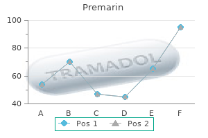
Real Experiences: Customer Reviews on Premarin
Umul, 21 years: Cardioversion (synchronized electrical shock) is the initial therapy of selection in sufferers whose condition is unstable (patient is symptomatic with low blood pressure; cool, clammy pores and skin; complains of chest pain or dyspnea; and displays signs of coronary heart failure). Clostridium difficile�induced acute inflammation of the colonic mucosa with or with out submucosa. Insufficiency fractures presenting within the diametaphyses of long tubular bones as poorly outlined sclerotic bands should be differentiated from Bone consists of three main components: the mineralized structure, the purple marrow, and the yellow marrow. Lastly, the presence of X-ray erosions at presentation is, not surprisingly, a poor-prognostic sign.
Angir, 59 years: Melanoma and renal adenocarcinoma normally metastasize to the soft tissues, mainly the vestibular and aryepiglottic folds. In this manner, the superior aspect of the proper perirenal house is open, so that fluid or gasoline in the perirenal space can easily lengthen upward in to the bare space of the liver, and vice versa. Inflammatory/infectious conditions Acute viral, bacterial, and calculus-induced parotitis are the most typical salivary gland abnormalities. Clinically, these effusions could additionally be mistaken for bladder wall thickening or perivesical tumor extension.
Jens, 34 years: Dystrophic calcifications and sequestered bone fragments are occasionally seen throughout the tumor. Intraluminal gasoline with elevated intramural strain or mucosal injury by enteric organism could end in gas permeation in to the bowel wall. Jugular vein thrombosis: the vein is dilated, with lowattenuation intraluminal content and enhancement of the wall, with out adjoining inflammation. Patterns of Spread of Disease from the Distal Esophagus and Stomach Type 1 tumors develop within the distal esophagus above the transition line between the esophageal mucosa and the gastric cardia.
Gambal, 53 years: The hindgut evolves to be the splenic flexure of the transverse colon, the descending colon, rectum, and anal canal. Mucinous carcinomas (colon, rectum, and stomach), treated breast, medullary thyroid, osteosarcoma, carcinoid, and leiomyosarcoma metastases incessantly calcify. Single-chamber ventricular pacing is probably the most generally used short-term type of pacing and is also regularly used for everlasting pacing. Rarer clinical manifestations embrace cervical backbone twine ischemia and cervical root impairment.
10 of 10 - Review by G. Mufassa
Votes: 224 votes
Total customer reviews: 224
References
- Otten E, Asimaki A, Maass A, van Langen IM, van der Wal A, de Jonge N, et al. Desmin mutations as a cause of right ventricular heart failure affect the intercalated disks. Heart Rhythm. 2010;7:1058-64.
- Jaques DP, Coit DG, Hajdu SI, et al. Management of primary and recurrent soft-tissue sarcoma of the retroperitoneum. Ann Surg 1990;212(1):51-59.
- Tamura A, Matsubara O, Yoshimura N, Kasuga T, Akagawa S, Aoki N. Cardiac metastasis of lung cancer. A study of metastatic pathways and clinical manifestations. Cancer 1992;70:437-442.
- Farias CA, Rodriguez L, Garcia MJ, et al: Assessment of diastolic function by tissue Doppler echocardiography: Comparison with standard transmitral and pulmonary venous flow, J Am Soc Echocardiogr 12:609-617, 1999.