
Montelukast dosages: 10 mg, 5 mg, 4 mg
Montelukast packs: 30 pills, 60 pills, 90 pills, 120 pills, 180 pills, 270 pills, 360 pills
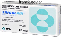
Buy cheap montelukast 10 mg online
An isoperistaltic afferent limb is created from a 10 cm phase of proximal ileum and the ureters are anastomosed to the afferent limb. Bowel continuity is restored by performing a side-to-side or end-to-end ileal anastomosis. The patient empties the pouch by rising belly stress and stress-free the pelvic ground musculature. Contrast infusion is restricted to 200 ml, to be able to keep away from stressing the model new anastomotic suture lines. Extravasation may happen from the suture lines within the reservoir, on the ureteral pouch anastomosis, or on the urethral pouch anastomosis. Reflux is often seen in to the ureters if the Studer pouch is filled to its capability. Anastomotic strictures can be dilated and stented, however long-term outcomes of ballon dilation tend to be disappointing in this group of sufferers. Pouchogram at three weeks after surgical procedure demonstrates a large extravasation on the inferior facet of the anastomosis which was drained percutaneously (long arrow). There is a drainage catheter inside the pouch to promote its therapeutic (short arrows). A section of the terminal ileum is isolated from the rest of the ileum, and brought to the skin floor because the catheterizing stoma. With current strategies, no makes an attempt are made to make the ureteral anastomoses to the pouch nonrefluxing. Diagnosis and management of colovesical fistulae: six-year experience of 90 consecutive circumstances. Diagnosis of blunt bladder injury: a prospective comparative examine of computed tomography cystography and standard retrograde cystography. Experience in one hundred sufferers with an ileal low stress bladder substitute mixed with an afferent tubular isoperistaltic phase. Evaluation of the urothelium of the pyelocalyceal methods and ureters is crucial in sufferers with a history of hematuria (gross or microscopic), and in sufferers with a history of urothelial most cancers. An additional indication is evaluation of the residual ureteral stump in a patient who has undergone a simple nephrectomy for benign disease, or radical nephrectomy for renal cell carcinoma; no ureteral stump is left behind in sufferers with urothelial carcinoma of the upper urinary tract, where the kidney and the complete ureter are eliminated along with a cuff of the urinary bladder at the ureteral insertion site. Technique There are two major methods used to carry out a retrograde research of the amassing system, both of which require cystoscopy, normally carried out by a urologist. Contrast may be injected either via a catheter positioned within the renal pelvis or proximal ureters (also often known as a catheter retrograde pyeloureterogram), or through a catheter positioned at the ureteral orifice (also known as an occlusive-tip or bulb retrograde ureterogram). Although there are stories of major ureteral catheterization by radiologists utilizing fluoroscopic guidance alone, or by cystoscopy carried out by radiologists,1,2 catheterization of the ureteral orifices through the urethra is most frequently carried out by urologists with cystoscopic guidance. An occlusive-tip or bulb retrograde ureterogram is carried out to consider the ureter alone, often when a earlier. However, small urothelial lesions within the pyelocalyceal system is probably not detectable on a single projection, which is the rationale a catheter retrograde pyelogram. There is a cystoscope within the bladder (thick arrow), and a catheter with an olive-shaped tip at the left ureteral orifice (arrow) which was used to inject the contrast. Note the externally rotated hips due to the lithotomy place necessary for cystoscopy. It is also the only methodology by which the urothelium of a ureteral stump could be evaluated. Evaluation of the pyelocalyceal methods requires a number of views for full and optimum visualization, and is finest carried out with a catheter passed in to the renal amassing system. Retrograde catheter placement usually requires common or spinal anesthesia, notably in male sufferers,3 and is done with cystoscopic guidance. Catheter retrograde pyelogram Patient preparation Since the retrograde catheter placement requires anesthesia or conscious sedation on the very minimum, the sufferers are advised to fast in a single day. In sufferers with a history of serious opposed response to radiographic distinction brokers, a standard steroid prep is prudent. Viscous lidocaine jelly applied to the urethral meatus may be useful, but sufferers may require intravenous or intramuscular pain drugs to successfully full the process. The position of the retrograde catheter at the start of the examine can also be assessed.
10 mg montelukast mastercard
Transient ischemia of the right colon has been associated with hypotension and numerous vascular reactions related to cocaine, penicillin, or oral contraceptive use. This form is normally reversible, therapeutic inside syndrome have a optimistic household historical past, the vast majority of PeutzJeghers sufferers may have just a few, large, pedunculated polyps within the colon, not a carpet of small lesions. Spot radiograph carried out throughout single distinction barium enema reveals smooth-surfaced hemispheric nodules all through the distal descending and sigmoid colon. However, a distinction enema, if carried out, will demonstrate indicators of acute ischemia in 80�90% of sufferers, as easy, round mucosal elevations, termed thumbprinting and thick, transversely oriented folds. Mucosal ischemia alone may be manifested as a colonic urticarial sample, with relatively flat, polygonal islands separated by skinny bariumfilled grooves. If a contrast enema is performed, small or large ulcers of punctate or longitudinal form may be demonstrated. Chronic radiation colitis is a type of continual ischemia, owing to progressive obliterative endarteritis. Barium research are usually carried out in the persistent part, to exclude other causes of bloody discharge, diarrhea, or decrease stomach ache. The mucosal atrophy and wall fibrosis of radiation colitis is manifested as tubular, featureless, narrow colon. This sometimes occurs within the rectum, as a outcome of most radiation is carried out for prostatic or cervical cancer. Spot radiograph of the transverse colon demonstrates quite a few 3�6 mm flat polygonal islands of mucosa separated by barium-filled grooves. Although originally described in urticaria, this radiographic pattern is normally seen in diseases causing mucosal ischemia associated with colonic dilatation due to obstruction or adynamic ileus or in sufferers with a variety of acute infections. Spot radiograph of the proximal sigmoid colon shows a 4 cm mild narrowing (arrow) with smooth, tapered margins and mildly nodular mucosa. They are sometimes manifested as clean, undulating, or lobulated folds extending as a lot as 3 cm from the anorectal junction. In different patients, inside hemorrhoids might appear as a bunch of multiple small, smooth, ovoid, submucusoal-appearing nodules in contiguity with anorectal junction, resembling a "bunch of grapes". Therefore, if nodules on the anorectal junction have an irregular contour or floor, or if lobulated folds lengthen higher than three cm from the anorectal junction, endoscopy with biopsy should be performed to exclude a rectal carcinoma. Other polypoid lesions may be seen at the anorectal junction, such as an inflammatory cloacogenic polyp. Image from overhead radiograph demonstrates a mildly narrow tubular rectosigmoid colon with finely granular mucosa within the rectum. Spot radiograph demonstrates four 4�6 mm, ovoid, smooth-surfaced elevations (arrows) grouped collectively just proximal to the anorectal junction. A smoothsurfaced mass is seen on the inferior wall of the sigmoid colon (arrowheads). Colonic wall thickening is due to hyperplasia and fibrosis of the muscularis propria because of infiltrating endometrial tissue. Endometriosis Ectopic endometrial tissue primarily entails the peritoneal surfaces of pelvic organs, in particular the ovaries, fallopian tubes, and rectouterine area (pouch of Douglas). The serosa and subserosal fats of the rectosigmoid junction and sigmoid colon is extra regularly involved than that of the terminal ileum. Endometrial tissue may burrow, nevertheless, in to the muscularis propria, submucosa, and even mucosa of pelvic bowel loops. As the endometrial tissue passes via the proliferative and secretory phases of the menstrual cycle, bleeding, necrosis, and regeneration of endometrial tissue leads to serosal puckering and extensive subserosal fibrosis. The findings are indistinguishable from intraperitoneal metastasis, however the age of the woman and clinical history are guides to the diagnosis. Rarely, deeper bowel wall invasion might end in a easy, polypoid mass or annular narrowing. Findings at defecography Rectal intussusception Asymmetric or concentric telescoping of a proximal portion of the rectum in to a extra distal portion of the rectum is termed intussusception. Invagination of rectal wall in to the anal canal is irregular, nevertheless, resulting in sensation of incomplete evacuation, obstructed defecation, and solitary rectal ulcer syndrome.
Diseases
- Dystonia musculorum deformans type 2
- Cat Rodrigues syndrome
- Bone tumor (generic term)
- Nevi flammei, familial multiple
- Human monocytic ehrlichiosis
- Physical urticaria
10 mg montelukast purchase free shipping
Signal abnormality related to hepatic dysfunction (alcoholic cirrhosis, hepatitis, portal-systemic shunts) probably related to elevated serum ammonia and manganese levels. Usually presents after age forty y with progressive movement problems (choreoathetosis, rigidity, hypokinesia); behavioral and progressive psychological dysfunction/dementia. Juvenile Huntington disease also happens in a small variety of sufferers in the second decade. Patients present with rigidity, hypokinesia, seizures, and/or progressive mental dysfunction. Axial picture reveals zones of decreased attenuation in the thalami and to a lesser extent in the basal ganglia. Axial image shows bilateral calcifications in the basal ganglia, thalami, and cerebral white matter. Severe disorder of neuronal migration (weeks 7�16 of gestation) with absent or incomplete formation of gyri, sulci, and sylvian fissures. Pachygyria (nonlissencephalic cortical dysplasia) Gray matter heterotopia Thick gyri with shallow sulci involving all or portions of the mind. Thickened cortex with relatively clean graywhite interface could have areas of decreased attenuation in the white matter (gliosis). Laminar heterotopia appears as a band or bands of grey matter inside the cerebral white matter. Cleft in mind extending from the ventricle to cortical surface lined by heterotopic gray matter. Ischemia or insult to portion of germinal matrix seen before hemisphere formation. Nonmalignant lesions in white matter related to tuberous sclerosis, consisting of areas of demyelination and/or dysplastic white matter along pathways of radial glial fibers during neuronal migration. Sporadic leukoencephalopathy, additionally referred to as fibrinoid leukoencephalopathy, presents in first year of life with macrocephaly, progressive psychomotor retardation, resulting usually in demise throughout early childhood; additionally juvenile and grownup forms. Axial postcontrast photographs in an 11-month-old woman present zones of low attenuation within the white matter of each cerebral hemispheres, most prominently in the frontal lobes. Heterogeneous or diffuse decreased attenuation in cerebral white matter; initially entails the subcortical white matter with progression to the other remaining white matter; with or with out involvement of cerebellum and brainstem; no contrast enhancement; progressive cerebral and cerebellar atrophy. Symmetric diffuse zones of decreased attenuation in deep cerebral/periventricular white matter with progression of abnormal attenuation peripherally to involve the subcortical white matter; decreased attenuation involving the cerebellar white matter; no distinction enhancement; progressive atrophy. Comments Autosomal recessive (usually happens in Ashkenazi Jews), spongy degeneration dysfunction of the brain attributable to deficiency of aspartoacylase (from abnormal locus on chromosome 17-short arm) resulting in N-acetylaspartic aciduria and deposits in mind and plasma; presents in infancy with macrocephaly, hypotonia, seizures, spasticity, and optic atrophy; dying typically happens in second year. X-linked (type 1) or autosomal recessive (type 2) leukodystrophy; 5 subtypes; deficiency of proteolipid part of myelin; abnormality on chromosome Xq22; presentation during neonatal period (type 2)/ infancy (type 1) with abnormal eye actions, nystagmus, and delayed psychomotor improvement; dying in first decade; males females. Three subtypes relying on onset: late childish form (80%), juvenile kind, and adult type. Progressive neurologic deterioration with peripheral neuropathy, gait problems, and cognitive dysfunction resulting in demise. X-linked recessive leukodystrophy (males) involving chromosome Xq28 with useful deficiency of the peroxisomal enzyme acyl- coenzyme A (CoA) synthetase, resulting in irregular metabolism and breakdown of very lengthy chain fatty acids. These fatty acids accumulate in many tissues, including mind, with resultant demyelination, irritation, gliosis, and necrosis. Onset three to 10 y, with psychomotor retardation, seizures, hypotonia, facial dysmorphism, progressive and deterioration. Also known as globoid cell leukodystrophy, autosomal recessive dysfunction involving chromosome 14q24. Seizures, psychomotor dysfunction, optic atrophy, and progressive neurologic deterioration leading to demise. Canavan�van Bogaert�Bertrand disease Pelizaeus�Merzbacher illness Metachromatic leukodystrophy Childhood adrenoleukodystrophy Zones with decreased attenuation normally in parietooccipital periventricular white matter and corpus callosum; progression of abnormality to the remaining cerebral white matter, with or with out distinction enhancement at areas of lively demyelination/ inflammation. Krabbe disease Symmetric confluent zones of decreased attenuation involving the periventricular white matter with progressive involvement towards the subcortical white matter; cerebral white matter concerned cerebellar white matter; no contrast enhancement; progressive cerebral atrophy.
Montelukast 4 mg purchase without prescription
The differential analysis for varices includes submucosally spreading tumors and esophagitis with thickened folds due to edema and inflammation extending in to the submucosa. When considered en face on double distinction esophagography, esophageal intramural pseudodiverticula are sometimes mistaken for tiny ulcers. When seen in profile, nevertheless, they usually appear to be floating or levitating exterior the wall of the esophagus without any obvious communication with the lumen. Downhill varices One of the more widespread causes of downhill varices is bronchogenic carcinoma with metastases to the mediastinum causing obstruction of the superior vena cava. Downhill varices sometimes appear as serpiginous filling defects which, unlike uphill esophageal varices, are confined to the higher or midesophagus. Uphill varices are caused by portal hypertension with elevated pressure in the portal venous system transmitted upward by way of dilated esophageal collaterals to the superior vena cava. Whether uphill or downhill, varices are important because of the chance of higher gastrointestinal bleeding. Esophageal food impactions In adults, esophageal foreign body impactions are most commonly brought on by inadequately chewed items of meat. Large serpiginous defects are seen within the lower third of the thoracic esophagus due to uphill esophageal varices associated with portal hypertension. Contrast studies are sometimes performed in patients with suspected meals impaction to verify the presence of obstruction, determine its location, and rule out esophageal perforation. An impacted meals bolus typically appears as a polypoid defect with an irregular meniscus superiorly. A follow-up barium examine may subsequently be of value 1 to 2 weeks after the impaction has resolved to decide whether or not a pathologic area of narrowing precipitated this impaction. Scalloped defects (arrows) are seen in the higher thoracic esophagus above the aortic arch secondary to downhill esophageal varices associated with obstruction of the superior vena cava. The varices are fully effaced on another view with larger distention of the higher thoracic esophagus. There is a sophisticated infiltrating carcinoma of the midesophagus (black arrows) with barium getting into the airway via an esophagobronchial fistula (white arrow). A linear collection of barium (arrows) is seen in profile on the proper lateral wall of the distal esophagus. A linear ulcer from reflux esophagitis may have an analogous look, however the appropriate diagnosis was instructed by the clinical historical past. When an esophageal-airway fistula is suspected on medical grounds, barium must be used as a substitute of water-soluble distinction brokers, because these hyperosmolar brokers might trigger extreme pulmonary edema if a fistula is present. When an esophagopleural fistula is suspected, the presence and placement of the fistula could be confirmed by research utilizing water-soluble distinction agents. Despite the dramatic scientific presentation, most esophageal hematomas resolve spontaneously in 1 to 2 weeks on conservative therapy. Such tears are usually attributable to violent retching or vomiting after an alcoholic binge. These tears could occasionally be recognized on esophagography as linear collections of barium in the distal esophagus at or near the gastroesophageal junction. Endoscopy is the most common explanation for esophageal perforation, accounting for up to 75% of circumstances. A large, ovoid assortment (white arrows) of water-soluble distinction materials is seen tracking in the wall of the esophagus with a skinny radiolucent stripe (small black arrows) separating contrast in the collection from distinction within the lumen. This intramural hematoma resulted from attempted endoscopic dilatation of a stricture in the upper esophagus. Also observe the presence of a small, sealed-off perforation (large black arrow) at the web site of the stricture. The hematoma is filling with barium from a laceration at the web site of the perforation. There is focal extravasation of water-soluble distinction material from a full-thickness perforation of the left lateral wall of the distal esophagus (black arrows) in to the left aspect of the mediastinum (white arrows). This affected person offered with sudden onset of acute substernal pain precipitated by extreme retching after an alcoholic binge. In distinction, thoracic esophageal perforation may be manifested on chest radiographs by mediastinal widening, pneumomediastinum, and a pleural effusion or hydropneumothorax. Though barium is the most delicate contrast agent for detecting small leaks, it can doubtlessly trigger a granulomatous reaction within the mediastinum and may persist indefinitely, compromising follow-up research to assess healing of the leak.

Purchase generic montelukast canada
Early debridement and operative fixation of fractures may be undertaken if the patient is sufficiently stable. Examination of the parotid duct, branches of the facial nerve, and main vessels is finest accomplished in the operating room. In uncommon instances, uncontrollable hemorrhage might require ligation of the ipsilateral exterior carotid artery. Massive facial injury can lead to airway obstruction either by lack of the supporting bony framework of the face or by accumulation of blood, particles such as fractured enamel, edema, or tissue flaps that occlude the larynx. Immediate restoration of a patent airway is the best precedence in trauma administration. Initially, simple airway maneuvers corresponding to chin raise, suctioning blood and secretions, and placement of an oral airway ought to be tried. If these are unsuccessful in restoring air flow, a definitive airway must be obtained rapidly. Orotracheal intubation is troublesome due to huge bleeding and edema, distorted anatomic landmarks, and particles, together with avulsed teeth, fragments of bone, and bullet fragments. Use of paralytic agents for orotracheal intubation could trigger lack of voluntary muscle maintenance of a patent airway, however however, will facilitate intubation in a struggling, hypoxic patient. Attempted awake orotracheal intubation, though typically recommended in these cases, is equally extraordinarily tough and usually unsuccessful. Consequently, use of a cricothyrotomy is usually the only viable option to establish a patent airway and must be employed early within the administration. Once the airway is secured, the spine is immobilized and the rest of the first survey and resuscitative interventions are accomplished. Massive facial injuries are dramatic and infrequently distract clinicians from a systematic major survey. Repair of facial lacerations can produce surprisingly good results, providing that tissue has not been avulsed and the arterial supply is intact. Debridement of tissue should be kept to a minimal, and tissues ought to be closed in layers, with individual muscle layers, subcutaneous tissues, and pores and skin closed individually. Airway administration is extremely difficult in these instances, as anatomic landmarks are severely distorted. Primary suturing is normally highly profitable because of the wonderful blood supply of the scalp. The cricothyroidotomy incision could additionally be transverse or vertical, about four fingers above the suprasternal notch (right). This patient ought to bear radiological analysis of the neck, chest, and stomach to find the lacking teeth. Oral Trauma Lacerations of the lips happen commonly because the lip is crushed between a hanging object and the underlying tooth. Because the blood supply to the lips is great, uncomplicated healing is the rule. However, care have to be taken to properly align lacerations that traverse the vermilion border as a outcome of even minor misalignment on this area is noticeable and disfiguring. With through-and-through lacerations involving the pores and skin and mucosal surfaces of the mouth, the mucosal laceration is repaired first, adopted by pores and skin closure. Lacerations of the tongue must be repaired utilizing absorbable sutures after elimination of clots and irrigation of the wound. Because of its rich vascularity, the tongue is able to huge swelling, and delayed airway compromise is possible. Missing enamel must be accounted for and x-rays of the neck, chest, and abdomen should be obtained in the applicable instances. In instances of aspiration in the trachebronchial tree endocopy and removal of the tooth ought to be carried out to be able to keep away from critical infectious complications within the lung. Lacerations within the vicinity of the tragus should immediate cautious examination of the muscular tissues of facial expression as a outcome of facial nerve harm is frequently missed through the preliminary examination of sufferers with multiple trauma. Injury of the smaller branches of the facial nerve may also advantage exploration and surgical restore depending on the severity of the deficit. Care must be taken to moisten the conjunctivae with artificial tears to keep away from desiccation and ulceration of the cornea because of incomplete closure of the eyelid. Lacerations of the parotid gland might lead to delayed sialocele or salivary cutaneous fistula.
Buy discount montelukast line
Benazepril (benazepril, Lotensin) Indication Hypertension Dosage Adults the usual initial dose is 10 mg once every day. Dosage-Diabetic nephropathy Adults 25 mg 3 occasions day by day in type 1 diabetes mellitus Dosage-Left ventricular dysfunction after myocardial infarction Adults Initiate with 6. A target dose of fifty mg 3 times per day could also be achieved over the subsequent several weeks. Fosinopril (fosinopril, Monopril) Indications Hypertension Heart failure Dosage-Hypertension Adults Initiate with 10 mg/d; dosage could additionally be elevated to the standard effective dose of 20-40 mg/d. The patient should be observed beneath medical supervision for a minimal of 2 h for the presence of hypotension or orthostasis following the preliminary dose of fosinopril. An initial dose of 5 mg could also be utilized in sufferers with average to severe renal impairment or in those that have been vigorously diuresed. Patients with a low systolic blood strain (120 mmHg) when therapy is initiated or in the course of the first three days after the infarct must be given a decrease dose of two. If hypotension occurs (systolic blood stress 100 mmHg), a every day maintenance dose of 5 mg may be given with short-term reductions to 2. Lisinopril (Prinivil, Zestril) Indications Hypertension Heart failure Acute myocardial infarction Dosage-Hypertension Adults Initiate with 10 mg/d; dosage could also be adjusted to the standard effective dose of 10-40 mg/d in accordance with response. Dosage-Heart failure Adults the usual initial dose is 5 mg/d administered underneath shut medical observation, particularly in sufferers with low blood strain. Another dose of 5 mg could additionally be given 24 h later, followed by 10 mg at 48 h, then 10 mg once every day thereafter for six weeks. Patients ought to obtain, as acceptable, the standard beneficial remedies, such as thrombolytics, aspirin, and 6. Moexipril (Univasc) Indication Hypertension Dosage Adults the standard preliminary dose is 7. Dosage could additionally be elevated progressively to a maximum of 30 mg/d (given in one or two divided doses) in accordance with response. Note: Moexipril should be taken on an empty abdomen, ideally 1 h prior to a meal. For patients older than 70 years and for sufferers with CrCl of 30-60 mL/min, provoke with 2 mg once every day and titrate in accordance with response to a maximum of 8 mg/d. Stable coronary artery disease the standard starting dose is 4 mg as quickly as daily for 2 weeks. The dose can then be increased as tolerated to a maintenance dose of 8 mg as quickly as day by day. In aged sufferers (> 70 yrs), provoke with a decrease dose of 2 mg daily for the first week, adopted by four mg once day by day in the second week and 8 mg once daily for upkeep dose if tolerated. Dosage-Heart failure Adults the standard initial dose is 5 mg twice daily titrated according to medical response. Preparations Accupril (Parke-Davis/Pfizer); quinapril (generic): 5, 10, 20, 40 mg tablets Fixed-Dose Combinations for Treatment of Hypertension: Accuretic-quinapril/hydrochlorothiazide mixture tablets: 10 mg/12. Ramipril (ramipril, Altace) Indications Hypertension Heart failure postmyocardial infarction Reduction in threat of myocardial infarction, stroke, and death from cardiovascular causes Dosage-Hypertension Adults the same old preliminary dose is 2. Dosage could additionally be titrated upward till blood stress is managed or to a maximum of 5 mg/d. Dosage-Heart failure post myocardial infarction Adults the standard preliminary dose is 2. Quinapril (quinapril, Accupril) Indications Hypertension Heart failure Dosage-Hypertension Adults the standard initial dose is 10 or 20 mg once daily in patients not receiving diuretics. This dose could additionally be increased, at intervals of no less than 2 weeks, to a most of eighty mg/d (given as a single dose or in two divided doses) according to response. For sufferers with creatinine clearance of 31-60 mL/min, the preliminary dose should be 5 mg every day. For patients with creatinine clearance of 10-30 mL/min, the preliminary dose must be 2. Candesartan (Atacand) Indication Hypertension Heart failure Dosage Adults Hypertension the standard preliminary dose is 16 mg once daily. Trandolapril (Mavik) Indications Hypertension Heart failure postmyocardial infarction Left ventricular dysfunction postmyocardial infarction Dosage-Hypertension Adults the usual preliminary dose is 1 mg/d in nonblack patients and a pair of mg/d in black patients.
Seven Barks (Hydrangea). Montelukast.
- Are there any interactions with medications?
- Are there safety concerns?
- How does Hydrangea work?
- Dosing considerations for Hydrangea.
- What is Hydrangea?
- Enlarged prostate, prostate and bladder infections, kidney stones, and hayfever.
Source: http://www.rxlist.com/script/main/art.asp?articlekey=96653
Buy generic montelukast 4 mg line
The Ponseti approach is lengthy established however has taken time to gain wide acceptance � from the mid Nineteen Nineties it has turn out to be the treatment of choice in all new instances. Dorsalis pedis artery could also be absent Examine foot creases: medial, plantar, posterior Affected limb could also be shortened, calf muscle is atrophic, and foot is brief compared to opposite side. Ponseti casting method Serial casts are utilized weekly for approximately the first 6 weeks of life. A three-incision approach allows harvesting, proximal pull-through and distal reimplantation (classically in to the ossified intermediate or lateral cuneiform). If patients are older than 5 years they may need bony procedures to straighten the lateral border of the foot. The envelope for successful treatment with Ponseti casting has been extending, and it might be tried even in late presenting or relapsing cases earlier than surgery. Bony surgical procedure adduction deformity Calcaneocuboid fusion (Dillwyn Evans procedure) Metatarsal osteotomy. Hindfoot deformity Varus heel Opening medial wedge or laterally primarily based closing wedge osteotomy of the calcaneum Residual cavus and adductus Wedge tarsectomy Triple arthrodesis salvage process for stiff, painful foot in sufferers >12 years old. Post-reduction abduction splinting is required to keep the place � for 23 hours per day for three months after which 12 hours per day until age 5 (or as close to this as can realistically be achieved). Surgery Posteromedial launch Incisions vary; nonetheless, the standard posteromedial release has been performed through a medial curvilinear incision, tracking the tibial neurovascular bundle from the calf behind the medial malleolus and in to the foot. Medial forefoot, lateral translation of midfoot with heel in valgus Natural historical past uncertain � might resolve spontaneously, response to casting uncertain owing to multiplanar deformity. Calcaneovalgus Dorsiflexion positioning of the feet is a standard (one in 1000 live births) result of uterine close-packing May be related to posteromedial bowing of tibia � both are benign Foot usually spontaneously resolves over a period of weeks after birth Serial casting may be considered if fails to appropriate to past plantigrade It is important to differentiate from congenital vertical talus: Congenital vertical talus is less versatile than calcaneovalgus Congenital vertical talus navicular has mounted dorsal dislocation on talus A plantarflexion radiograph is diagnostic (see section on congenital vertical talus). Congenital vertical talus this uncommon situation is a crucial exclusion when evaluating a paediatric foot deformity. Its principal features are: the navicular is dislocated dorsally off the talar head There is equinus of the hindfoot the cuboid is displaced dorsally the dorsal gentle tissues are tight. A lateral compelled plantarflexion radiograph provides a pathognomonic appearance of the forefoot remaining dorsal to and misaligned with the long axis of the talus. Traditionally this has concerned single or two-stage releases; however, K-wire fixation of an aligned plantarflexed foot followed by further serial casting has recently been described and is a promising various strategy. Child could sometimes complain of discomfort; their toe could catch when placing their socks on, callosity of the dorsum of the toe with footwear. It is technically difficult and sometimes produces stiff toes in extension with a rotational element. Examination corner Paeds oral 1: Clinical picture of kid with curly toes Management is conservative and operative. Variable prevalence � approximately 25% of adolescents 80% female Strong family historical past � X-linked dominant/autosomal dominant with variable penetrance Metatarsus primus varus is a risk issue (first to second intermetatarsal ray angle of >10�). Epidemiology from valgus to neutral and even in to varus (demonstrating normal subtalar function). This is a standard variant in kids and is almost universally present in infants. Rigid pes planus Is a rare downside, however when it happens is usually brought on by a tarsal coalition (also generally recognized as peroneal spastic flat foot), however necessary differentials embody: Congenital vertical talus Juvenile idiopathic arthritis Trauma to subtalar joint Requires treatment when symptomatic. Pes planus Pes planus (flat foot) describes melancholy of the medial longitudinal arch of the foot. There is associated valgus hindfoot and supination/abduction of the forefoot on the hindfoot. In a compliant child this is done by observing the toes from behind and asking them to stand on tiptoe. At the identical time the heel will correct Tarsal coalition Definition An autosomal dominant dysfunction of primitive mesenchymal segmentation and differentiation leading to fusion of tarsal bones and rigid flat foot. The coalitions can develop from fibrous (syndesmosis), through cartilaginous (synchondrosis), to osseous (synostosis). Epidemiology Prevalence between 2 and 6% relying on diagnostic methodology (clinical, radiographic) Bilateral in 50% 385 Section 6: the paediatric oral Multiple coalitions occur in a single in five cases Three in 4 are said to be asymptomatic. However, many youngsters who current with ache have proof of degenerative modifications within the hindfoot.
Cheap montelukast 4 mg with mastercard
Urethra In females, the urethra is short, of comparatively uniform caliber, and programs obliquely downward and forward from the bladder neck to the urethral meatus. The posterior urethra is greatest imaged with a voiding cystourethrogram, as it has smooth muscle in its partitions (referred to as the posterior urethral sphincter complex) which is constricted within the resting state and completely relaxed only during voiding. The posterior urethra extends from the bladder neck to the exterior sphincter and consists of the prostatic urethra and the membranous urethra. The prostatic urethra passes via the prostate and is identified by an impression on its posterior facet, the verumontanum; the utricle (a vestige of the mullerian ducts) opens in the middle of the verumontanum and the prostatic ducts and the ejaculatory ducts open on either side of the verumontanum. The membranous urethra is approximately 1 cm long and is located instantly distal to the verumontanum. The bulbomembranous junction has a cone-shaped appearance and the urethral caliber changes on the stage of the membranous urethra (thick arrow) where the external sphincter is located. The posterior urethra is opacified because of retrograde flow of contrast but the prostatic and membranous urethra (both parts of the posterior urethra) totally distend only when the patient voids (B). The bladder is trabeculated with several small diverticulae as a outcome of bladder outlet obstruction attributable to prostate enlargement. There is retrograde filling of the prostatic ducts (arrows), giving a "prostatogram. Evaluation for leaks and fistulas Bladder leaks Evaluation of the urinary bladder for leak or extravasation could additionally be required in the following scientific conditions: blunt or penetrating abdominopelvic trauma; following surgical procedure corresponding to partial cystectomy for neoplasm, nephroureterectomy with bladder cuff excision for upper observe urothelial carcinoma, ureteral reimplantation, renal transplant with anastomosis of the donor ureter to urinary bladder (ureteroneocystostomy), 196 Chapter 10: Fluoroscopic analysis of the bladder, urethra, and urinary diversions A B. When the bladder was stuffed additional till a detrusor contraction occurred, intraperitoneal extravasation is seen, with contrast outlining bowel loops within the pelvis. Intraperitoneal accidents as a end result of blunt trauma usually require operative repair but biopsy associated leaks will usually heal with Foley catheter drainage alone. Multiple websites of extravasation are seen in to the perivesical extraperitoneal house. Images ought to be obtained in multiple obliques for optimum evaluation and confident exclusion of a leak. Bladder trauma Motor car crashes are the most common explanation for injuries to the bladder. The majority of patients with bladder injuries as a outcome of blunt trauma have related pelvic fractures (60�90% of patients), whereas one-third of sufferers with pelvic fractures may have an associated bladder injury. The bladder can be injured by penetrating trauma because of stab wounds or gunshot wounds, or be concerned in iatrogenic accidents similar to after deep bladder biopsies. This distinction is crucial factor in the management of bladder trauma as most intraperitoneal injuries require operative restore of the laceration, while extraperitoneal accidents largely heal with bladder drainage alone. Patients who current with pelvic fractures and have gross hematuria have a excessive chance of bladder harm, ranging from 32 to 85% in numerous sequence,eight,9 and may bear imaging to consider the bladder. In the absence of pelvic fractures, microscopic or gross hematuria are considered to be relative indications for imaging evaluation, with the need for imaging determined by clinical symptoms corresponding to suprapubic pain or voiding difficulties. Extraperitoneal ruptures may be either easy or advanced,6 relying on whether or not the distinction extravasation is confined to the pelvic extraperitoneal house alone (simple), or associated with disruption of fascial planes in order that the extravasated distinction extends exterior the pelvis to involve the anterior abdominal wall, perineum, or the exterior genitalia (complex) 6�8. One-third of bladder accidents are intraperitoneal and outcome from damage to the lower stomach when the urinary bladder is distended. These accidents contain the dome of the bladder, which is roofed by peritoneum, and so an injury at this web site causes intraperitoneal extravasation; there could also be no related pelvic fractures. The presence of radiographic contrast materials exterior the urinary bladder signifies bladder rupture. There is intensive extraperitoneal extravasation which is complex and extends in to the perineum. Most urothelial neoplasms of the bladder are treated with a radical cystectomy for an extirpative remedy. A Foley catheter is positioned to promote bladder therapeutic and a cystogram may be requested to verify full therapeutic of the bladder prior to removal of the Foley catheter. Since the ureter inserts posteriorly on the urinary bladder, pictures in steep contralateral indirect projection are important to evaluate the surgical web site. Small quantity of reflux is seen round the proper ureteral stent in to the right pelvic ureter (arrow) (C). The ureter is anastomosed to the superior dome of the bladder, and the bladder has a tented look on the aspect of the reimplant. The surgical procedures utilized for ureteral reimplantation are either a psoas hitch or a Boari flap combined with a psoas hitch, which are properly described in Rassweiler et al (2007).
Purchase montelukast 5 mg overnight delivery
Detection of poor anal sphincter tone signifies a single distinction enema could also be a simpler examination. Patient mobility may be examined by having the affected person activate the radiographic table top. Non-latex gloves are most well-liked for rectal examination, as anaphylactic reactions to latex in gloves or in rectal tip balloons have been reported. Colonic rest Use of a colonic stress-free agent makes contrast enema more comfortable. Intravenous glucagon permits better colonic distention, bettering radiographic detection of lesions. Rapid injection of glucagon may cause colonic cramps, nausea, or even dry-retching. Contraindications to glucagon embrace a historical past of prior hypersensitivity response to glucagon, pheochromocytoma, or insulinoma. A single contrast barium examination is preferred in patients with colonic obstruction suspected by plain radiograph. Some surgeons choose that a patient with a suspected high-grade obstruction receive a water-soluble contrast enema to avoid having residual barium within the colon prior to emergent surgery. A barium study is contraindicated in a affected person with suspected colonic perforation. Therefore, sufferers with suspected proximal perforation within the proximal colon ought to endure a single contrast enema utilizing an iodinated distinction agent. Patients with a suspected rectal or sigmoid leak could receive a fullstrength water-soluble contrast enema via a Foley catheter. Patients with poor rectal tone or unable to roll 360 degrees ought to endure a single distinction examination, with either barium or water-soluble distinction, depending on the medical history. Patients with redundant transverse or proper colons are tougher to research with double distinction approach. Choice of examination A double distinction (air contrast) barium enema is carried out in outpatients with suspected polyps, non-obstructing carcinoma, diverticular disease, or mild inflammatory bowel illness. A double contrast examination photographs the luminal contour in profile and the mucosal floor en face. The radiologist appears for alteration of the luminal contour, including disappearance of the contour, or protrusion of the contour in to or out of the bowel. Alterations of the Enema tip insertion After being coated with lubricant, a wide bore enema tip is gently inserted in to the rectum. This tip has an connected non-latex enema balloon/insufflator sponge pad to aid retention and separate wider bore blue-colored tube with air bulb to pump air in to the rectum (an air tip named for Roscoe Miller, M. We use this tip for both double and single contrast examinations and discard the big blue insufflator bulb after each use. Spot radiograph of the sigmoid colon reveals scattered diverticula (arrowhead) and delicate round muscle thickening. Spot radiograph of the sigmoid colon obtained with a shallow barium pool demonstrates a 1. For single contrast examinations of the postoperative rectosigmoid colon, rectosigmoid stump, or ileoanal anastomosis/ J pouch, a small bore catheter corresponding to a 22 French Foley catheter is used. A Foley catheter can be used if an anovaginal or rectovaginal fistula is suspected. The radiologist enters the fluoroscopic suite with a "game plan" primarily based on the radiology request slip and another info gleaned from the pc database of endoscopy, pathology, and operative stories and a evaluate of old films, if available. The examination is then tailored to info acquired during a quick patient interview. The affected person turns in to a place that directs the barium column toward the dependent space of curiosity. The radiologist directs the patient to turn so the barium column scrubs and coats the area of curiosity, then instructs the patient to roll so the dense barium column is removed by gravity from the world of curiosity earlier than a spot radiograph is obtained. Other lesions are best demonstrated when the barium pool is totally removed from the picture.
10 mg montelukast otc
Obstructing tumors may cause nausea and vomiting, ulcerated tumors may trigger higher gastrointestinal bleeding, scirrhous tumors could trigger early satiety, and cardiac tumors may cause dysphagia. Concern about lacking gastric most cancers on barium research has sometimes been used as the rationale for performing endoscopy because the initial diagnostic take a look at in sufferers with higher gastrointestinal signs. In one research, nonetheless, double contrast barium examinations confirmed the lesion in 99% of patients with gastric most cancers, and malignant tumor was diagnosed or suspected on the basis of the radiographic findings in 96%. Thus, a high sensitivity may be achieved within the radiographic diagnosis of gastric carcinoma without exposing an inordinate number of sufferers to unnecessary endoscopy. Advanced gastric cancers may be manifested on barium studies as polypoid, ulcerative, infiltrative, or scirrhous tumors. On double contrast research, polypoid lesions on the dependent or posterior wall are seen as filling defects within the barium pool, whereas polypoid lesions on the non-dependent or anterior wall are etched in white by a skinny layer of barium trapped between the edge of the mass and the adjacent mucosa. Occasionally, polypoid carcinomas of the distal gastric antrum might prolapse by way of the pylorus in to the duodenal bulb, appearing as mass lesions on the base of the bulb. Ulcerated carcinomas are these by which a lot of the tumor mass has been replaced by ulceration because of necrosis. The radiographic look of malignant gastric ulcers and their differentiation from benign gastric ulcers have been discussed elsewhere (see earlier part, Benign versus malignant gastric ulcers, and see. Scirrhous gastric carcinomas usually come up within the distal abdomen, gradually extending from the antrum proximally in to the physique and fundus. These tumors may be manifested on barium research by narrowing and rigidity of the stomach associated with mucosal nodularity, thickened folds, and a grossly irregular contour, producing a basic linitis plastica or leather bottle look secondary to a marked desmoplastic response incited by the tumor. This circumferentially infiltrating tumor causes irregular narrowing and ulceration of the gastric antrum. There is marked narrowing of the gastric antrum and body with mucosal nodularity, thickened folds, and an irregular luminal contour. These findings are attribute of a scirrhous tumor with a linitis plastica appearance. With double distinction technique, nonetheless, 40% of all scirrhous carcinomas are found to be localized lesions involving the gastric fundus or physique with sparing of the antrum. Conversely, different patients with scirrhous tumors may have solely minimal loss of distensibility in the abdomen, so the prognosis can be missed if the radiologist relies too closely on the diploma of luminal narrowing as the most important standards for diagnosing these lesions. It is important to be conscious of the constraints of endoscopy in diagnosing scirrhous carcinomas of the abdomen. There is irregular narrowing of the fundus and physique of the abdomen with sparing of the antrum. Also notice speedy emptying of ingested barium from this rigid, noncompliant abdomen in to multiple loops of small bowel. There is focal narrowing of the gastric physique with mucosal nodularity, ulceration, and irregular luminal narrowing due to lymphomatous infiltration of the wall. This look is unimaginable to differentiate from that of a major scirrhous gastric carcinoma. There is loss of distensibility of the gastric antrum with appreciable nodularity of the mucosa because of infiltration of the wall by metastatic tumor. While most sufferers with malignant linitis plastica are discovered to have scirrhous carcinomas, metastatic breast cancer and lymphoma involving the abdomen may produce similar radiographic findings due to a dense infiltrate of metastatic tumor or lymphomatous tissue within the gastric wall. Metastatic breast cancer and lymphoma should due to this fact be included within the differential prognosis of linitis plastica. With the altering distribution of gastric most cancers over the previous half century, carcinoma of the cardia has been identified with elevated frequency. Affected people often current with referred dysphagia to the higher chest, the thoracic inlet, or even the pharynx, so the cardia and fundus should be fastidiously evaluated on barium studies in all patients with dysphagia, regardless of its subjective localization. Some tumors on the cardia may be recognized solely by distortion or obliteration of the cardiac rosette, with relatively refined nodularity, mass effect, or ulceration on this area. A proper lateral double contrast spot image of the gastric fundus reveals obliteration of the conventional anatomic landmarks on the cardia with nodularity and ulceration (white arrows) in this region. Conversely, esophageal carcinomas have a higher degree of esophageal involvement. There is a mass (black arrows) on the higher curvature of the abdomen, containing a large area of central necrosis and cavitation (white arrows).
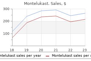
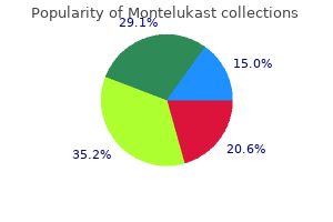
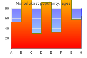
Real Experiences: Customer Reviews on Montelukast
Denpok, 51 years: Subchondral bone adjustments include thickening of the subchondral bone by the laying down of recent bone on existing trabeculae and the formation of bone cysts. Appendix 5 Dose Adjustment in Patients with Renal Insufficiency Drug CrCl:30to60mL/min a-Adrenergic Antagonists Doxazosin Use ordinary dose. Severe hypotension or shock the usual dose for a single direct intravenous injection ranges from 0.
Myxir, 65 years: Plain chest and neck films: They can diagnose international our bodies, fractures, pneumothorax, subcutaneous emphysema, and hematomas. Endophytic progress sample of retinoblastoma: Irregular, strong, and heterogeneous peripheral intraocular mass, usually posterior to the equator, with inward protrusion in to vitreous; related to vitreous seeding. This section of the e-book will take you through areas which are generally examined from the above listing.
Karrypto, 44 years: Spot radiograph of the ileum from enteroclysis shows a big, irregularly contoured barium-filled cavity (arrowheads) increasing beyond the anticipated luminal contour of the bowel. Image obtained during defecation demonstrates regular flattening of the puborectalis muscle (thin arrow), extensive opening of the anal sphincter (thick arrow), and a reasonable anterior rectocele (R) deviating the decrease vagina (V) anteriorly. Superficial dissection the incision is deepened through subcutaneous fat to the fascia overlying the deltoid and pectoralis major muscular tissues.
Karmok, 62 years: Split fractures tend to happen in younger sufferers with stronger bone, and depressed fractures more often happen in the aged patient with osteoporosis. Positive findings are increased subluxation of the vertebrae in either flexion (posterior ligaments disrupted) or extension (anterior ligaments disrupted). Colobomas appear as outpouchings of variable dimension and form arising from the posterior globe.
Rathgar, 21 years: Anterior cord syndrome is often seen in elderly sufferers in conjunction with medical circumstances, significantly arterial embolization from the guts. Autosomal recessive illness in youngsters with cutaneous photosensitivity, progressive neurologic impairment, optic atrophy, cataracts, dwarfism, and thoracic kyphosis. It reaches the thigh by operating beneath the inguinal ligament lateral to the femoral artery mendacity on the psoas and iliacus tendon.
Fasim, 46 years: This realistically provides the candidate round three minutes to assess the patient and provide a prognosis. The physical properties of the steel are tremendously influenced by the grain measurement and the variety of dislocations. Open sucking wounds must be coated with a clear sq. gauze taped only on three sides to keep away from a pressure pneumothorax.
Vibald, 53 years: Position the patient is positioned supine with a sandbag under the buttock of the affected facet. With the patient lying supine, the blood is much less visible than in an upright position, when it forms a clearly seen layer of blood in the dependent portion of the anterior chamber. The loading dose should be adopted by 20 mg of conivaptan administered in a steady intravenous infusion over 24 hours.
Rakus, 26 years: Orthopaedic ceramics could additionally be bioinert (alumina, zirconia) or bioactive (hydroxyapatite, glass ceramic). Compartment stress measurement is a useful adjunct in borderline cases and may simply be carried out by inserting a needle strain transducer inside the compartment in question. Cord) Notes Suprascapular nerve passes beneath transverse ligament of scapula Partly lined by deltoid and trapezius Often not clearly differentiated from the infraspinatus.
10 of 10 - Review by Z. Murat
Votes: 203 votes
Total customer reviews: 203
References
- Androne AS, Hryniewicz K, Hudaihed A, et al. Relation of unrecognized hypervolemia in chronic heart failure to clinical status, hemodynamics, and patient outcomes. Am J Cardiol 2004;93:1254.
- Cappelleri JC, Rosen RC: The Sexual Health Inventory for Men (SHIM): a 5-year review of research and clinical experience, Int J Impotence Res 17:307n319, 2005.
- Ljungman P, Gleaves CA, Meyers JD. Respiratory virus infection in immunocompromised patients. Bone Marrow Transplant. 1989; 4:35-40.
- Rajabally YA, Martey J. Neuropathy in Parkinson disease: Prevalence and determinants. Neurology 2011;77:1947-1950.
- Lemanske RF Jr, Jackson DJ, Gangnon RE, et al. Rhinovirus illnesses during infancy predict subsequent childhood wheezing. J Allergy Clin Immunol 2005; 116: 571-577.
- Feindel CM, David TE: Aortic valve sparing operations: Basic concepts, Int J Cardiol 97:61, 2004.
- Makoul G, Clayman ML. An integrative model of shared decision making in medical encounters. Patient Educ Couns 2006; 60(3):301-12.
- Coriat P, Richer C, Douraki T et al. Influence of chronic angiotensin-converting enzyme inhibition on anesthetic induction. Anesthesiology 1994;81(2):299-307.