Triamterene dosages: 75 mg
Triamterene packs: 30 pills, 60 pills, 90 pills, 120 pills, 180 pills, 270 pills, 360 pills
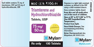
Purchase 75 mg triamterene mastercard
Innate immunity: this second line of protection (if the nonspeciic barrier is breached) is composed of a variety of cells and antimicrobial secretions, and manifests itself by producing inlammation and fever. Functionally, the respiratory system performs ive fundamental capabilities: Filters and humidiies the air and moves it in and out of the lungs. Neurons Nerve cells are known as neurons, and their construction relects the useful traits of a person neuron. Information comes to the neuron largely by way of treelike processes called axons, which terminate on the neuron at specialized junctions known as synapses. Synapses can happen on neuronal processes called dendrites or on the neuronal cell body, called a soma or perikaryon. Neurons convey eferent (motor or output) information via motion potentials that course along a single axon arising from the soma that then synapses on a selective target, normally one other neuron or target cell, such as muscle cells. Common types of neurons embody the next: Unipolar (often referred to as pseudounipolar): a neuron with one axon that divides into two lengthy processes (sensory neurons found in the spinal ganglia of a spinal nerve). Bipolar: a neuron that possesses one axon and one dendrite (rare but found within the retina and olfactory epithelium). Multipolar: a neuron that possesses one axon and two or extra dendrites (the most typical type). Chapter 1 Introduction to the Human Body 23 1 Clinical Focus 1-7 Asthma Asthma could be intrinsic (no clearly defined environmental trigger) or extrinsic (has an outlined trigger). Asthma often outcomes from a hypersensitivity reaction to an allergen (dust, pollen, mold), which leads to irritation of the respiratory passages and easy muscle contraction (narrowing of the passages), swelling (edema) of the epithelium, and increased manufacturing of mucus. Presenting symptoms are often wheezing, shortness of breath, coughing, tachycardia, and emotions of chest tightness. Asthma is a pathologic irritation of the airways and occurs in each children and adults. Normal bronchus Mucosal floor Epithelium Basement membrane Opening of submucosal gland Blood vessel Smooth m. Cartilage Submucosal gland Increased mucus production Early asthmatic response Smooth m. Neurons can range significantly in measurement, starting from a quantity of micrometers to greater than 100 �m in diameter. Neurons might possess quite a few branching dendrites, studded with dendritic spines that improve the receptive area of the neuron many-fold. Ependymal cells: these cells line the ventricles of the brain and the central canal of the spinal wire, which contains cerebrospinal luid. Perineurium: a dense layer of connective tissue that encircles a bundle (fascicle) of nerve ibers. Epineurium: an outer thick connective tissue sheath that encircles bundles of fascicles; that is the "nerve" typically seen grossly coursing all through the human body. Peripheral nerves embrace the 12 pairs of cranial nerves arising from the mind or brainstem and the 31 pairs of spinal nerves arising from the spinal twine. Meninges he mind and spinal cord are surrounded by three membranous connective tissue layers referred to as the meninges. Arachnoid mater: the ine, weblike avascular membrane instantly beneath the dural floor. Pia mater: the fragile membrane of connective tissue that intimately envelops the mind and spinal cord. Somatic and visceral: associated to skin and skeletal muscle (somatic) or to clean muscle, cardiac muscle, and glands (visceral). V Trigeminal Sensory-face sinuses, tooth h Op M tha unwell lm ic ax ary nd Ma ibu lar Motor-mm. Features of the somatic nervous system embrace the following: It is a one-neuron motor system. Anterior ramus Gray ramus communicans Anterior root Sympathetic chain ganglion Free endings Splanchnic n. Sensory neuron of abdominal viscera Sensory Motor Preganglionic sympathetic White ramus communicans Sympathetic chain Preganglionic sympathetic neurons passing to synapse in one other sympathetic chain ganglion Neuroeffector junctions on easy m. Clinically, dermatome maps of the body may be useful in localizing spinal cord or peripheral nerve lesions (see Chapter 2).
Triamterene 75 mg purchase on line
During each step, the practitioner should "look, pay attention, and really feel" to run via the differential analysis. During stabilization of these patients it is necessary to keep in mind the original pathology that necessitated intubation. Sudden decompensation in an asthmatic is a common example of doctor intervention worsening the situation unless the pathophysiology of the arrest is appreciated. A multitrauma patient could have an intrathoracic or intraabdominal disaster, and a septic patient may be deteriorating clinically from lack of source management. Tension pneumothorax can lead to marked hypotension on account of decreased cardiac output and marked hypoxia from V/Q mismatch. This can cause hypotension and decreased cardiac output from decreased venous return and marked hypoxia from V/Q mismatch. Step 1: Disconnect the Patient From the Ventilator that is maybe the easiest step to carry out. In some hospitals, a dedicated intensivist will take cost of those sufferers, but in others, this kind of protection will not be available. This part offers a framework for managing crashing, mechanically ventilated sufferers. The patient can fall anywhere on the spectrum from being ventilated for airway safety, with normal important indicators, blood strain, and oxygen saturation, to being ventilated and in cardiac arrest. Determining where the affected person is on this spectrum, including assessment for hypotension and hypoxia, will dictate how a lot time that the practitioner has to implement rescue strategies. Step 2: Breathing-Hand-Ventilate With 100% Oxygen Ensure that one hundred pc oxygen is being delivered and limit the respiratory price to 8 to 10 breaths/min. Decreased breath sounds could present clues regarding primary stem intubation, pneumothorax, or an atelectatic lung. Feel for subcutaneous crepitus (a sign of pneumothorax) and assess for difficulty in hand ventilating (a signal of low dynamic or static respiratory system compliance). Patients are frequently moved out and in of emergency medical service vehicles, transferred to and from stretchers for imaging research, and turned for procedures or bathing, all of that are capable of dislodging the tube. Determine the power to repair the pilot balloon mechanism with a commercially available equipment. Another fast and readily available technique is to cross an intubating stylet (gum elastic bougie or Eschmann introducer). At least one of these methods to determine proper positioning must be used early sufficient in the resuscitation to correct any airway issues. If time permits, a focused history from the bedside nurse, respiratory therapist, or paramedic and a focused bodily examination will point out which facet of the chest to decompress. It is essential to do not forget that chest tube placement is required in sufferers after needle decompression. Step 2: Perform a Focused Physical Examination Take a basic survey of the affected person. Step three: Assess Gas Exchange Hypoxia can be identified with pulse oximetry if the waveform is reliable. Step four: Check Respiratory Mechanics Determine whether or not peak pressure and plateau stress have changed from their previous values. Peak pressure is a function of volume, resistance to airflow, and respiratory system compliance. Plateau pressure is obtained throughout an inspiratory pause, thus eliminating airflow, and therefore displays only respiratory system compliance. An isolated enhance in peak pressure is indicative of elevated resistance to airflow. An isolated improve in plateau strain is indicative of a lower in respiratory system compliance. Note that plateau stress can never be greater than peak stress and that if plateau pressure rises, so will peak strain. It is important to keep in mind the relationship (peak pressure - plateau pressure). The pressure-time curve can be utilized to decide plateau pressure with an inspiratory maintain. In this situation, the patient desires a higher circulate price than the ventilator is delivering.
Cheap triamterene 75 mg buy on-line
Patients with orthostatic hypotension as the cause for syncope have been older, had extra comorbid situations, and have been found to be extra frequently taking antihypertensive drugs. The investigators concluded that an orthostatic increase in pulse of larger than 25 beats/min constitutes a optimistic tilt take a look at and fewer than 20 beats/min constitutes a unfavorable test (sensitivity of 75%, specificity of 95%, and predictive value of 92% when utilizing near-syncope or a rise in coronary heart rate higher than 25 beats/min). Bradycardia in the face of hemorrhage has generally been considered a preterminal discovering of irreversible shock, and bradycardia has been documented in hypovolemic, acutely aware trauma patients as nicely. This paradoxical bradycardia may be extra regularly related to rapid and massive bleeding. Indications and Contraindications When the volume standing of a patient is assessed with the usage of orthostatic important signs, several factors ought to be remembered. Many components affect orthostatic blood pressure, together with age, preexisting medical situations, medications, and autonomic dysfunction. It should be emphasised that knowledge relating the effect of blood loss to orthostatic important signs are limited to phlebotomized healthy volunteers. Orthostatic important signs could be considered an adjunct for the analysis of any patient with known or suspected loss of blood quantity or a historical past of syncope. Contraindications to orthostatic measures include supine hypotension, the medical syndrome of shock, severely altered psychological status, the setting of attainable spinal accidents, and lower extremity or pelvic fractures. The use of medications that block the normal vasomotor and chronotropic response to orthostatic checks can be a contraindication to using this check for the evaluation of volume status. In these conditions, the primary discovering will be the sensation of near-syncope with little or no change in important signs. In sufferers receiving intravenous rehydration therapy, serial orthostatic very important indicators are widely used to decide the tip point of remedy before launch. In one research, individual orthostatic important signal response to saline infusion in women with hyperemesis gravidarum was related to different measures of rehydration, together with weight achieve and decreased urine particular gravity. A variety of research have been conducted on normotensive, normovolemic patients to assess finish factors for orthostatic vital signal parameters. Such studies have included sitting-tostanding strategies and varying rates of postural modifications,181 together with mendacity times of 5 to 10 minutes and standing occasions of 0 to 2 minutes. Interpretation essentially the most sensitive criteria for orthostasis are tachycardia or symptoms of cerebral hypoperfusion. Specific population-based thresholds for adjustments in pulse fee and blood strain have some value in figuring out patients at high risk for important lack of blood volume, but nice individual variability limits the usage of this system as a screening check. That is, a loss of 500 ml, and occasionally extra, could additionally be associated with a unfavorable orthostatic vital sign assessment. The clinical interpretation of orthostatic changes in blood pressure and pulse varies extensively. Technique To get hold of orthostatic important signs, record the blood stress and pulse after the patient has been within the supine position for two to three minutes (see Box 1. Next, ask the patient to stand and be prepared to help if severe symptoms or syncope develop. [newline]If extreme symptoms develop on standing, defined as syncope or excessive dizziness requiring the affected person to lie down, the take a look at is considered positive and should be terminated. If not symptomatic, allow the affected person to stand for 1 minute and then report the blood stress and pulse. A 1-minute interval resulted within the biggest difference between management and 1000-ml phlebotomy groups in a single research. Delayed capillary refill is a sign of lowered pores and skin turgor, often on account of quantity depletion or limited perfusion. The minimal strain essential to produce blanching yields probably the most reproducible values. Release the nail mattress and start timing with a stopwatch or simply by counting out "one-thousand-one, one-thousand-two" for an approximation of the interval. Interobserver reliability has been shown to be average, with kappa values of lower than zero. An lack of ability to preserve normal physique temperature is indicative of an unlimited number of doubtlessly severe disorders, including infections, neoplasms, shock, toxic reactions, and environmental exposures. Whether taken by the oral, rectal, or tympanic routes, reports of fever at home are very troublesome to interpret in the scientific setting.
Order triamterene 75 mg overnight delivery
A 23-gauge butterfly needle will usually suffice for venipuncture, no matter affected person dimension. Technique As in adults, the standard site for venipuncture in infants and children is the antecubital fossa. These devices are mentioned later on this chapter (see section on Vascular line Placement: Venous and Arterial). Assemble all necessary equipment, particularly needles, out of sight of the child and have equipment within simple attain earlier than beginning. Ask an assistant to help immobilize the affected person when drawing blood from infants and babies. If an extremity vein is to be used, apply a tourniquet proximal to the chosen vein. Cleanse the area surrounding the chosen website of pores and skin penetration with antiseptic answer and allow it to dry. Insert the needle rapidly by way of the skin and advance it slowly into the vein at an angle of roughly 30 levels with the bevel dealing with up. Successful vessel penetration is heralded by a flashback, or move, of blood into the needle hub or butterfly tubing. If the required quantity of blood is bigger than the capability of the attached syringe, pinch off the tubing, remove the stuffed syringe, connect a brand new syringe, and apply gentle suction again after releasing the pinched tubing. After the required quantity of blood is withdrawn, take away the needle and apply a sterile dressing and direct strain to the puncture website. Although peripheral sites for venous blood sampling are preferable in infants, the external jugular and femoral veins may be used for venipuncture throughout resuscitation or when peripheral sites are insufficient. Immobilize the foot in a dependent place, cleanse with antiseptic, and allow to dry. Place a heparinized capillary tube in the drop of blood and permit it to fill by capillary action. Microtainer Tube Method 6 If utilizing a Microtainer tube, contact the collector finish to the drop of blood and allow the blood to move down the wall of the tube to the underside. Do not squeeze foot Capillary tube Microtainer four Fill the tube till the blood reaches the demarcation line on the tube. Maintain the blood in the capillary tube using your index finger to preserve capillary rigidity on the tip of the tube. If venous entry is unavailable, arterial blood may be used for most laboratory exams, together with blood cultures. Venipuncture Using a Butterfly Needle Tourniquet Syringe or Vacutainer connected right here 21- to 25-gauge straight needle or a 21- to 25-gauge butterfly needle connected to a syringe. Puncture the skin after which advance the needle slowly till the jugular vein is entered and a flashback of blood is observed. After the appropriate quantity of blood is obtained, withdraw the needle and apply slight stress to the vessel. Place the kid in an upright position if potential after the needle is removed, and maintain pressure over the puncture web site for three to 5 minutes. In most sufferers, the femoral vein lies medial to the femoral artery and inferior to the inguinal ligament. Identify its location by inserting a mark on the pores and skin simply superior to the femoral triangle. Puncture the skin after which direct the needle or catheter towards the umbilicus at a 30- to 45-degree angle to the pores and skin and simply medial to the pulsation of the femoral artery. Apply pressure over the puncture site in the femoral triangle for at least 5 minutes. The anatomic considerations and technique are mentioned later (see sections on Peripheral Venous Catheterization: Percutaneous and Peripheral Venous Catheterization: Venous Cutdown). Complications Complications of venipuncture embrace hematoma formation, arterial puncture, native infection, injury to adjoining constructions, and phlebitis. Use special care when trying to puncture the external jugular or femoral vein.
Diseases
- Chromosome 10 ring
- CDG syndrome
- Microinfarct
- Kyasanur forest disease
- Wt limb blood syndrome
- Candidiasis
- Finnish type amyloidosis
- Stomach cancer, familial
- Hepatic fibrosis
Cheap 75 mg triamterene with amex
The over-the-needle catheter follows the self-contained guidewire throughout cannulation. Numerous commercially available sets characteristic types of guidewire and reservoir attachments which might be different from an over-the-needle catheter meeting. These kits are extraordinarily sensible for smaller vessels, particularly the radial, brachial, and axillary arteries, and have excellent success charges for first-time placement. Although some authors have suggested that guidewire-based methods will enhance arterial cannulation success rates in sure patients,forty one it seems that success is extra a function of operator expertise and personal desire. Then place a guidewire via the needle into the vessel lumen, and remove the needle. Although most kits have vessel dilators, particularly with bigger catheter sizes, warning is suggested. Dilate the tract only and never the artery to keep away from pointless blood loss and extreme arterial harm. With the rising use of ultrasound-assisted catheter placement, this method should seldom be required. Monitor for blood flashback in the hub of the needle to confirm intraarterial placement. Stabilize the needle and advance the guidewire into the vessel by using the actuating lever. When the lever reaches the reference mark (black arrow) on the system, the wire begins to exit the needle. Firmly maintain the needle in place, and advance the catheter over the wire and into the vessel. Remove the needle and guidewire assembly and attach the transducer tubing to the catheter hub. Use the wing clip (arrow) to suture the catheter to the skin, and then cowl with a sterile dressing. A cutdown can be carried out on any artery but is mostly reserved for distal decrease limb arteries and, rarely, the brachial artery. After a web site has been chosen, prepare the overlying pores and skin with an antiseptic answer. Using sterile approach, inject local anesthetic solution subcutaneously in a horizontal line 2 to three cm lengthy and perpendicular to the artery. Omit this step if the affected person is unconscious or in any other case anesthetized at the cutdown site. Once the encompassing gentle tissue has been retracted and after exposing roughly 1 cm of the artery, isolate the artery by passing two silk sutures beneath it with the hemostat. Introduce an over-the-needle catheter device, such as the sort used within the percutaneous method, and introduce it by way of the skin simply distal to the incision. When this has been accomplished, take away the 2 silk sutures, which have solely been used to control the vessel, and close the skin incision. Do not tie off the artery the best way that a vein is tied off during a venous cutdown. Local Puncture Site and Catheter Care Once the catheter has been positioned efficiently, advance it until the hub is in contact with the skin. Note that the catheter enters the surgical wound percutaneously to decrease entry of bacteria into the healing wound and allow higher stabilization of the catheter. Entry of the catheter into the vessel is extra parallel to the vessel than illustrated. To accomplish this, take a reasonable chew of pores and skin with the needle, and tie a knot in the suture while leaving both tails of the suture long. Then, after laying two ties, place a second set of knots on the back portion without occluding the lumen by constriction. Another option to safe these strains is to apply commercially available sutureless securement units. According to one examine, sutured lines are related to a 10% price of catheter-related bloodstream an infection. In comparability, lines that had been secured with a sutureless method had an infection price of lower than 1% and eliminated the potential for unintended needlestick from suturing.
Cheap 75 mg triamterene mastercard
Take care to not reposition the affected person after preliminary evaluation because this can alter the position and trajectory. Use a 16-gauge catheter with a retractable needle to minimize potential harm to the underlying structures. Advance the needle within the predetermined location Complications could happen when a pericardial effusion is misdiagnosed. The most common issue causing misdiagnosis is the presence of a fat pad anterior to the heart. Unless the effusion is loculated, it should lie inside the most dependent portion of the guts and should be circumferential, relying on its dimension. Considering these components and evaluating the pericardium in multiple views will aid in reducing this misdiagnosis. Additionally, take care when performing pericardiocentesis in steady sufferers, notably when the effusion is small. Smaller effusions may be tougher to entry and subsequently lead to elevated problems. Salem K, Mulji A, Lonn E: Echocardiographically guided pericardiocentesis-the gold commonplace for the management of pericardial effusion and cardiac tamponade. Vayre F, Lardoux H, Pezzano M, et al: Subxiphoid pericardiocentesis guided against this two-dimensional echocardiography in cardiac tamponade: expertise of one hundred ten consecutive patients. First, epicardial fat pads tend to happen anteriorly, unlike circumferential effusions, which occur posteriorly. If a fats pad is suspected, a quantity of ultrasound views should be obtained to rule out a posterior effusion. Second, the echocardiographic appearance of an epicardial fat pad is isoechoic and homogeneous. This differs from blood within the pericardial area, which may seem like fronds of clot waving within an anechoic (black) pericardial area. It can also be useful when concomitant diseases of the lung or mediastinum are thought-about. Thus, patient safety and cardiopulmonary stability should be thought of before the choice is made to transport the affected person. In sure circumstances, it even offers a more definitive diagnosis than echocardiography does as a outcome of it may reveal the kind of pericardial fluid (by differences in tissue density) and pericardial disease. Fluid boluses may enhance hemodynamics quickly, especially in patients with concomitant hypovolemia. Therapeutic pericardiocentesis may be an urgent or emergency process based on affected person hemodynamics. In the semi-stable affected person, ultrasound steerage should be utilized for pericardial fluid aspiration; nonetheless, within the case of cardiovascular collapse or cardiac arrest, a blind method could also be warranted. Diagnostic Pericardiocentesis the utilization of pericardiocentesis to determine the purpose for nonhemorrhagic effusions is frequent apply despite varying opinions on its utility. Measurement of pericardial fluid pH can also be helpful as a result of inflammatory fluid is significantly more acidotic than noninflammatory fluid. In one massive sequence, fluid samples have been obtained in 90% of aspirations, but the particular trigger was determined from solely 24% of those specimens. A pericardial window is often employed to acquire each pericardial fluid and a pericardial biopsy specimen, because once a tissue pattern is obtained, a definitive diagnosis is much extra likely. It is uncertain whether or not this system is safer than Treating Pericardial Effusions and Tamponade Treatment of pericardial effusions within the Ed is determined by the degree of hemodynamic compromise. Patients with stable effusions should be handled supportively while the underlying cause (known or suspected) is addressed. For example, stable sufferers with pericardial effusions secondary to uremia may greatest be treated with dialysis, statement, and serial echocardiograms. When used diagnostically after trauma to assess for the presence of pericardial bleeding, the procedure has a false-negative fee. Patients with a pericardial effusion who remain hypotensive despite fluid resuscitation require pressing therapeutic drainage. The choice to wait for consultants is best made by the emergency doctor on the bedside and should be based on clinical judgment. In 2012, Halpern and colleagues introduced a scoring system based on effusion measurement, echocardiography derangements, and scientific standards. Tamponade Caused by Nonhemorrhagic Effusions Most nonhemorrhagic effusions are liquid.
Buy discount triamterene line
Brady W, Adams M, Perron A, et al: the impression of the 12-lead electrocardiogram within the analysis of the emergency division patient [abstract]. Surawicz B, uhley H, Borun R, et al: Task Force I: standardization of terminology and interpretation. American Heart Association Committee Report: Recommendations for standardization of leads and of specs for devices in electrocardiography and vectorcardiography. Lapostolle F, Petrovic T, Bernot B, et al: Comparison of using typical and prewired electrodes for electrocardiography in an emergency setting: the Spaghetti Study. Thaler T, Tempelmann V, Maggiorini M, et al: the frequency of electrocardiographic errors as a end result of electrode cable switches: a before and after research. Transcutaneous pacing is generally a temporizing measure that may precede transvenous cardiac pacing. Emergency cardiac pacing may be instituted either prophylactically or therapeutically. Therapeutic indications embrace symptomatic bradyarrhythmias and overdrive pacing. Pacing for asystole has very minimal success however it has been used for this condition. Several approaches to pacing can be taken, including transcutaneous, transvenous, transthoracic, epicardial, endocardial, and esophageal. Transvenous pacing ought to be reserved for patients who require prolonged pacing T the transvenous method of endocardial pacing is usually used and is both protected and effective. However, in some instances, anatomic, logistic, and hemodynamic impediments can prohibit successful pacing by even the most skilled clinician. Transvenous Cardiac Pacing Indications Bradycardias Symptomatic sinus node dysfunction Second- and third-degree coronary heart block Atrial fibrillation with a gradual ventricular response With myocardial infarction: new left bundle branch block, bifasicular block, alternating bundle branch block Malfunction of an implanted pacemaker Tachycardias Supraventricular dysrhythmias Ventricular dysrhythmias Equipment Sheath introducer Connecting cable Sterile sleeve Adapter pins Alligator clamp Pacing generator Contraindications Prosthetic tricuspid valve Severe hypothermia Complications Inadvertent arterial puncture Venous thrombosis/thrombophlebitis Pneumothorax/other anatomic harm Ventricular arrhythmia Misplacement of the pacing catheter Myocardial/pericardial perforation Entanglement of the pacing catheter Transvenous pacing catheter 3-mL syringe the contents of a typical transvenous pacemaker equipment are proven right here. Additional tools required for insertion of the sheath introducer is reviewed in Chapter 22. Individual kits may differ by producer; be acquainted with the equipment available at your establishment before performing the procedure. Initial efforts focused on the transcutaneous approach (see later on this section). Over the succeeding years several scattered experiments have been reported, and in 1951 Callaghan and Bigelow first used the transvenous strategy to stimulate asystolic hearts in hypothermic canines. In 1964 Vogel and coworkers demonstrated the use of a flexible catheter passed without fluoroscopic guidance for intracardiac electrocardiography. During the same yr, Harris and colleagues confirmed the ease and speed with which this procedure could presumably be completed. Asynchronous pacing frequently resulted in the pacemaker firing in the course of the susceptible period of an intrinsic depolarization; this occasionally brought on ventricular tachycardia or fibrillation. In 1967 a requirement pacemaker generator that sensed intrinsic depolarizations and inhibited the pacemaker for a predetermined period was used efficiently by Zuckerman and associates in six patients. Rosenberg and coworkers introduced an improved pacing catheter known as the Elecath semifloating pacing wire. Rosenberg and coworkers1 achieved pacing in 72% of their sufferers with a median process time of 18 minutes. In 1970, Swan and Ganz launched the strategy of coronary heart catheterization with a flowdirected balloon-tipped catheter. They concluded that the balloon-tipped catheter was the method of choice for temporary transvenous pacing. In most cases the particular indications for cardiac pacing are clear; nonetheless, some areas are nonetheless controversial. In common, the indications can be grouped into those who trigger either tachycardias or bradycardias (see Review Box 15. Sinus node dysfunction may be manifested as sinus arrest, tachybrady (sick sinus) syndrome, or sinus bradycardia. Early pacing is essential when carried out for this purpose if success is to be achieved20 (see later on this section). Because these sufferers are inclined to progress to high-degree block without warning, a pacemaker is usually placed prophylactically. Some sufferers are prophylactically Sinus Node Dysfunction Bradycardias paced on a temporary basis, even in the absence of hemodynamic compromise.
Purchase triamterene with american express
Displacement of 1 cm or angulation of 45 degrees of one or more fragments necessitates open discount and internal fixation or prosthetic replacement. Clavicular fracture usually outcomes from a fall on an outstretched hand or from direct trauma to the shoulder. Fractures of the medial third of the clavicle are uncommon (about 5%), but fractures of the center third are common (about 80%). Fracture with tear of coracoclavicular ligament and upward displacement of medial fragment. Fracture via acromioclavicular joint; no displacement Fractures of middle third of clavicle (most common). Fracture of middle third of clavicle Fractures of middle third of clavicle best treatment with cosy figure-of-8. Subscapularis tendon (fused to capsule) Infraspinatus tendon (fused to capsule) Glenoid fossa (cavity) (articular cartilage) Middle glenohumeral lig. Teres minor tendon (fused to capsule) Synovial membrane (cut edge) Inferior glenohumeral lig. Anterior view Acromioclavicular joint capsule (incorporating acromioclavicular lig. Coronal section by way of joint Synovial membrane Acromioclavicular joint Capsular lig. Muscles Muscles of the shoulder embody the supericial back muscles, the deltoid and teres main muscular tissues, the four rotator cuf muscles, and the supericial muscle tissue of the pectoral area (anterior chest wall). It is essential to note that 16 different muscular tissues connect to the scapula (back, limb, and neck muscles) and account for the vary of motion of the scapula because the higher limb is abducted (the scapula rotates), adducted, lexed, prolonged, and rotated. Note that abduction at the shoulder is initiated by the supraspinatus muscle as a lot as about 15 levels of abduction and then abduction to 90 degrees is achieved largely by the motion of the deltoid muscle. For full elevation to a hundred and eighty levels, the scapula should laterally rotate upward (the inferior angle swings laterally) by the motion of the superior portion of the trapezius muscle, levator scapulae muscle, and serratus anterior muscle. In reality, abduction on the shoulder is a easy movement, and even as one initiates abduction, the scapula begins to rotate laterally as well. During adduction at the shoulder, the scapula is introduced again into its resting place primarily by the actions of the medial and decrease ibers of the trapezius muscle, the pectoralis minor muscle, and the rhomboid muscle tissue. Because of its broad encapsulation of the shoulder, the deltoid muscle features primarily as an abductor of the shoulder, though its posterior muscle ibers also help in extension and lateral rotation. The muscles of the rotator cuff group are as follows: Subscapularis Infraspinatus Supraspinatus Teres minor Repeated abduction and flexion. To bring about abduction, deltoid muscle contracts strongly however only pulls humerus upward towards acromion while scapula rotates and shoulder girdle is elevated. Test for partial tear of cuff is inability to maintain ninety levels of abduction towards gentle resistance. The open rotator cuff repair view demonstrates a big tear of the supraspinatus and infraspinatus tendons. Chapter 7 Upper Limb 377 7 Clinical Focus 7-5 Shoulder Tendinitis and Bursitis Movement at the shoulder joint (or virtually any joint) can result in irritation of the tendons surrounding that joint and secondary irritation of the bursa that cushions the joint from the overlying muscle or tendon. A painful joint may result, probably even with calcification inside the degenerated tendon. The supraspinatus muscle tendon is particularly weak as a outcome of it could become pinched by the higher tubercle of the humerus, the acromion, and the coracoacromial ligament. Acromion Supraspinatus tendon Capsule Scapula Abduction of arm causes repeated impingement of higher tubercle of humerus on acromion, resulting in degeneration and irritation of supraspinatus tendon, secondary inflammation of bursa, and ache on abduction of arm. Irrigation of bursa with saline solution using two needles usually accomplished to take away extra calcific material. Apex (inlet): passageway for buildings coming into or leaving the shoulder and arm; bounded by the irst rib, clavicle, and superior part of the scapula. Posterior wall: subscapularis, teres major, latissimus dorsi, and long head of the triceps muscle. Important structures in the axilla embody the next: Axillary artery: divided into three elements for descriptive functions. Axillary lymph nodes: ive major collections of nodes embedded in a substantial quantity of fats. Axillary fasciae embrace the following: Pectoral fascia: invests the pectoralis main muscle; attaches to the sternum and clavicle.
Real Experiences: Customer Reviews on Triamterene
Arokkh, 51 years: The thyroid gland appears wholesome, so we are ready to assume that the C cells of the thyroid gland developed normally, together with the superior parathyroid glands of the fourth pouch.
Masil, 44 years: In sufferers receiving intravenous rehydration remedy, serial orthostatic vital signs are broadly used to choose the top level of remedy before release.
Avogadro, 48 years: Thompson T, Stanford K, Dick R, et al: Triage evaluation in pediatric emergency departments: a national survey.
Navaras, 61 years: Parasympathetic eferents (pelvic splanchnics) induce a reflex contraction of the detrusor muscle and rest of the inner sphincter (males only), enhancing the urge to void.
Karrypto, 60 years: Serial examination is critical as the prognosis of rigidity pneumothorax by physical examination can be very subtle.
Porgan, 55 years: The wire will appear as a hyperechoic (white) linear echo as it enters the right atrium and it could be seen to advance in actual time.
Gamal, 34 years: B the patient must be reclining at a 30� to 45� angle and instructed to breathe normally.
Dimitar, 28 years: Associations embrace: � Short stature � Webbed neck (appears in 20�25% of instances; see.
10 of 10 - Review by K. Mezir
Votes: 63 votes
Total customer reviews: 63
References
- Barber L, Egan JJ, Lomax J, et al. A prospective study of a quantitative PCR ELISA assay for the diagnosis of CMV pneumonia in lung and heart-transplant recipients. J Heart Lung Transplant. 2000;19:771-780.
- Montalescot G, White HD, Gallo R, et al. Enoxaparin versus unfractionated heparin in elective percutaneous coronary intervention. N Engl J Med. 2006;355:1006-1017.
- Morgan GW, Freeman AP, McLean RG, et al. Late cardiac, thyroid, and pulmonary sequelae of mantle radiotherapy for Hodgkin's disease. Int J Radiat Oncol Biol Phys 1985;11:1925-1931.
- Toole JF, Malinow MR, Chambless LE, et al. Lowering homocysteine in patients with ischemic stroke to prevent recurrent stroke, myocardial infarction, and death: the Vitamin Intervention for Stroke Prevention (VISP) randomized controlled trial. JAMA 2004;291:565-75.
- Rudkin SE, Kahn CA, Oman JA, et al: Prospective correlation of arterial vs venous blood gas measurements in trauma patients. Am J Emerg Med 30:1371, 2012.
- Rice TW: Benign esophageal tumors: Esophagoscopy and endoscopic esophageal ultrasound. Semin Thorac Cardiovasc Surg 15:20, 2003.
- Mitchell RS, Miller DC, Dake MD, et al. Thoracic aortic aneurysm repair with an endovascular stent graft: the "first generation". Ann Thorac Surg 1999; 67:1971-1974.
- Smith MJ, Stiefel MF, Magge S, et al. Packed red blood cell transfusion increases local cerebral oxygenation. Crit Care Med. 2005;33:1104-8.

