
Atorlip-5 dosages: 5 mg
Atorlip-5 packs: 60 pills, 90 pills, 120 pills, 180 pills, 270 pills, 360 pills

Discount atorlip-5 5 mg buy
The arrangement within the synapse is such that the neurotransmitter launched from presynaptic nerve ending acts on a wide space of the postsynaptic membrane and activates a lot of receptors (ion channels) to activate postsynaptic potential (Application Box 116. There are neurexin receptors in the postsynaptic membrane to which neurexins bind. Thus, neurexins that hyperlink presynaptic and postsynaptic membranes present structural stability to the synaptic structure. In many vertebrates, Postsynaptic Membrane the postsynaptic membrane normally is a part of a dendritic spine, but could also be part of a cell body (soma) or a part of an axon, which accommodates receptors for the neurotransmitters. The space of the postsynaptic membrane modified for synaptic transmission is known as postsynaptic density. It is proposed that neurexins not solely bind the synapses collectively, but additionally present basis for synaptic specificity. This occurs as a end result of presence of particular binding proteins for the receptors on the postsynaptic membrane. Transmitter is launched into the synaptic cleft in a quantized quantity that diffuses passively across the cleft to the postsynaptic membrane. Role of Membrane Proteins Normally, small synaptic vesicles recycle within the presynaptic nerve terminal. When action potential arrives, calcium inflow facilitates fusion of vesicles with presynaptic membrane that causes discharge of granular content material into the synaptic cleft. Fusion of synaptic vesicle with cell membrane is facilitated by synaptobrevin, a v-snare protein current in vesicular membrane, and syntaxin, a t-snare protein current in the cell membrane. In truth, synaptobrevin attaches and interacts with syntaxin for docking and priming of vesicles. Steps of Synaptic Transmission Synaptic transmission is the process by which data from presynaptic neuron passes to the postsynaptic neuron through the synapse. In a chemical synapse, it happens as a result of launch of neurotransmitter from presynaptic nerve terminal that initiates action potential in the postsynaptic neuron. The mechanism of synaptic transmission can be divided into presynaptic and postsynaptic mechanisms. Presynaptic Mechanisms Five major steps are concerned in the presynaptic mechanism of synaptic transmission. Vesicles containing neurotransmitter molecules which are concentrated at energetic zone of the presynaptic axon terminal bear docking and priming. Docking is the process by which vesicles connect with the membrane and priming is the process by which the vesicles turn into able to discharge their content in response to a stimulus. The motion potential that arrives at presynaptic axon terminal depolarizes the presynaptic membrane. Depolarization of membrane causes opening of voltage-gated calcium channels that allows calcium to enter the axon terminal by way of the active zone. Increase in calcium focus within the presynaptic terminal will increase calcium-mediated exocytosis of the vesicles. Calcium causes fusion of vesicles to the presynaptic membrane by inflicting contraction of microfilaments in the dense tuft that facilitates their movement, and then assist to discharge their content into the cleft. Kiss and Run Discharge: Discharge of synaptic vesicular contents takes place by way of a small hole within the cell membrane, which instantly closes rapidly. Clinical Significance Many neurotoxins inhibit release of neurotransmitters by stopping attachment of synaptobrevin and syntaxin. For example, tetanus and botulinum toxins act on synaptobrevin and syntaxin that in turn prevents fusion of vesicles with membrane that blocks launch of neurotransmitters from presynaptic terminals. Botulinum toxin C acts on syntaxin and prevents its attachment with synaptobrevin. Botulinum toxin B, D, F and G act on synaptobrevin and prevents its attachment with syntaxin. Thus, botulinum toxins produce flaccid paralysis by inhibiting launch of acetylcholine at neuromuscular junction. When the responsiveness decreases to that particular ligand, the method known as homologous desensitization. For example, continuous secretion of catecholamines in excess causes desensitization of receptors to catecholamines.

Order atorlip-5 on line amex
The switch of enormous amount (bulk flow) of water helps in transport of ions like K+ and Ca++ which would possibly be carried along with water. Moreover, the hydrostatic strain can be much less in peritubular capillaries as blood has passed via the upstream resistance vessels earlier than getting into these capillaries. Thus, excessive oncotic and low hydrostatic pressures favor uptake of water from the interstitial tissue area surrounding tubules. This transfer of water from peritubular space into peritubular capillaries maintains the gradient for water reabsorption from tubular lumen. Glucose Reabsorption Glucose is reabsorbed completely from tubular fluid in the proximal tubule. Na+ is pumped out of the tubular cells by Na+-K+ pump located on the basolateral membrane. This decreases intracellular Na+ focus and creates gradient for Na+ entry into the cell from the tubular fluid. The provider protein that transports Na+ additionally reabsorbs glucose (sodiumglucose cotransporter). Thus, reabsorption of glucose from the luminal membrane is secondary transport to the lively process positioned on the basolateral membrane of the cells and, subsequently, is a typical instance of secondary energetic transport. Role of Peritubular Capillaries the peritubular capillaries play and essential role in absorption of solutes and water from the tubule. Peritubular capillaries are derived from efferent arteriole and due to this fact receive blood from the glomerulus. As the blood draining from glomerulus has already been filtered within the glomerular capillary and protein has not been filtered via the filtration barrier within the renal corpuscle, blood in peritubular capillary has excessive oncotic stress. This helps in reabsorption of glucose together with Na+, when Na+ is reabsorbed into the tubular cell from the tubular fluid. However, almost all of the filtered glucose is reabsorbed in the proximal tubule, so that in regular situation, urine is essentially glucose free. The fee of reabsorption of glucose is proportional to the amount of glucose filtered. Therefore, plasma glucose level determines the transport maximum for glucose (TmG). Therefore, when the graph of TmG is plotted, the actual curve deviates from the best curve. Note, as quickly as the TmG for glucose is reached, glucose reabsorption saturates and glucose seems in urine. However, saturation of glucose reabsorp tion occurs earlier than the expected plasma degree, which known as renal splay. Amino acids are then transported from the cell throughout the basolateral membrane into the interstitial tissue house and from there into the blood. By these mechanisms nearly all the filtered proteins and amino acids are reabsorbed in proximal tubule. But, when quantity of protein filtered is more or protein reabsorption is less, proteinuria happens (Clinical Box 78. Usually proteinuria occurs as a outcome of the disruption of the glomerular filtering membrane as happens in glomerulonephritis that increases the tubular load of proteins. The major source of protein in the urine is the excess filtration by the glomerular membrane or less reabsorption within the proximal tubule. The different source will be the protein synthesized by the cells of thick ascending limb of loop of Henle, which known as Tamm-Horsfall protein. This protein is often excreted in urine because the mechanism for protein reabsorption is primarily situated within the proximal tubule. Transport of Organic Solutes the proximal tubule secretes numerous natural cations and anions. Usually, these natural compounds are bound to plasma proteins that stop filtration of the substances via the glomerular membrane. Therefore, the excretion of this organic compound in urine by way of their secretion by the tubular cells (rather than by way of filtration), constitutes an essential mechanism for their elimination from the physique.
5 mg atorlip-5 order
Fertilization in Vitro the sperm used for insemination in vitro is ready by the wash and swim up or density gradient centrifugation (preferred) approach. Approximately 50,000 to one hundred,000 capacitated sperm are placed within the culture media containing the oocyte within 4�6 hours of retrieval. Its concentration increases quickly to attain a peak in about 10�12 weeks of gestation, when the focus is about 5 mg/mL. Then, the focus decreases to 75% at about twenty fifth weeks and remains at that stage until term. Embryo Transfer the fertilized ova at the 6�8 blastomere stage are placed into the uterine cavity near the fundus about 3 days after fertilization through a fine flexible delicate catheter transcervically. Not more than three embryo are transferred per cycle to decrease a quantity of being pregnant. Once placenta is totally fashioned and starts secreting hormones (usually after sixth weeks of pregnancy), functions of corpus luteum slowly decline. Therefore, morning sickness (sense of nausea and vomiting within the morning) is an early function of being pregnant. Morning illness is common in the first being pregnant, and usually disappears after first trimester. Clinical Significance Human chorionic gonadotropin is detected in plasma as early as 6 days after conception. The subunit incorporates 92 amino acids and has molecular weight 642 Section 7: Reproductive System avoided. Source Human chorionic gonadotropin is secreted from syncytiotrophoblast of placenta. Note, plasma quantity will increase early and attains about 40% improve, whereas, pink cell quantity will increase steadily attains about 30% enhance. Functions Human chorionic gonadotropin has features similar to growth hormone and prolactin. It alters gasoline availability for the fetus by antagonizing maternal glucose consumption and enhancing fat mobilization. The main estrogen secreted in pregnancy is estriol, which normally secreted in very much less amount from the ovary of a nonpregnant girl. The major maternal changes are increase in blood volume and cardiac output, hyperventilation, increased renal blood move and glomerular filtration, and appreciable weight gain. Changes in Blood Volume There is fast and significant improve in complete blood volume in pregnancy. The enhance is about 40% of the prepregnant stage, which occurs as a outcome of enhance in both plasma and cell components. The improve in plasma volume occurs on the earliest, as early as first month of gestation. Relaxin secreted from placenta causes uterine relaxation in the early part of being pregnant like progesterone to facilitate implantation and forestall expulsion of fetus. Toward time period, it causes relaxation of pubic symphysis and pelvic ligaments to facilitate delivery of fetus. Chapter seventy two: Pregnancy and Parturition 643 within the early phase of being pregnant and is considerably high by the end of first trimester. The enhance in cardiac output is due to the increase in each stroke quantity and heart rate. Stroke Volume Stoke quantity will increase by about 30%, which peaks at about 24 weeks of pregnancy. Increase in pink cell mass happens slowly after sixteen weeks of gestation and the rise is usually about 20�30%. Erythropoiesis is stimulated in pregnancy as a result of increased erythropoietin production. Pulse strain is wide due to improve in systolic and reduce in diastolic pressures.
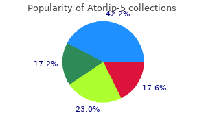
Discount atorlip-5 5 mg without a prescription
For instance, a affected person on mechanical ventilator, alveolar ventilation decreases as a end result of elevated lifeless area quantity (by tubing and so on. In such patients, if minute ventilation remains fixed, alveolar gas exchange suffers. Total quantity is estimated and equal dead space volume is proportionately calculated from the total. Therefore, patients with speedy and shallow respiration develop hypoxia and hypercapnea. Such topics have alveolar ventilation even larger than topics with regular respiration. Thus, to enhance alveolar air flow, you will need to enhance the depth of breathing than to increase the frequency. In reality, during average to extreme exercise, a educated athlete achieves the goal alveolar air flow by mainly rising the depth somewhat than the frequency of respiration (Application Box one hundred and five. Construction of Equivalent Dead Space-Alveolar Air Boundary Dead space is full of pure oxygen prior to expiration. The boundary between dead area air and alveolar air is S-shaped due to a point of blending of alveolar gas with dead area air. However, an equivalent sharp boundary is constructed in such a way that the amount of N2 within the dead house space is the same as the quantity of N2 within the alveolar air part. The dead house volume is then calculated as the volume expired as a lot as this line, and is learn from the quantity recording. Hence, the lifeless area quantity from the corresponding area is determined by easy proportion. To know the quantity of air that takes half in alveolar ventilation, first the lifeless space volume is subtracted from the tidal volume and then the volume is multiplied by respiration (respiratory) frequency. To say, when tidal quantity is 500 ml, lifeless space quantity is a hundred and fifty ml and respiratory rate is 12 /min: Alveolar ventilation is = (500 ml �150 ml) � 12 = 350 ml � 12 = 4200 ml / min Measurement of Alveolar Ventilation Alveolar air flow is easy to calculate if useless area quantity is known. Alveolar air flow is calculated in the pulmonary operate laboratory from the quantity of expired carbon dioxide per minute and fractional concentration of carbon dioxide in the alveolar gasoline. Because no gas exchange occurs in the conducting airways and the inspired air contains basically no carbon dioxide, all the expired carbon dioxide originates from alveoli. Exchange of fuel between the alveoli and the capillary blood occurs throughout the alveolar-capillary membrane by diffusion in response to partial stress gradients of the gases. For instance, oxygen uptake from alveoli into the pulmonary capillary blood happens due to partial pressure gradient of oxygen across the alveolocapillary membrane. Scientist contributed Antoine Laurent de Lavoisier (1743�1794) French chemist and biologist was the first scientist to present the significance of oxygen in combustion and in the gaseous exchange within the lungs. With the help of Pierre Simon de Laplace (1749-1827) he had devised a calorimeter and measured the respiratory quotient. Exacerbation of chronic lung illness corresponding to bronchial asthma, continual bronchitis and emphysema. Depression of respiratory centers as occurs in head accidents or by drugs corresponding to barbiturates and opiates. Pulmonary capillary blood move Alveolar-Capillary Membrane Alveolar-capillary membrane (also known as respiratory membrane) types the blood-gas interface that separates blood in the pulmonary capillaries from the gasoline in the alveoli. Diffusion of gases between alveolar air and pulmonary capillary blood takes place via alveolar-capillary membrane. The alveolar-capillary membrane is exceedingly skinny, and is principally composed of alveolar epithelium, interstitial fluid layer, and capillary endothelium. As the blood perfuses the alveolar capillaries and air ventilates the alveoli, oxygen and carbon dioxide move across the blood-gas interface by diffusion. Layers of Alveolar-Capillary Membrane From interior of the alveoli to the capillary blood, the alveolar-capillary interface consists of six layers. However, O2 from hemoglobin molecule in the purple cells of pulmonary capillaries to enter into alveolar lumen (or the transport within the reverse direction), passes by way of 10 layers. Hemoglobin molecule Hyperventilation Hyperventilation often happens as a outcome of stimulation of respiratory centers. However, voluntary hyperventilation and train induced hyperventilation are frequent physiological causes of hyperventilation.
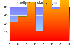
Purchase atorlip-5 visa
Chapter 49: Small Intestinal Motility 415 Intestinointestinal Reflex When a half of the gut is over-distended, the remainder of the gut relaxes. For instance rest of the sphincter occurs by vagal stimulation as seen in gastroileal reflex. This decreases the intestinal motility, sometimes even leading to paralysis of the intestine. Peristalsis first begins in the small intestine (6�8 hours later) adopted by in the abdomen (8�12 hours) and eventually in the colon (2�3 days). This happens because of elevated discharge of non-adrenergic fibers within the splanchnic nerves. Gastroileal Reflex When meals enters the stomach (stretching of stomach), the motility of the terminal a half of the ileum is enhanced. This will increase entry of contents of ileum into the colon by way of ileocecal sphincter. Law of the Intestine When a bolus of chyme enters the intestine, the a half of the intestine behind the bolus contracts and the portion of the gut ahead of it relaxes. This is meant to propel the intestinal content material within the ahead path as occurs in peristalsis. Normally, the ileocecal sphincter is tonically contracted, and therefore the sphincter stays closed more often than not and prevents small intestinal emptying. When a peristaltic wave reaches the terminal part of the ileum, the sphincter relaxes so that the ileal content material enters the cecum. Intestinal Colic Severe belly cramps are skilled in localized obstruction of small intestine. The phase proximal to the obstruction dilates and will get crammed with fluid and gasoline. This increases the stress inside the lumen that causes compression of blood vessels within the intestinal wall. Abdominal cramps are also skilled in other diseases that result in distention of the gut. The major function of small intestine is to adequately mix the chyme with intestinal and pancreatic juice. In examinations, "Describe the mechanism and significance of intestinal motitlites" could come as a Long Question. Understand the physiology of colonic actions, colonic reflexes, and their capabilities. Learn the physiological foundation of Hirschsprung, illness, irritable bowel syndrome, diarrhea, and constipation. Colon constitutes about 90% of enormous intestine and consists of ascending, transverse, descending, and sigmoid colons. Small intestine receives chyme of meals sequentially with no mixing of individual meals, whereas large gut accommodates mixture of chymes of many meals of 1 to three days. On average the entire transit time of chyme of a meal through large intestine as recorded from passage of radiopaque markers is about 30 to forty eight hours. From pelvic colon to rectum, the transit could be very slow, which may take 2 to three days. Transit time is much less in excessive fiber food plan, generally might even be lowered to 6 hours through the whole intestine. Therefore, though colon receives about 2 liters of chyme per day from small gut, its output is only about 200 mL. The objectives of colonic contractions are to combine the chyme and flow into it throughout the mucosal surface of the colon in order that maximum contact occurs between the chyme and the mucosal epithelium. This, plus the sluggish motion of the chyme throughout the colon, which is about 5�10 cm/hour allows most absorption of salt and water. Longitudinal muscle layer of muscularis externa is concentrated into three bands, known as as tenia coli. Colonic Movements Colonic movements embody haustral contractions, propulsive movements, mass peristalsis, and colonic reflexes.
Syndromes
- Occur many times a day
- Ultrasound of the abdomen
- Numbness in the hands, feet, or other areas
- Lightheadedness
- Taking certain over-the-counter medicines, vitamins, supplements, cold medicines, antibiotics, or other drugs
- Mouth sores, including yeast infection (thrush)
- Low-grade fever
- Often occurring at rest
- Swelling of the feet or ankles
- Percutaneous transhepatic cholangiogram (PTC)

Discount generic atorlip-5 canada
The hormone binds with the receptor, which has intrinsic tyrosine kinase exercise. The receptor has three domains: extracellular, membrane and intracellular domains. The extracellular area possesses the binding website for hormone and the intracellular area possesses tyrosine kinase activity. The binding of hormone with the receptor causes conformational change within the receptor that exposes the intracellular websites of the receptor for autophosphorylation. The kinase autophosphorylates tyrosine residue within the receptor and tyrosine residues on intracellular protein substrates. The phosphorylation of tyrosine residue initiates cascade of phosphorylation reactions that phosphorylates varied enzymes like serine and threonine kinases, and phosphatases. They have wonderful resemblance with the receptors for 1,25-dihydroxycholecalciferol, thyroid hormones, and retinoic acids. Receptors for all these various hormones are considered to be a part of a single gene superfamily. They have five domains (A to E), and the homology is especially seen for the C area, particularly C1 subdomain. Steps of Signal Transduction Binding of hormone with the receptor triggers following sequence of occasions: 1. The binding of hormone with the receptor causes conformational change in the receptor protein. Heat shock Calcium�Calmodulin System this is the system of transduction of the hormone signal during which binding of hormone to a receptor on the cell 450 Section 6: Endocrine Physiology. Calcium can also be mobilized from the intracellular storage websites like mitochondria and endoplasmic reticulum. In many tissues, the secretion of calcium from the intracellular storage websites triggers opening of calcium channels within the cell membrane. This further will increase the calcium concentration in the cell and replaces calcium in endoplasmic reticulum and mitochondria. Calmodulin-dependent Kinases Calmodulin is a polypeptide containing 148 amino acids and has four calcium binding domains. When calcium binds with calmodulin, it activates different calmodulin-dependent kinases. Calcium Binding Proteins the calcium binding proteins in the cells are calmodulin, troponin, and calbindin. The troponin is the calcium binding protein present within the skeletal muscle involved in muscle contraction. Calcineurin, a calmodulin-activated protein, is a phosphatase, which inactivates calcium channels via dephosphorylation. However, some actions of steroid hormones manifest more rapidly than the transcription course of. Thus, steroids also act via intracellular second messengers that are activated by different hormones. This forms the physiological foundation of interaction of steroid hormones with other hormones. For instance, estrogen and dopamine work together on the second messenger degree for nongenomic actions of estrogen. Few hormones act instantly by altering ion channels within the membrane and plenty of by way of the G proteins. Steroid hormones and thyroid hormones act by altering cellular transcription and translation mechanisms. Binding with the receptor, activate the intrinsic tyrosine kinase activity of the receptor that triggers the induction of intracellular signaling pathway. Up-regulation and down-regulation of receptors occurs with sustained decrease or improve within the degree of hormone in the blood.
Atorlip-5 5 mg low cost
Understand the mucosal modifications in intestinal epithelium to improve the floor area for absorption. Each villus is a fingerlike projection covered by a layer of columnar epithelium and incorporates a community of capillaries and lacteals (lymphatics). The intestinal mucosa is supported externally by thin layer of smooth muscle fibres, muscularis mucosae. Scientist contributed Johann Nathanael Lieberk�hn (1711�1756) was a German doctor and physiologist. Besides his physiological work, Lieberk�hn was most recognized for his preparation of medical specimens-these have been still presented as much as the nineteenth century, especially in Moscow, as masterpieces. Intestinal Glands As mentioned in chapter 36, the intestinal wall has all of the layers of the gut (Refer to . Throughout the size of small intestine, the mucous membrane is covered by villi. The mucous membrane of the intestine contains many valve-like folds referred to as valvulae conniventes, which add to the floor space for absorption. In intestine, the surface area for absorption is increased by about 600 fold by villi, brush border and valvulae conniventes. Paneth cells are endocrine cells current in the crypts of Lieberk�hn of their deeper half. They secrete defensins, the naturally occurring antibiotics that defend developing enterocytes in opposition to infections. The undifferentiated cells are the progenitor cells in the mucosa present in the crypts of Lieberk�hn that. There are additionally enterochromaffin cells, Paneth cells and undifferentiated cells in the intestinal mucosa. After the life span of about 2�5 days, enterocytes are sloughed together with mucosal cells. Shedding of those epithelial cells accounts for day by day excretion of about 30 g of protein because the cells are protein-rich. Enzyme Enterokinase -dextrinase Maltase Sucrase Lactase Peptidases Nucleotidases Substrate Trypsinogen -dextrins Maltose Sucrose Lactose Terminal amino acids Nucleotides Product Trypsin Glucose Glucose Glucose and fructose Glucose and galactose Peptides and amino acids at amino finish of peptides Nitrogenous bases, pentoses, and phosphates Types of Cells in Villi the absorptive floor of intestinal mucosa is elevated by the intestinal villi. Simple columnar cells: They perform absorptive function due to the presence of brush border consisting of large number of villi. Goblet cells: these are mucous secreting cells and are interspersed between the columnar cells. Endocrine cells: these are scattered in the villi as nicely as are widely distributed throughout the gastrointestinal tract. Enterochromaffin cells: Due to their resemblance to chromaffin cells of the adrenal medulla c. Argentaffin cells: As the intracytoplasmic granules stain positively with silver salts by discount reaction (agyrophil cells, on the other hand, require the addition of exogenous reducing substances for staining) d. Enterokinase is present in the brush border of enterocytes and is extruded with denudation of mucosal epithelium. The cations are secreted by energetic transport and anions are transported along with cations to preserve electroneutrality. Mucin is the main element of mucous that varieties a gel to cowl the mucosal epithelium. Mucous protects the intestinal epithelium and helps in clean passage of chyme via the intestinal lumen. Experiment to Study Intestinal Secretion In animal models, experiments are performed to study the rate and composition of intestinal secretion. In these animals, a loop of gut is resected and both ends of the loop are related to anterior belly wall in such a method that they open to exterior. Thereafter, varied stimuli are utilized on the loop and their results are studied. This provides suitable environment for digestion and absorption of food materials in the intestine.
Buy atorlip-5 5 mg amex
Epinephrine is injected in anaphylactic shock that will increase blood strain by causing vasoconstriction and by rising cardiac output. Dopamine is the drug of selection in traumatic and cardiogenic shock for 3 causes: i. It causes vasoconstriction in the systemic blood vessels that will increase blood pressure. Over-warming of the body must be prevented because it causes cutaneous vasodilation and precipitates shock (Clinical Box a hundred and one. There is a basic notion to cover the physique by blankets in order that skin remains heat. But, it should by no means be done, as elevated skin temperature causes cutaneous vasodilation and additional precipitates shock. Dopamine is often most popular in cardiogenic shock, because it maintain renal perfusion. Refractory shock, Compensatory mechanisms of shock, Reversible shock, irreversible shock, Refractory shock, Reperfusion-induced injury, Traumatic shock, Distributive shock, Physiological foundation of administration of shock could also be requested as Short Questions in exam. Understand the physiological foundation of cardiac modifications because of strain and quantity overload in heart failure. Give the physiological basis of orthopnea, paroxysmal nocturnal dyspnea and dependent edema in heart failure. Many suppose heart failure means fainting, and a few think it because the stage simply earlier than dying. However, coronary heart failure is a specific entity of cardiac abnormality that leads to decreased pumping of the center. Acute vs Chronic Failure Acute heart failure is seen following acute myocardial infarction or rupture of a coronary heart valve. In persistent failure, which happens due to a slowly pro gressive illness like cardiomyopathy, or continual valvular disease, blood pressure may be maintained (edema in the dependent parts is a feature). Definition it is a pathophysiologic state in which an abnormality of cardiac perform ends in inability of the center to pump blood at a price sufficient for the requirements of the tissues of the body. It occurs both because of decreased myocardial contractility or due to an increased strain or quantity overload. The usual physiological alterations are a decreased stroke quantity (in ahead failure) and damming of blood within the venous compartment (in backward failure). Both these pathophysiologic mechanisms coexist and each are due to a single dysfunction, i. In left ventricular failure, the congestion occurs in the pulmonary circulation; subsequently, dyspnea and orthopnea are common options. High Output vs Low Output Failure In excessive output failure, coronary heart pumps abnormally giant portions of blood to deliver enough oxygen to the tissues. This happens in following circumstances: � Severe anemia � Hyperthyroidism Types of Heart Failure Heart failure has been categorised in numerous ways. Pressure Overload In pressure overload, elevated blood strain increases the afterload. This leads to concentric hypertrophy of the ventri cles (increased ventricular wall thickening compared to the rise in ventricular cavity dimension. The useful capacity of ventricle in such a state of affairs depends on the stress exerted on the ven tricle. According to Laplace regulation, stress within the ventricular wall is the product of pressure and radius of ventricular cavity divided by ventricular wall thickness. In the compensatory stage (stage of concentric hypertrophy) of stress overload, wall stress normalizes as a end result of improve in wall thickness and decrease in cavity radius of ventricle. But in the stage of hypertrophy and dilatation, ventricular stress will increase as a outcome of proportionate will increase in radius as a outcome of chamber dilation, which leads to failure of the myocardium to pump blood, causing systolic failure. In backward heart failure, the stress and volume in the atrium and venous compartment behind the failed ventricle are more, due to this fact, edema happens. In ahead coronary heart failure, lack of ability of the ventricle to pump blood causes tissue hypoxia that increases renin secretion (due to renal hypoxia). This activates renninangiotensin-aldosterone axis, therefore, edema (due to water retention) may be seen.
Discount 5 mg atorlip-5 overnight delivery
Note, intrapleural stress is negative in both the phases of respiration, and alveolar stress is negative during inspiration and positive during expiration. A decrease in intrapleural pressure helps the lungs to increase, which in turn ends in influx of air into the lungs. Intrapleural strain maintains normal form of chest wall and lungs because it resists the recoiling of those struc tures. Loss of normal intrapleural pressure ends in lung collapse and barrel formed chest. Clinical Significance the clinical significance of intrapleural pressure is noticed when the chest wall is perforated and air enters the pleu ral space. Thus, when a connection is made between the atmosphere and the pleural space, air strikes into the pleural house (air moves from regions of excessive to low pressure) and intrapleural pressure becomes atmospheric. This con dition, during which air accumulates in the pleural area, is named pneumothorax. Due to loss of normal intrapleural pressure in this con dition, the lungs collapse instantly due to inward recoiling, and the rib cage expands outward because of its outward recoiling. In such a situation, the transpulmonary pressure is zero because the strain difference throughout the lung is eradicated. In standing position, the vacuum in intrapleural house is greater in the apex and fewer in the base of the lung, as a result of gravity pulls the lungs downward (away from the apex of the thoracic cage). Significance of Intrapleural Pressure Physiological Significance It has two physiological significances. Therefore, expansion of the thoracic cage as happens throughout inspi ration causes the intrapleural pressure (the strain between the lung and chest wall) to fall (to turn into Chapter 104: Mechanics of Breathing 891 inside and outdoors the airway. Transairway stress is necessary in preserving the airways open throughout pressured expiration. During inspiration, alveolar stress decreases (becomes subatmospheric) that sucks air into the lungs, and, throughout expiration, the alveolar stress will increase that removes air from the lungs. Transpulmonary pressure, which is the dif ference in alveolar pressure and pleural strain, is there fore +5 cm H2O [0 � (�5) = +5] on the finish of expiration. Transpulmonary strain is also referred to as dis tending strain as it helps in inflation of lungs. During aspiration of pleural fluid for analysis, air enters into the pleural cavity as a outcome of defective technique. For such purposes, nitrogen is injected into the pleural house to provide relaxation to the diseased lung. Summary of Mechanics of Inspiration and Expiration Sequence of Events Leading to Inspiration 1. Diaphragm and other inspiratory muscle tissue contract that cause enlargement of thoracic cage which in turn makes intrapleural stress more unfavorable. This will increase transpulmonary stress on account of which inflation of lungs occurs. Alveolar strain decreases (becomes subatmos pheric) and therefore, air flows into airways and lungs. Inflow of air stops when alveolar pressure equalizes with atmospheric strain (Flowchart 104. Therefore, intrapleural stress turns into much less nega tive which decreases the transpulmonary stress. Lungs that were stretched in inspiration start defla ting, as a outcome of which alveolar diameter decreases. The alveolar stress becomes larger than the atmospheric stress that pushes air out of the lungs (Flowchart 104. Airflow out of lungs continues till alveolar strain equalizes atmospheric strain. Transmural Pressure Transmural strain is the pressure distinction throughout an airway or across the lung wall. There are two major trans mural pressures concerned in breathing: transpulmonary stress, and transairway pressure. Transpulmonary Pressure Transpulmonary strain is the pressure difference across the lung wall.
Purchase atorlip-5 from india
Nocturia happens because of loss of concentric ability of kidney and increased osmotic load on nephron. They current with anorexia, nausea, vomiting, hiccough, pruritus, muscular twitching, fits and drowsiness. Treatment Consists of identifying the renal illness, identify the precipitating factors and preventing the further renal harm. Physiologically, kidney operate checks are done to assess glomerular filtration and tubular capabilities. Tubular perform exams, Glomerular function tests, Clearance tests, Urine evaluation, Acute renal failure, Chronic renal failure could also be requested as Short Questions in exam. Understand the relation of functional anatomy of urinary bladder with its capabilities. It accumulates urine with out much rise in stress in it and empties its content material at acceptable time following appropriate rise in strain. Urine from kidneys flows all the way down to the bladder via ureters because of the motion of gravity, which is aided by peristaltic actions of ureter. Peristaltic waves in ureters originate by a pacemaker tissue situated close to the calyces at a frequency of about one per minute. Once in the bladder, regurgitation of urine back into the ureter is prevented passively by a valvular flap present on the entry level of the ureter at the base of the bladder. Detrusor muscles are organized in three bundles: spi ral, longitudinal and round. The epithelium of the bladder is fashioned by a superfi cial layer of flat cells and a deep layer of cuboidal cells. Innervation Bladder is innervated by both sympathetic and parasym pathetic fibers. Parasympathetic fibers originate from the sacral 2, 3 and four segments of the spinal twine and attain the blad der by way of pelvic nerves, Chapter eighty three: Physiology of Micturition and Bladder Dysfunctions 723. The sympathetic fibers originate from lumbar 1, 2 and three segments of the spinal twine and attain the bladder by way of hypogastric nerve. The somatic fibers originate from S2, S3 and S4 and innervate bladder and external sphincter by way of pudendal nerve. Filling of bladder, desire to cross urine and painful dis tension, all these sensations of bladder are mediated by afferents in the pelvic nerve. The inside sphincter, which is situated at the neck of the bladder, is made up of a bundle of easy muscle (sphincter vesicae), and innervated by sympathetic (hypogastric) and parasympathetic (pelvic) nerves. The exterior sphincter is made up of a flap of skele tal muscle, which is current across the urethra in its proximal half (sphincter urethrae). The external sphincter is innervated by somatic (pudendal) nerve and therefore underneath voluntary management. The sensation of bladder filling is skilled at the blad der volume of about 150 mL and the feeling to cross urine is experienced at bladder quantity of 150�250 mL. At quantity of about 400 mL, the feeling to move urine becomes uncomfortable and at about seven hundred mL, it turns into very painful resulting in loss of management. This is primarily a reflex phenomenon, which is usually integrated in the spinal wire. Unless the bladder is crammed, urine accumulates in uri nary bladder with out much improve within the intravesical strain, as bladder wall is made up of clean mus cles that exhibit the property of plasticity. This relationship between the bladder quantity and pressure is best studied by cystometry. Cystometry: Cystometry is the procedure to research the relationship between the bladder quantity and pressure. For this purpose, a catheter is inserted into the bladder and bladder is totally emptied. Then, as bladder vol ume is gradually increased by slowly pushing water to fill bladder, intravesical strain is recorded at completely different blad der volumes. The preliminary rise in intravesical pressure happens when bladder is crammed with 50 ml of water (at point A. The intravesical stress increases steeply when the intravesical volume exceeds four hundred mL (between the factors C and D. Urinary bladder being a spherical viscus, the stress will increase as the organ fills. Therefore, increase in intravesical pressure is minimal except the viscus is comparatively full.
Real Experiences: Customer Reviews on Atorlip-5
Innostian, 34 years: However, in circumstances of increased demand for cardiac output as occurs in train, the atrial systole contributes considerably to the tip diastolic filling of the ventricle. An instance is the Na+�H+ change in the proximal tubule that reabsorbs Na+ from the tubular fluid in trade for secretion of H+ into it. Anatomical sphincters are situated at gastroesophageal, gastroduodenal and ileocecal junctions, and at the opening of bile duct in the duodenum and opening of rectum into anus.
Kliff, 38 years: Rapidly Adapting Receptors the quickly adapting receptors are sensory terminals of myelinated afferent fibers which might be found within the larger conducting airways. Source Estrogen is secreted from granulosa cells of ovarian follicles, corpus luteum, and placenta. Also, sudden discount in exterior pressure from atmospheric to subatmospheric degree can cause fast expansion of fuel within the lungs and produce air embolism, which generally happens when a pressurized cabin of an aeroplane or a rocket ruptures at high altitude.
Aschnu, 63 years: Thus, the size of the luteal part stays constant no matter the cycle length. On activation, the tubulovesicular membranes fuse with the cell membrane and microvilli that project into the canaliculi. However, virtually all the filtered glucose is reabsorbed in the proximal tubule, so that in regular condition, urine is basically glucose free.
Merdarion, 26 years: In lungs, increased change of blood gases occurs due to stimulation of the respiratory fee and dilation of bronchiolar tree. Endothelin 1 is fashioned from bigendothelin 1, which additionally possesses endothelin activity. Therefore, decreased oncotic strain or elevated hydrostatic strain causes edema formation.
9 of 10 - Review by U. Norris
Votes: 142 votes
Total customer reviews: 142
References
- Choyke P, Kobayashi H. Functional magnetic resonance imaging of the kidney using macromolecular contrast agents. Abdom Imaging. 2006;31:224-231.
- Winterbauer RH, Elfenbein IB, Ball WC Jr. Incidence and clinical significance of tumor embolization to the lungs. Am J Med 1968;45:271-90.
- Doggweiler R, Jasmin L, Schmidt RA: Neurogenically mediated cystitis in rats: an animal model, J Urol 160(4):1551n1556, 1998.
- Waghmare A, Campbell AP, Xie H, et al. Respiratory syncytial virus lower respiratory disease in hematopoietic cell transplant recipients: viral RNA detection in blood, antiviral treatment, and clinical outcomes. Clin Infect Dis 2013;57(12):1731-1741.
- Jin J, Rosen MJ: Laparoscopic versus open ventral hernia repair. Surg Clin North Am 88:1083, 2008.
- Halken S, Host A, Niklassen U, et al. Effect of mattress and pillow encasings on children with asthma and house dust mite allergy. J Allergy Clin Immunol 2003; 111: 169-176.
- Hunstad DA, Justice SS, Hung CS, et al: Suppression of bladder epithelial cytokine responses by uropathogenic Escherichia coli, Infect Immun 73(7):3999n4006, 2005.