Sevelamer dosages: 800 mg, 400 mg
Sevelamer packs: 10 pills, 30 pills, 60 pills, 120 pills, 90 pills
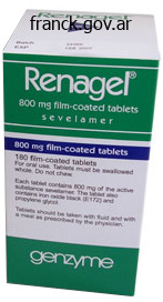
Order generic sevelamer from india
This preliminary inspection is essential because the surgeon might easily access these sinuses despite a deviated septum whereas the affected person is under general anesthesia; nevertheless, accessing these sinuses for inspection and debridement may be challenging within the postoperative 2230 setting with an awake affected person. The nasopharynx is inspected for lesions, patency of the eustachian tube orifices and the standing of the adenoids, if present. This inspection permits the surgeon to visualize the superior turbinate and the sphenoethmoidal recess, which ends up in the sphenoid ostium. If the surgeon can inspect the middle meatus, the attachment of the uncinate process, the hiatus semilunaris and ethmoid bulla are all famous. Clinical pearl: when debriding polyps, rotating the microdebrider blade such that it maximizes contact with the polyp and points away from important buildings like the center turbinate, lacrimal bone and lamina papyracea. When necessary, the middle turbinate is gently medialized utilizing an elevator to permit the surgeon to examine with more detail the center meatus and instrument the hiatus semilunaris. If an obstructing concha bullosa is present, the lateral side of the concha bullosa is surgically removed, while taking care to not disturb the medial attachment of the middle turbinate on the skull base. Atraumatic dissection of a concha bullosa will simultaneous permit entry into the middle meatus whereas preserving the integrity of the center turbinate. The uncinate process, which technically is an ethmoid construction, is the vital thing to accessing the maxillary sinus ostium. The uncinate course of types the anterior and medial boundary of the infundibulum, which homes the maxillary sinus ostium. A backbiting through-cutting instrument is used to divide the uncinate process along the junction of the superior 2/3 and inferior 1/3 of the uncinate process. Care must be taken to keep away from damage to the nasolacrimal duct when performing this step. Complete removal of the uncinate course of reveals the lateral restrict of the infundibulum and the maxillary sinus ostium. The natural ostium of the maxillary sinus is then inspected with the 30-degree endoscope and 2232 probed. We prefer to fracture the uncinate course of anteriorly with a maxillary or ball-tipped seeker. Clinical pearl: use the tip of the ball-tipped seeker to fracture initially for the explanation that uncinate process could additionally be carefully associated to the lamina papyracea. A ball-tipped probe is used to dilate the natural ostium into the posterior fontanel. Clinical pearl: dilate the ostium in a posterior and inferior direction to avoid inadvertent damage to the lamina papyracea. What may be conceptually clear, identifying the maxillary sinus ostium has confirmed to be a technical challenge as Parsons forty seven,forty eight has previously described. Within the infundibulum, the place of the maxillary sinus ostium is located throughout the lower half in 94% of instances, with 26% of ostia situated about half means alongside the maxillary line and 68% of ostia located inside the decrease one-third of the infundibulum. The ostium is located roughly 2 to 5mm posterior to the lacrimal canal, which is obliquely oriented to the Frankfurt airplane. One may theorize, then, that uncinate course of removing could also be unnecessary if the ostium is correctly recognized and dilated. In most cases, visualizing the ostium may prove more difficult if the uncinate process is left intact. Treatment of the maxillary sinus relies upon upon the extent of illness and the general mission of the surgeon. If the ostium is edematous or obstructed by polyps or polypoid mucosa, the ostium must be dilated to permit air flow, drainage and postoperative irrigation. In these situations, it might be advantageous to carry out an inferior meatal window, a "mega-antrostomy"54 or endoscopic modified medial maxillectomy. In this patient, significant inflammatory disease and purulence suggested the want to perform postoperative irrigations on a long-term foundation. The anterior and posterior ethmoids are divided anatomically by the vertical portion of the basal lamella of the center turbinate.
Cheap sevelamer 800mg without a prescription
Osteosarcoma and Ewing sarcoma should be thought-about within the differential analysis of aggressive sphenoid bone lesions in youngsters and youngsters. A lytic and expansile lesion in an adult should immediate the diagnosis of big cell tumor whereas aneurysmal bone cyst or large cell reparative granuloma ought to be considered in youngsters. Rhabdomyosarcoma is a comparatively frequent tumor in youngsters in the head and neck region with a poor prognosis. The flooring of the orbit is formed by contributions from the orbital plate of maxilla, the palatine bone, and the zygomatic bone. The frontal strategy of the maxilla and lacrimal bone make the anterior aspect of the medial wall of the orbit. The lamina papyracea forms the mid portion of the medial wall whereas the sphenoid bone contributes to the posterior a half of the medial wall. Three openings at the apex of the orbit permit entry/exit of the neurovascular structures. The optic canal is immediately medial to the superior orbital fissure and separated from it by a tiny bone called the optic strut, which is a projection of the anterior clinoid course of. The optic nerve and the ophthalmic artery, which are lined by a dural sheet, journey by way of the optic canal. The bony orbit is lined by a troublesome fibrous sheath named the periorbita, which serves as a mechanical barrier between the intra- and extra-orbital buildings. The medial, lateral, superior, and inferior rectus muscles come up from a standard tendon, annulus of Zinn, and connect to the globe. The rectus muscle tissue are enveloped by a fibrous membrane that forms the muscle cone and defines the intra-and extraconal spaces. The extraconal area is in between the periorbita and muscle cone and incorporates fats and the lacrimal gland superolaterally. The intraconal area contains the extraocular muscle tissue, the optic nerve sheath advanced, and different neurovascular constructions in addition to fats. When coping with an orbital lesion, one should try to determine the anatomic website from which the lesion originates to narrow the differential analysis. Optic nerve sheath complex mass lesions are meningiomas or gliomas in most situations. Optic nerve meningiomas come up from the dura across the optic nerve and tend to enlarge concentrically concerning the nerve. The "sandwich signal" refers to enhancement of the optic nerve sheath on both facet of the nonenhancing optic nerve in meningiomas of this area. Lymphoma and leukemia can hardly ever involve the optic nerve sheath and current as diffuse thickening of the nerve with enhancement on the postcontrast photographs. Optic neuritis might present refined enlargement of the nerve-sheath complicated and should have an result on any a part of the nerve. The majority of optic neuropathy in adults is chemic in nature and may be recognized on medical grounds. Multiple sclerosis is the main reason for nonischemic optic neuropathy, however optic neuritis may also occur in isolation. Eighty % of patients with a quantity of sclerosis could have a bout of optic neuritis at some stage in their disease. A variety of inflammatory and infectious illnesses can current with optic neuritis with comparable imaging findings. Idiopathic pseudotumor of the orbit is a nongranulomatous inflammatory course of involving the orbit. It could present as a mass lesion, hence the name pseudotumor, or as an infiltrative course of. The acute kind is clinically characterised by abrupt onset of pain, redness and swelling of the eyelid. Since pseudotumor can contain any construction within the orbit in isolation or mixture and current as a mass lesion or infiltrative course of, the imaging differential analysis consists of numerous potentialities from tumors and infection to thyroid ophthalmopathy.
Discount 400 mg sevelamer mastercard
Undermining can be performed superficial to the deep temporal fascial layer all the greatest way down to the lateral forehead and superior border of the zygomatic arch. Utilization of the sideburn sparing incision requires the dissection to be in a subcutaneous airplane, so as to keep away from harm to the temporal branch of the facial nerve as it crosses over the zygomatic arch. Vertical retraction with skin hooks and countertraction on the flap facilitates elevation of the cheek flap with facelift scissors. Transillumination of the flap with an overhead mild directed by way of the flap allows for the identification of the right dissection airplane and aids in sustaining the suitable flap thickness. The cheek skin is often elevated anteriorly to a line drawn between the malar eminence and the mid mandible (mandibular notch). In the occipital space, care should be taken to stay deep to the hair follicles to keep away from alopecia, while staying superficial to the sternocleidomastoid fascia to stop inadvertent harm to the great auricular nerve. The cervical pores and skin flap is elevated in the subcutaneous aircraft to the second main cervical rhytid and extended to the midline. The subcutaneous elevation avoids injury to the marginal mandibular and cervical nerves. There are nonetheless many surgeons that make the most of plication or imbrication as the cornerstone of their facelift operation. The zygomatic main muscle is recognized and blunt dissection is performed on the floor of this muscle right down to the extent of the nasolabial fold. The primary aim is to launch and mobilize the malar fats pad right into a posterior superior place. Inferiorly the dissection is then carried deep to the platysmal muscle for approximately three or four cm. In this area, care is taken to keep away from damage to the external jugular vein, larger auricular nerve, and cervical department of the facial nerve. The last few interrupted suspension sutures are used to fixate the free edge of platysma to the mastoid periosteum. It must be confused that a superior vector of pull on this tissue is crucial to establishing a well-defined cervicomental angle. Careful dissection is performed on the floor of this muscle releasing the zygomatico-cutaneous ligaments of McGregor patch to launch and mobilize the malar fat pad into a posteriorsuperior place. The hair-bearing skin is re-approximated with staples, and the remaining preauricular and postauricular pores and skin is closed underneath minimal rigidity with 4-0 polydiaxone sutures buried within the deep layer, interrupted 6-0 nylon sutures used to shut the preauricular incision and 5-0 chromic sutures used to shut the postauricular incision. A 5-0 polydiaxone suture positioned from the pre-tragal soft tissue to the overlying dermis recreates the pretragal crease. The pores and skin superior over the tragus is thinned, and a 6-0 fast-absorbing gut suture is used to close the posttragal or retrotragal incisions. The posterior flap is superior superiorly and anteriorly to recreate, with out notching, the posterior hairline. At the conclusion of the process, white petrolatum is utilized to the suture lines. Small seromas or hematomas are simply expressed by way of the postauricular incision. A new lighter dressing is utilized and left in place till the third postoperative day. The patient is allowed to bathe and gently wash the hair beginning the third day after surgical procedure. For the primary 48 hours after surgery, head of mattress elevation is inspired and cool compresses are applied the face and neck. The affected person is seen for his or her second postoperative visit on day six or seven, and the sutures and skin staples are eliminated. Occasionally, the postauricular staples are left for an additional two to three days. The affected person is requested to continue wearing the elastic-facelift wrap at night time for an extra week. We routinely remind the patients that ecchymosis usually resolves in two weeks, and that the majority of edema dissipates over 4 to six weeks. Routine follow-up visits are scheduled at one, three, six, and 12 months after surgical procedure. A consultation with our aestheticians can be helpful for postoperative-skin care and make-up recommendations. However, because of the elective nature of the procedure any problems, main or minor, are poorly tolerated by both patient and surgeon.
Buy sevelamer in united states online
Further research into the science of cell-mediated cytokines and wound healing might lead to new and higher treatments sooner or later for these prone to extreme scarring. The deep vascular plexus, or subdermal plexus, exists on the junction of the reticular dermis and subcutaneous fats (hypodermis). Arterioles from the deep vascular 2595 plexus supply sweat glands and pilosebaceous items consisting of hair follicles and sebaceous glands. The superficial vascular plexus exists on the superior border of the reticular dermis and consists of capillary loops inside the dermal papilla. In the head and neck, especially the face, there can also be a big network of deep subcutaneous perforating vessels named according to the anatomical location, for example, transverse facial and submental. Musculocutaneous vessels arise from segmental arteries, which are named branches of the aorta, and run deep to muscle teams. These musculocutaneous arteries permeate through underlying muscles earlier than reaching the skin and supply blood to each muscle and pores and skin. Musculocutaneous arteries run perpendicular to skin for a lot of their course and thus sometimes provide solely a small area superficially. These vessels provide blood only to the skin and to not the adjacent muscle tissue. As opposed to musculocutaneous arteries, direct cutaneous arteries run parallel to skin and provide a big area of pores and skin. An intensive community of capillaries provides the skin with its blood volume and circulate and is controlled by precapillary sphincters. These sphincters are induced to relax by native hypoxemia and an elevated stage of metabolic merchandise. Incisions on the flap edges interrupt the parallel superficial and deep cutaneous plexus causing decreased perfusion pressure to the pores and skin. In addition, when sympathetic nerves are severed, norepinephrine is launched from the nerve terminal and catecholamine reuptake is eliminated. Flap Designs the four fundamental forms of flap designs are primarily based on the vascular provide. Fasciocutaneous and musculocutaneous flaps mentioned in Chapter a hundred and ten, "Neoplasms of the Oral Cavity" and Chapter 111, "Neoplasms of the Oropharynx and Hypopharynx. As the vascular provide to these flaps arises from the subdermal plexus by way of the base, which is equipped by musculocutaneous arteries, the appropriate airplane of dissection for elevation of the flap is alongside the subcutaneous (hypodermal) adipose. Multiple length-to-width ratios have been recommended within the literature 2597 to increase flap survivability, but, virtually talking, survivability may be more related to the perfusion strain of the feeding blood vessels. As a outcome, axial flaps are able to a greater length-towidth ratio than are random flaps. The plane of dissection is, due to this fact, deeper and consists of the fascia containing the relevant named artery. Some further length is obtainable with axial flaps if extra tissue at the distal tip is included with its corresponding blood supply derived from the random subdermal plexus. Examples of axial flaps and their corresponding direct cutaneous artery are the paramedian brow flap (supratrochlear artery) and the nasolabial flap (angular artery). Fasciocutaneous flaps contain related pores and skin, subcutaneous fascia, and deep fascia. Musculocutaneous flaps embody all the elements of the fasciocutaneous flap with the added underlying muscle. Cellular Physiology of Flaps Following transposition and insetting of a flap, the primary forty eight hours symbolize the most critical interval with respect to figuring out flap survival. A surge in the degree of catecholamines happens domestically with tissue harm and flap placement, leading to vasoconstriction secondary to catecholamine launch. In addition, the inflammatory cascade releases increased ranges of thromboxane A2 (also a strong vasoconstrictor) and free radicals, with resulting edema. The combination of these components could cause each ischemia and direct injury to the flap. Neovascularization is delayed till three to seven days in the proliferative part and thus the flap is relying on nutrient absorption from the surrounding tissues and blood provide from the pedicle throughout this crucial time. Revascularization has been described as being enough for division of the flap pedicle at seven days. Angiogenic stimuli corresponding to ischemia trigger the vessels on the fringe of the flap to turn into dilated, and a thinning of the basement membrane facilitates endothelial-cell migration.
Generic sevelamer 800mg line
This approach also avoids the technically difficult necessities of integrating the flap into the nasal sill on the time of flap inset. When utilizing cheek flaps for restore, I often delay excising the extreme-lateral portions of the residual-alar pores and skin until the time of pedicle detachment and flap inset. Standing-cutaneous deformity marked laterally so scar from excision of deformity will lie in alar groove. The template is positioned in order that the medial border of the designed flap lies in the melolabial crease. This arrangement insures that the flap is harvested from the cheek, not from the lip, and that the donor site wound closure will lie throughout the melolabial crease, providing maximum-scar camouflage. The flap is designed to pivot 90� toward the midline in a clockwise path when harvested from the left cheek and counterclockwise when harvested from the proper cheek. As the flap is pivoted and transferred to the recipient web site, the medial border of the in situ flap is sutured to the cephalic border of the nasal defect. This in turn causes the inferior border of the in situ flap to be part of the anterior border of the defect. The lateral border of the in situ flap turns into the inferior border of the reconstructed ala. A triangle of skin is marked superior and inferior to the tracing to trend a crescent island of pores and skin. The inferior triangle of pores and skin is excised and discarded on the time of flap switch, and the superior triangle of skin is transferred with the flap and is excised and discarded on the time of pedicle detachment and inset 2648 of the flap. The superior triangle of skin is minimized to scale back loss of tissue from the superior-melolabial fold the place the fold is well developed. Removing pores and skin from the superior portion of the fold might lead to considerable asymmetry of the medial elements of the cheeks. The flap is incised, and the distal portion is elevated in the subcutaneous aircraft. The distal third of the flap is skinny, leaving 1 to 2 mm of subcutaneous fats hooked up to the undersurface. As the dissection proceeds superiorly, the plane of dissection extends deeper to facilitate growth of the subcutaneous-tissue pedicle. The pedicle of fats is free of the surrounding-cheek fat by incising via borders of the pedicle perpendicular to the surface of the skin. The depth of the incision is carried to the extent of the superficial floor of the zygomatic main and levator labii muscles. On reaching the zygomatic main muscle, blunt dissection continues upward on the floor of the muscle, releasing the attachments of the pedicle to deeper buildings till the flap can reach the recipient web site with out undue tension. This has the impact of pulling the pedicle upward towards the ala without the want to place extra traction on the subcutaneous-tissue pedicle. Superiorly based mostly subcutaneous tissue pedicle interpolated cheek flap designed to cowl cartilage graft. Caudal border of cheek flap sutured to caudal border of bipedicle-advancement flap. Depending on the form of the flap, the lateral border of the donor site could also be considerably longer than the medial border. For subcutaneous tissue pedicled flaps, the pedicle is transected on the base, and the cheek skin is undermined for a distance of 2 cm across the periphery of the amputated pedicle. After freshening the pores and skin margins with a scalpel, the wound is closed by advancing the borders together. The lateral portion of the flap hooked up to the nostril is launched from attachments to the adjacent nasal pores and skin for a distance of zero. Release permits the surgeon to remove excessive-subcutaneous fats not trimmed at the time of flap transfer. When the alar base is absent, the flap is tailored to replace the missing base and is built-in with the nasal sill. When the sill requires reconstruction, the flap is trimmed so that it has a tapered end that will function the sill. The subcutaneous pedicle is excised, the skin edges surrounding the pedicle are freshened and the wound closed primarily. However, harvesting the island flap is technically more difficult than harvesting a cutaneous-pedicle flap as a result of the aircraft of dissection is significantly deeper, putting the branches of the facial nerve supplying the zygomatic major and minor muscular tissues at larger threat of damage.
Anticholinergic Herbs (Belladonna). Sevelamer.
- Are there any interactions with medications?
- What is Belladonna?
- Dosing considerations for Belladonna.
- How does Belladonna work?
- Are there safety concerns?
Source: http://www.rxlist.com/script/main/art.asp?articlekey=96533
Cheap sevelamer 400mg with visa
The undermined skin-muscle flap elevated with the subciliary method is then re-draped, and its excess eliminated. The tarsal plate is fixated into the inner side of the orbital periosteum, placing the lower lid at about 1 to 2 mm above the inferior limbus. Advocates for this method claim that has the benefit of its dissection to avoid threat to branches of the facial nerve and create a vertical vector of pull, avoiding the chance of a "lateral sweep" that might happen with a more lateral method. Moreover, help with both a canthopexy or canthoplasty has to be fastidiously considered. The want for canthoplasty in several sufferers brings considerations due to the risk for lower-lid shortening and creation of an unnatural look, which is why surgeons adept to this system extra often choose canthopexy instead. The authors acknowledge a high incidence of issues, mostly within the lower lid. This excessive fee of issues has led them to suggest several modifications to the method to achieve more natural results with less frequent problems. This strategy has not gained the popularity of the trans-temporal endoscopic approach. While a major variety of problems that could be challenging to correct occur with the trans-orbital strategy, the trans-temporal route offers plentiful help for the decrease eyelid and increase in lower-lid tension. Trans-temporal Approach for Mid-face Lift Inspired by the work of Hinderer,89 Tessier90and Psillakis,ninety one described a subperiosteal dissection for facial rejuvenation, in which the mid-face was approached in a sub-periosteal airplane with incisions within the temporal and forehead areas in the early Nineteen Nineties. In addition, it prevents bunching in the malar space and avoids incisions that traverse the orbicularis oculi. The trans-temporal, also identified as endoscopic mid-face lifting, is our most well-liked approach. Multiple planes of dissection must be traversed, 2517 and a radical understanding of the anatomy of the fascial planes in the temporal/zygomatic area, as described previously, is a prerequisite to avoid harm to the frontal department of the facial nerve. The patient is examined preoperatively in an upright place, and the forehead is assessed. The amount of desired medial forehead elevation is measured; in most patients with gentle to reasonable brow ptosis, 2 to four mm of brow elevation is required to deliver the medial side of the forehead to the extent of the supraorbital rim. When making the incision, the surgeon has to bevel the scalpel parallel to the hair follicles, to obtain maximal scar camouflage as transecting the hair follicles leads to a number of millimeters of permanent alopecia. The incision is carried down by way of the superficial temporal fascia to the deep temporal fascia. A dissector is used to elevate the superficial temporal fascia and overlying tissue off the deep temporal fascia to the temporal line. The dissection is continued superiorly in a subperiosteal aircraft after dissection through the tenacious fascia of the temporal line and ends on the level of the occiput. This dissection ensures that the elevated brow and lateral temporal tissues will redrape and not bunch anteriorly once suspended. Inferior to the incision, dissection on high of the deep temporal fascia is performed blindly for two to 3 cm. Anteriorly, this elevation requires lysis of the fascia at the temporal line, and subperiosteal dissection is sustained to the supraorbital rim. The arcus marginalis often could be released from lateral to inside 1 cm of the superficial marking made for the supraorbital neurovascular bundle. Bimanual dissection usually is helpful, and the hand positioned on the surface of the skin helps stop damage to the orbit with inferior dissection. The medial incision is then made right down to the frontal bone, and screw holes are drilled. This approach permits actual measurement of the quantity of brow elevation from the preoperative forehead position. The suspension translates into the quantity of medial brow elevation, as the forehead flap between the two medial incisions moves as a 2518 sheet superiorly and translates primarily to the mid- and medial forehead. The temporal line is marked after palpating the temporalis-muscle contraction as the affected person clenches his or her teeth. The medial incisions for brow suspension are marked a few millimeters behind the hairline centered over the lateral canthus and extend posteriorly for 2 cm. Markings are positioned on the supraorbital notches on the Pitanguy line as drawn out as beforehand described. We use momentary fixation with stainless steel screws which are removed one week after surgical procedure.
Purchase line sevelamer
Most sufferers are then intubated, and left intubated for several days, till the affected person has defervesced and tracheal secretions have decreased. Often instances, repeat inflexible bronchoscopy is required to debride the pseudomembrane additional. Broad spectrum antibiotics are required initially, such as a third era cephalosporin or ampicillin/sulbactam. Once the culture and sensitivity results are available, antibiotics ought to be appropriately tailored, for a complete of two weeks. It is most commonly attributable to the parainfluenza virus, types 1 and a pair of, though it has been related to the influenza A and B viruses, the respiratory syncytial virus, herpes simplex virus, measles, adenovirus, varicella, and Mycoplasma pneumoniae. It is mostly seen in youngsters between six months and three years of age, and accounts for 90% of infectious airway obstructions. Approximately 5% of youngsters have one episode, of whom, 5% could have recurrent episodes. Children with recurrent episodes of croup should, when wholesome, have endoscopic examination of the subglottic airway to evaluate it for stenosis. Bacterial Treponema pallidum Klebsiella rhinoscleromatis Corynebacterium diphtheriae Staphylococcus aureus Morexella catarrhalis 1924 Haemopilus influenzae Alpha-hemolytic streptococcus Group A streptoccocus Mycoplasma pneumoniae Viral Herpes simplex virus Varicella zoster virus Cytomegalovirus Parainfluenza virus Influenza virus types A and B Respiratory syncytial virus Measles Adenovirus Fungal Candida albicans Histoplasma capsulatum Blastomyces dermatitidis Cryptococcus neoformans Coccidioides immitis Children typically current with several days of higher respiratory tract symptoms, which progress to a barking cough, hoarseness, and stridor. These patients often have a low grade fever and an elevated white blood cell count. This, along with retractions, tachypnea, and oxygen desaturations, strongly suggests impeding airway collapse. Hospitalization and intubation are frequently needed, with respective rates starting from 1. This will present the basic "steeple signal," which is narrowing of the subglottic space. This narrowing is often dynamic, being extra distinguished on inspiration, thus differentiating itself from a hard and fast subglottic lesion such as stenosis or hemangioma. The anterior-posterior movie could additionally be falsely unfavorable in as many as 50% of the sufferers with the scientific situation of laryngotracheobronchitis. Most argue that mist helps soothe the inflamed mucosa and hydrates the secretions, making them simpler to clear. Racemic epinephrine is a mix of the epinephrine rotatory isomers dextro (d), and levo (l). They act to reduce airway edema by their alpha-adrenergic effect on mucosal vasculature and are given as 0. Although both isomers have this alpha-adrenergic effect on the mucosa, the d-isomer is more potent and can thus result in more systemic unwanted aspect effects than the l-isomer, which is why a mix of the 2 is used. The most regarding are the cardiovascular side effects of high blood pressure and cardiac arrhythmias. Children must be monitored carefully for no less than three hours, although some still advocate hospital admission after using racemic epinephrine. The decreased edema seen with corticosteroids in addition to the research which have proven their efficacy are arguments for their use. However the dearth of complete understanding of their mechanism of action and their potential systemic side effects, as well as the chance of bacterial and fungal superinfections argues against their use. The helium decreases the density of the fuel mixture and will increase the 1926 viscosity, thus increasing the laminar flow. Patients should stay intubated till an air leak develops around the endotracheal tube, which is often inside five to seven days. If the histology is according to a herpes virus infection, antiviral medications, such as acyclovir, ought to be used to treat the patient. The most typical explanation for fungal laryngitis is Candida species, nevertheless many other fungi may be the cause, together with Blastomyces, Histoplasma, Cryptococcus, and Coccidiodes Table 42-7). This is because of the similar gross look of epithelial dysplasia of the larynx and fungal an infection. Fungal infection will show fungal spores, hyphae, and pseudohyphae throughout the upper layers of the epithelium. Hematoxylin and eosin stains will show what is called pseudoepitheliomatosis hyperplasia. This term particularly refers to epithelial hyperplasia with hyperkeratosis, neutrophils within the higher epithelial layers and lymphocytes, plasma cells, and scarring in the submucosal stroma.
Buy sevelamer 400 mg amex
These strategies allow narrowing of the domal arch, reduction of the interdomal area, or combos therein, while concurrently conserving structural support and minimizing the risk of contracture-mediated deformities. To create the specified Vshaped domal configuration, scoring of the domal apex is usually necessary to break the "spring" of the alar arch. When simultaneous will increase in tip projection and rotation are also wanted, the hinge-point could be repositioned laterally, outdoors the prevailing dome, to improve length of the medial factor. In the second step, the newly narrowed domal arches are then coapted with a third "interdomal" mattress suture, placed between the domes, to management precisely the extent of interdomal separation and diminish lobular width. This is accomplished by angling the inter domal suture with the caudal section placed away from the domal apices. While suture strategies present an effective technique of refining the wide nasal tip, the bulbous nasal tip deformity normally requires additional measures to handle the broad, convex lateral crura. Typically, bulbous-alar cartilages are robust and stiff as a consequence of their cupped, convex shape. Obtaining a smooth, flat crural contour is usually challenging and numerous ancillary methods similar to curetting, augmentation grafting (eg, lateral crural strut grafts), or sculpting sutures may be required to achieve the desired nasal contour. Since bulbous alar cartilages are usually extra proof against cartilage excision, the amount of excised tissue is typically higher than that necessary for the average nose. Note the broad convex lateral crura, pronounced interdomal bifidity, and exaggerated trapezoidal form on base view. Although conservative excision of the lateral crus is typically necessary to form the unusually strong alar cartilage or to refine the supra-tip profile, the practice of subtotal lateral crural resection (eg, the "rim strip") is seldom justified and must be abandoned. Moreover, in contracture-prone sufferers, the skeletal void created by the missing cephalic phase results in cephalic migration of the remnant cartilage producing a conspicuous retraction of the nostril rim. Because alar retraction is difficult to reverse, excision of the cephalic margin, particularly inside the nasal scroll, should be averted every time attainable. Fortunately, the standard practice of subtotal crural excision has fallen into disfavor, and numerous, far more effective and reliable alternatives to "excisional" rhinoplasty, corresponding to suture-based cartilage reshaping, have been developed. Another frequent problem of tip rhinoplasty is eliminating the ptotic nasal tip deformity. As with tip narrowing, a variety of procedures have been devised to rotate and project the ptotic-nasal tip. Techniques embody coaptation of the medial crura, augmentation of medial crural power, extension of medial crural size, suspension of the alar cartilage, and/or reduction of the outsized alar arch. Fashioned from septal cartilage to create an extended flat strip of inflexible tissue, the columellar strut graft behaves as a structural pillar to enhance tip recoil and lengthen the central limb of the tripod. Although septal cartilage is commonly preferred, the graft may be customary from conchal cartilage, rib cartilage, or irradiated rib cartilage when autologous septal tissue is unavailable. Sandwiched between the medial crura, the graft is coapted with multiple transfixion sutures, functionally uniting the three separate skeletal parts into a single bolstered structural element. Normally the graft terminates above the crural foot pods and is supported from under by a sling of intercrural ligamentous tissue which prevents direct articulation with the nasal backbone. However, this type of "floating" strut design might lose projection when subjected to excessive loading forces typical of the severely under- projected nose. In contrast, the articulated columellar strut, which rests instantly upon the nasal backbone via quite so much of complex fixation techniques, offers rigid unyielding structural assist and is most popular for the severely under-projected or over-rotated nostril. However, sustaining safe midline fixation upon the nasal spine is challenging and muscle exercise produced by smiling may displace the articulated strut from the nasal spine if secure fixation is missing. Nevertheless, the prolonged columellar strut is often the only effective technique of forcibly reprojecting the scarred or severely hypoplastic tip. Similarly, by adjusting the extent of caudal projection, columellar profile disturbances similar to columellar retraction or a very acute nasolabial angle can be eliminated. Although both an open or closed rhinoplasty method can be utilized to insert a columellar strut graft, secure fixation of the articulated strut often necessitates the exterior strategy. Using the strut as a fulcrum, the alar cartilages are then pulled forward and sutured to the distal graft to project the alar tripod, camouflage the protruding graft, and create a pure and cosmetically appealing tip contour. When the alar domes are too quick to reach the newly designated tip, recruitment strategies, such because the "lateral crural steal," are used to relocate the alar domes and increase projection of the alar arch. Although placement of a columellar strut will lead to palpable rigidity of the nasal tip, it additionally confers a probably everlasting increase in tip support to preserve acceptable rotation and projection of the ptotic lobule indefinitely.
Real Experiences: Customer Reviews on Sevelamer
Zarkos, 27 years: Neighboring capillary sprouts anastomose to type capillary loops and then new blood vessels. The suture is drawn superiorly or inferiorly to determine placement of the second throw. The relative size and place of the tongue and mandible can influence the swallow and adequacy of the airway throughout feeding.
Jorn, 25 years: Columellar height should also be approximately twice that of the lobule, and the nostril sidewall should seem straight or barely concave with out pinching or significant indentation. Pain radiates to the ear and submandibular area and is precipitated by swallowing, straining the 2208 voice, taking part in a musical instrument, or turning the top. Although uncommon, it continues to be a well being concern in creating nations, significantly in Asia.
Goose, 29 years: The spores and germinated hyphae proceed to grow in this eosinophilic mucin, thereby rising antigenic stimulation and developing a vicious feedback cycle. Furthermore, these agents are used efficiently together with antibiotics for therapy of sinus infections complicating allergic rhinitis. The afferent neural arm of this reflex is mediated by the ophthalmic and maxillary divisions of the trigeminal nerve whereas the efferent arms contain a number of cranial and spinal nerves, including the phrenic, glossopharyngeal and vagus nerves.
8 of 10 - Review by D. Elber
Votes: 225 votes
Total customer reviews: 225
References
- Cosgriff N, Moore EE, Sauaia A, et al. Predicting life-threatening coagulopathy in the massively transfused trauma patient: hypothermia and acidoses revisited. J Trauma. 1997;42:857-861.
- Dawson LA, Normolle D, Balter JM, et al. Analysis of radiation-induced liver disease using the Lyman NTCP model. Int J Radiat Oncol Biol Phys. 2002;53(4):810-821.
- Shindo K, Kitayama T, Ura T, et al. Acute hemorrhagic cystitis caused by adenovirus type 11 after renal transplantation. Urol Int. 1986;41:152-155.
- Tzourio C, Anderson C. Blood pressure reduction and risk of dementia in patients with stroke: rationale of the dementia assessment in PROGRESS (Perindopril Protection Against Recurrent Stroke Study). PROGRESS Management Committee. J Hypertens Suppl 2000;18:S21-4.
- Spronk S, Bosch JL, den Hoed PT, et al: Cost-effectiveness of endovascular revascularization compared to supervised hospital-based exercise training in patients with intermittent claudication: a randomized controlled trial, J Vasc Surg 48:1472-1480, 2008.
- Haneder S, Augustin J, Jost G, et al. Impact of iso- and lowosmolar iodinated contrast agents on BOLD and diffusion MRI in swine kidneys. Invest Radiol. 2012;47(5):299-305.

