Cozaar dosages: 50 mg, 25 mg
Cozaar packs: 28 pills, 56 pills, 112 pills, 224 pills, 30 pills, 60 pills, 90 pills, 120 pills, 180 pills, 270 pills, 360 pills
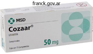
Purchase generic cozaar from india
In such cases, concomitant remedy of the brow depresscm (corrugators, procerus) ought to at the very least be mentioned with the patient Effective tteatment of the transverse brow rhytids entails subcutaneous or intramuscular injections into the frontalis muscle. Simultaneous tteatment of forehead depressoD, if undertaken, can involve the procerus and/or cor:rugatoD andfor the lateral orbicularis oculi above the orbital rim and slightly below the tail of the brow. As this muscle is vay skinny and superficial, inttadermal or instantly subdermal injections are suf:fi. Having the affected person smile or squint will help identify the particular space or lines in need of remedy. Chapter 197: Cosmetic Uses of Neurotoxins and Injectable Fillers 3243 (15), and the modifications are most considerable with dynamic contraction of the platy! Caution must be exercised to avoid extreme doses and to inject into the muscle but not deep to il Overly deep injections can improve the risk of dysphagia and/or neck wealmess. Elevation of the complete brow will require consideration to each the medial and the lateral forehead depressors. Frequently, elevation of only one end of the brow or the opposite might be desired by which case it suffices to handle those muscle tissue solely. For elevation of the medial brow, therapy of the corrugators and procerus as properly as the depreasor supercilii. Elevation of the lateral forehead requires a subdermal injection of the oibicularis oculi muscle just under the eyebrow lateral to the excessive point of the forehead, as soon as once more remaining exterior the orbital rim. Very low doses ofbotulinum toxin injected into the orbicularis oris muscle of the upper lip might help efface or soften vertical upper lip traces. In order to avoid issues with oral incompetence the injections are kept very superficial (subdermal), and very small doses are used initially. Nonetheless, professional public speakers and wind instrument musicians will not be best candidates for this treatment. Four evenly spaced injections are given across the upper lip at or immediately above the vermilion border. In addition to decreasing the depth of obvious vertical lip lines, a point of lip eversion can be incessantly famous. Botulinum Toxin Complications As noted earliet the safety profile of neurotoxins when used for beauty applications is quite favorable. Sound knowledge of the regional anatomy, consideration to proper doses and method and using high concentrationlow volume dilutions all help reduce the incidence of undesirable unwanted facet effects. LaJger volumes can result in inadvertent and undesirable unfold to adjacent nontargeted ~ sues. There have been no deaths reported or adverse long-term results from the cosmetic use of neurotoxins (16). Medications that may potentiate the exercise of botulinum toxin include aminoglycosides, cyclosporins, neuromusrular blockers, calcium channel blockers, quinidine. Injection pain could be minimized by using small gauge needles as properly as slowly inserting the needle and injecting slowly. Best outcomes are seen with delicate to reasonable banding 3244 Section X: Facial Plastic and Reconstn. Headaches happen in about 1% of patients, and patients ought to be knowledgeable of thiJ risk (17). Site-specific complications ought to be rare if appropriate consideration is paid to technical detail. Ptosia outcomes from migration of the injected toxin deep to the orbital septum where it could have an result on the levator palpebrae superioris of the upper eyelid. Staying outside the Oibital rim with all injections (as indicated earlier) ought to stop thiJ complication from growing. Upper eyelid ptosis can develop up to 1 week after injections and normally resolves within 2 to 6 weeks. Should it develop, ptosis may be handled with alpha-adrenetgic ophthalmic drops similar to lepidine zero. For this reason, you will need to keep at least 1 crn outside the lateral Oibital rim always.
Bird Bread (Common Stonecrop). Cozaar.
- How does Common Stonecrop work?
- Are there safety concerns?
- What is Common Stonecrop?
- Dosing considerations for Common Stonecrop.
- High blood pressure, coughs, wounds, burns, hemorrhoids, warts, eczema, and mouth ulcers.
Source: http://www.rxlist.com/script/main/art.asp?articlekey=96063
50 mg cozaar visa
Sutures may just tear via thinned tissue and desired tension should be achieved with the usage of an elevated number of stitches. Some surgeons have reported that particularly in these revision circumstances a deep plane lift generally permits for higher planes of dissection. The surgeon must be honest with himself or herself and with the patient about his or her capacity to enhance the aesthetic outcome. Any different prior neck surgery or irradiation may additionally adversely affect the benefit of dissection and progress of therapeutic or, as within the case of neck dissection, might expose deep neck constructions to catastrophic injury. Incorporating these concerns into the surgical plan will help keep away from a grave outcome. After surgical procedure, the patient and family are knowledgeable once more of issues which may come up and instructed to call the doctor for any questions. The authors use a large cumbersome dressing proper after surgical procedure, partly to discourage turning of the head, which might result in hematoma formation. Postoperative orders are geared towards preserving the patient calm and as pain-free as possible. Increased ache leads to elevated blood stress, which might increase the probability of complications. The affected person also ought to be seen on the first postoperative day; drains are often removed at this time, relying on the quantity of drainage noted. Before releasing the strain of the bandage and removing the drains, fluffs are held over the area from which the drains are going to be eliminated, and pressure is utilized. Before placing the new dressing on the first postoperative day, antibiotic ointment or cream is applied on all incisions (we prefer gentamicin cream), maldng sure to apply some at the exterior auditory canal to forestall any Pseudomonas from the external canal from contaminating the incisions. Usually by the second or third postoperative day, the massive cumbersome dressing could be totally eliminated, and an elasticized facial sling can be utilized. This helps the sldn flap to maintain shut contact with the subcutaneous tissue and provides sooner revascularization and even contraction. It is necessary to pad the ears as a outcome of these elasticized bandages can rub the thin sldn of the pinna and thus cause irritation. Patients are cautioned to not use hair dryers in the quick postoperative period as a outcome of some numbness of the periauricular and scalp areas will be current, possibly causing sufferers to bum their pores and skin with the hair dryer as a result of lack of sensation. Over ensuing visits, all of the sutures are removed and the incisions examined beneath the microscope, especially within the hair-bearing areas to check for ingrown hairs, which might occur up to several months after surgical procedure. After performing beveled incisions, this step additionally ensures that the hair will really grow through the incision for camouflage. Chapter 188: Rhytidectomy (Face-Lift) 3125 Almost all patients have significant swelling or bruising postoperatively, and sometimes that is diffirult for the affected person to accept. Patients have no idea how they need to look and whether issues had been done properly except the doctor says so. For that reason, giving them prints of their preoperative photographs on the first or second postoperative visit is extremely useful. Although variable in its innervation sample, Pantaloni discovered that in additional than half of the circumstances, it innervated the superior third of the auricle and the mastoid space. Often, the skin flap is adherent within the space of each nerves and sharp dissection or traction can injure the nerve, resulting in permanent sensory deficit of the ear and periaurirular pores and skin. The motor nerve department most weak to direct, technical injury is the frontal department of the zygomatic-temporal division of the facial nerve. The authors keep away from deep plane dissection completely in the region superior to the arch and anterior to the temporal hairline to negate the risk of frontal nerve branch damage. Anatomic concerns are necessary for the marginal mandibular nerve and buccal nerve divisions as well. Buccal motor nerve branches are injured with aggressive dissection medial to the anterior border of the parotid gland. An uneven flattening of the midfacial contour might accompany buccal division injury. Transient paralysis of a number of facial nerve branches usually is expounded to local action imposed by infiltrative anesthesia. The most commonly injured nerve is the sensory greater aurirular nerve because of its intimate affiliation with the firm fascia surrounding the sternocleidomastoid muscle With a reported incidence as excessive as eight. Expanding hematomas require instant reoperation to control the bleeding source and to evacuate and drain the newly dissected pores and skin flaps.
Purchase cozaar uk
Peripheral vestibular nynugmus is enhanced and central nystagmus is suppressed when visual fixation is eliminated using Frenzel lenses. Ho~ observing eye movements allows the examiner to acquire important information concerning vestibular function. Characterizing nystagmus components usually permits the examiner to infer the etiology of the attention movement and/or location ofthe lesion. Vestibular control of eye actions ends in the sluggish phase, whereas the quick phase is attributable to the speedy resetting of the eyes in the oibit. Does the nystagmus have a fast part and slow section (jerk nystagmus), or are the actions equal in both instructions (pendular nystagmus) In circumstances of horizontal or vertical jerk nystagmus, by which path does the fast part beat. Test Performance the affected person is seated in an upright position and fixates on a stationary target in primar:y gaze place with bestcorrected imaginative and prescient (with glasses or contact lenses in place if applicable). The affected person must be queried about monovision correction because monovision may be related to the lack to verge usually. This input asymmetry usually leads to a spontaneous, direction-fixed, horizontal-rotary jerk nystagmus with Chapter a hundred sixty five: Clinical Evaluation of the Patient with Vertigo fast-phase movement towards the wholesome ear (away from the damaged side). Firstdegree nystagmus is the least intense and is simply observed with gaze toward the fast part. Second-degree nystagmus is more intense and is noticed with the eyes in primary gaze place or when gazing towards the quick phase. Thirddegree nystagmus is probably the most intense and is current when the eyes are in primary gaze place, gazing towards the fast section or, gazing in the path of the slow part. If second- and third-degree nystagmus are current, the finding represents an acute situation or a greater disparity between the good and bad sides. Characterizing the vestibular nystagmus degree is necessary in determining the time course and degree of compensation for a peripheral vestibular lesion. Over the following several days, the nystagmus intensity declines passing by way of the stages of second- and then first-degree nystagmus. It has been postulated that the neural integrator, which is liable for gaze-holding, is disabled when the nervous system is introduced with a sudden sustained asymmetric vestibular enter. The nystagmus intensity declines as the central system compensates (for a thorough description, see Ref. In contrast, central lesions of the brainstem and cerebellum might cause direction-changing horizontal, vertical, torsional, or pendular nystagmus that may appear diminished under Frenzel lenses. The commonest form of central nystagmus is ccmgenital nystagmus, which has been current since early childhood. This type of nystagmus is direction altering and waveform altering with the direction of gaze. Acquired forms of central nystagmus represent a failure of the central gaze-holding mechanisms within the brainstem and cerebellum or intrusions of inappropriate fast eye actions (saccades) throughout tried fixation. Normally, coordinated contraction of the extraocular muscle tissue beneath the control of the central neural integrator is required to maintain the eccentric gaze. However, any rhythmic eye motion with attempts to maintain the eyes in an eccentric position inside zero to 30 levels off midline is abnormal. Test Performance With the affected person seated using best-corrected VlSton (glasses or contacts), the examiner holds his/her finger 20 to 30 degrees from the nasal root, first within the horizontal airplane after which within the vertical plane, for 10 to 20 seconds in every position. Test Performance Saccadic eye movements are examined by having the affected person alternatively fixate on two stationary targets without moving their head. Observe for the next eye movement traits: accuracy of target acquisition, conjugate movement. Abnormalities in saccadic eye actions may be tough to detect in the clinic examination. There are several types of saccadic intrusions, all of which are indicators of an irregular central course of.
Buy cheap cozaar online
Four widespread anatomic variants that predispose to unfavorable rhinoplasty results: a research based on a hundred and fifty consecutive secondary rhinoplasties. Rhinoplasty within the African-American affected person Plast Reconstr Surg 2003;111:1322-1339. Anatomic basis and scientific implications for nasal tip assist in open versus dosed rhinoplasty. Topology of the lateral nasal cartilages: the anatomical relationship of the lateral nasal to the greater alar cartilage. GillffUin A thorough and considerate method to nasal evaluation is the muse upon which a profitable aesthetic and practical rhinoplasty consequence is constructed. Nonetheless, the "guidelines" contained in this chapter are meant to serve as a helpful, sensible framework upon which to consider nasal aesthetics and from which to move ahead to surgical planning. Terminology Trichion-the most anterior midline level on the hairline Glabella-the most anterior or prominent point on the forehead (lateral view) Nasion (aka radix)-the start line. The midline junction between the frontal bone and the nasal dorsum is also referred to as the radix. Rhinion-the midline junction of the nasal bones and the dorsal septum (osseocartilaginous junction) Supratip-the point alongside the nasal dorsum that lies just cephalic to the nasal tip Tip-the most anterior projecting part of the nasal profile (ideally). The tip "lobule" refers more broadly to the region bounded by the supratip superiorly, the anterior finish of the alar crease laterally, and the anterior nostril margin posteroinferiorly. Infratip lobule-the half ofthe nasal tip seen from the nasal base view that sits anterior to the columella and forefront of the nares and extends to the tip-defining point Soft tissue triangle (or soft tissue facet)-seen from the nasal base. Collectivdy these genemte data on features that have some bearing on nasal-fadal proportion such. Perllaps extra important than the actual angle measurement itselt the position (superiorvs. Chapter one hundred eighty: Rhinoplasty: Incisions, Approaches and Analysis 2943 from the nasion via the nasal tip-defining level. A larger (more obtuse) angle re:fiects a relati~ enhance in tip projection whereas the nasal tip would seem much less projected with a more acute nasofadal angle. Males usually have a extra acute nasolabial angle (90 to 105 degrees) consistent with much less tip rotation, whereas the perfect nasolabial angle in females ranges from a hundred to a hundred and fifteen degrees consistent with more tip rotation. The surgical strategy chosen for nasal swge:ry will range with the training, comfon level, experience. While there are a selection of swgical approaches and the preferred strategy may vary from surgeon to swgeon, as a common rule. Surgical approaches may be broadly categorized into exterior or endonasal approaches. Although no absolute indication exists for both an exterior or endonasal method and multiple components will play into the selection of the working surgeon as famous earli~ an inexpensive set of guidelines for every, which can be of profit to the less experienced swgeon, is printed in Table one hundred eighty. The swgical incisions mostly used or referred to in the literature embody � the mmginal (aka infracartilaginous) incision (Pig. It is typically sited over the medial crw:a about midway back along the columella and is generally irregularized (nonlinear) in some trend for bettf! Alone it could provide entry to the higher two-thirds of the nasal vault (bony and cartilaginous dorsum, nasal sidewall) in addition to retrograde entry to the lateral crw:a of the decrease lateral cartilages. Owing to its proximity to the rim of the nares, any Wltoward healing or scar contracture with this incision carries a better threat of visibility, retraction, notching. Effectively this divides the lateral aus into a superior (cephalic) and inferior (caudal) segment enabling removing of the cephalic strip for wlume reduction of the tip cartilage. This allows access to one or each side of the nasal septum and when combined with either an intercartilaginous or trarulcartilaginous incision dissection can facilitate publicity of the nasal dorsum proper as much as the nasion. This incision separates the attachment of the medial crural feet from the caudal septum. Like the hemitransfixion incision, the whole transfixion incision permits entry to either facet of the septum, the anterior nasal spine. It may be made alone or as a continuation of either an intercartilaginous incision (to facilitate alar supply together with. By virtue of its location, this incision preserves the mucosal attachment and blood supply to probably the most caudal aspect of the nasal septum.
Order cozaar 25 mg on-line
A well constructed and though1fully accomplished questionnaire can be a valuable tool for the ex. Alternatively, a medical assistant can conduct a cellphone interriew and document the data the disadvantages of this strategy include that this method could be extra time consuming and costly, and the resulting knowledge are leas complete than the info from a written questionnaire. Components of the History Distinction Between Vertigo, Disequilibrium, and Light-Headedness the tint essential piece of knowledge in the history is the character or quality of the sensation skilled throughout their episodes. Types of dizziness can be roughly divided into three broad categories: (a) vertigo, (b) disequilibrium. In most situations, patients will sense that the setting is in movement round them (objective vertigo) and should describe the phantasm that objects within their visible world. Such sturdy sensations of environmental and/or self-motion are most frequently generated by a sudden asymmetry within the peripheral vatibular system (labyrinth or eighth cranial nerve) and are valuable indicators of peripheral internal ear or nerve dysfunction. Key Historical Features Vertigo, disequilibrium, or light-headedness Continuous or episodic Seconds, minutes, hours, days, or longer Head/inner ear trauma, barotrauma, upper respiratory an infection, ear infection, systemic sickness or an infection, ototoxic treatment Hearing loss, tinnitus (continuous vs. Patients with disequilibrium might use synonyms like "imbalanced, clumsy, uncoordinated, or worry of falling. The third category-light-headedness-comprises both a variety of sensations (such as "wooziness, giddiness, feeling faint, or as if one is about to cross out") and of etiologies (including migraine, vaswlar, metabolic, druginduced, endocrine, or primary psychogenic causes) (Table one hundred sixty five. Light-headedness, although, is much less commonly a major criticism of patients with damage of the peripheral vestibular pathways. Instead, light-headedness is most often comorbid with vertigo and/or disequilibrium. Finally, patients who develop vertigo secondary to vestibular neuritis describe attack(s) of steady spinning lasting as much as 24 hours. In sufferers with transient ischemic assaults involving the vertebrobasilar circulation and brainstem, attacks usually last quarter-hour, whereas brainstem infarcts and cerebellar hemorrhages exhibit acute, severe vertigo lasting hours and impact the lack to stand. The time course of attacks in patients with disequilibrium is considerably much less stereotypical of their disease process. Similarly, sufferers with light-headedness could have a variable symptom image, with sensations lasting wherever from seconds to days. Time Course Once the nature and quality of the sensation is elucidated, the examiner then makes an attempt to decide the length of the symptoms (Table one hundred sixty five. When patients complain of vertigo, this time course distinction is exceedingly priceless since the commonest peripheral vestibular disease processes produce stereotypical assaults of constant period. Posttraumatic vertigo can be the outcomes of (a) direct mechanical trauma to the pinnacle or inner ear constructions. Furthermore, trauma may cause a dehiscence of the superior sec if the bone overlaying the canal was thin prior to the damage (15-17). Determining a historical past of latest infections is important For instance, vestibular neuritis is thought to be brought on by viral infection, and an higher respiratory viral prodrome could occur prior to vertigo onset (18). Other infections that can cause vertigo embrace herpes zoster oticus (Ramsay Hunt syndrome), suppurative otitis media, human immunodeficiency virus, syphilis, Lyme disease, and tuberculosis. Additionally, recent systemic infections or cancers may have necessitated the administration of ototoxic medication that can trigger vertigo. Accompanying Symptoms problems of the inner ear are regularly aggravated by head movements and modifications of the pinnacle and/or physique vis-a-vis gravity (Table one hundred sixty five. Patients with vestibular problems are most likely to keep their head as still as potential and keep away from sudden movements. In cases of bilateral and uncompensated unilateral vestibular dysfunction, sufferers have issue walking at midnight or on uneven surfaces and avoid quick head movements because of bobbing or blurring of their visible area (oscillopsia). Patients with orthostasis turn out to be extra symptomatic with modifications in place towards gravity such as arising from a mattress or chair and procure relief by lying flat. Patients with superior sec dehiscence may current with sound-induced vertigo (Tullio phenomenon) or pressureinduced vertigo (Hennebert sign) (17, 19). Medications and Comorbidity In many cases, accompanying signs help the examiner in determining if the site of lesion is positioned in the labyrinth, eighth cranial nerve. On the opposite hand, vertigo with out listening to loss may be generated by the labyrinth.
Cheap cozaar 50 mg free shipping
The septal branches of the superior labial artery enter the nose on each side of the nasal spine and form the major blood supply to the anterior septum. Large septal mucosal flaps may be pedicled on the nasal spine space with these branches for reconstructive functions. The dorsal nasal artery, an exterior branch of the ophthalmic artery, anastomoses with the lateral nasal branch of the angular artery, forming an axial arterial community for the dorsal nasal pores and skin. Arterial supply to the nasal tip derives from branches of the anterior ethmoid and angular arteries. The external nasal department of the anterior ethmoid artery perforates the transverse nasalis muscle of the nasal sidewall and descends toward the nasal tip. The lateral nasal branch of the angular artery sends off branches from the ala anteriorly toward the nasal tip. Remaining in the plane just above the perichondrium and periosteum throughout dissection minimizes injury to these vessels. Because of its excessive vascularity, the vestibular epithelium and intranasal mucosa are glorious sources of local flaps for reconstruction of the inning lining in fullthickness nasal defects. In such procedures, a free cartilage graft is commonly used to rebuild structure for the nose. The rich vascular intranasal lining flap nourishes the cartilage graft from its undersurface. During routine rhinoplasty, care have to be taken to protect vestibular skin within the internal nasal valve space to avoid internet formation and nasal valve stenosis (14). Sensory Nerve Supply Sensation to the external nasal skin is provided by branches of the ophthalmic and maxillary divisions of the trigeminal nerve. Twigs from the supratrochlear and infratrochlear branches of the ophthalmic nerve provide sensation to the skin of the radix, the rhinion, and the cephalic portion of the nasal sidewalls (11). The external nasal branch of the anterior ethmoidal nerve supplies the pores and skin over the dorsum of the caudal nostril down to and including the nasal tip. Injury to this nerve during intercartilaginous or cartilage-splitting incisions can lead to tip numbness (11). Branches of the infraorbital nerve present sensation to the facet of the lower half of the nostril and the lateral vestibule. The nasopalatine nerve, a branch of V2 that enters the nose by way of the incisive foramen, supplies the main sensory provide to the posterior two-thirds of the nasal septal mucosa, maxillary gingiva, and anterior palatal mucosa. This nerve may be injured during surgical procedure involving the maxillary crest or nasal floor and may find yourself in momentary numbness near the incisors (12). Noses with thin, less sebaceous skin assume a shape that closely matches the underlying cartilaginous and osseous structure. In such cases, overall proportion of the nostril turns into extra important than absolute size. An instance is the gentle tissue,/scar pollybeak that outcomes after aggressive supratip hump discount in a thick-skinned individual. In sufferers with thin pores and skin, even small irregularities of the underlying structure may turn into evident after surgery because the delicate tissue envelope redrapes very closely to the framework below. In these circumstances, care should be taken to camouflage grafts, edges of bone and cartilage, and another contour irregularities. Graft have to be placed in a exact manner, and any edges which will transmit by way of the skin ought to be beveled or crushed to mix seamlessly into surrounding structures. Soft tissue or crushed cartilage onlay grafts could assist in camouflaging irregularities. The nasal septum, consisting of a bony and a cartilaginous portion, provides assist in all three sections Inner Lining the nasal vestibule is lined with keratinizing squamous epithelium. The surfaces of the nasal cavities, with the exception of the superior olfactory epithelium, are lined 2926 Section X: Facial Plastic and Reconstn. Septum 1he nasal septum is a sagittal midline structure that divides the nostril into two cavities and provides structural help to the osseous and cartilaginous vaults 15). The pe~pendicular plate of the ethmoid types the dorsal facet of the osseous septum.
Syndromes
- Surgery (especially orthopedic or neurological surgery)
- Average red blood cell size (MCV)
- Abnormal urine color
- MAO inhibitors
- Acne-like skin sores that may ooze or crust
- Inability to deal with physical stress
- Time between periods changes each month
- Methyl salicylate
Purchase cozaar with paypal
Furthermore, the flexibility to electronically fade between edited pictures and the corresponding unedited image in real time greatly enhances the analytical energy of pc imaging for surgeon and affected person alike. For the patient, real-time transformation provides instant intuitive understanding of the beauty deformity and larger reassurance regarding the proposed surgical correction. For the surgeon, the morphed image offers improved diagnostic insights and a mutually agreeable nasal contour from which an individualized therapy plan may be developed and quantified. Although the benefits of laptop imaging are irrefutable, pc imaging is topic to potential abuse and/or misinterpretation. Consequently, care must be taken to keep away from overoptimistic simulations and to advise all patients that computer-enhanced photographs symbolize a best-case state of affairs, not a guaranteed end result (5, 16). Nevertheless, whereas precise recreation of the simulated image is just about unimaginable, in sldlled hands the ultimate outcome typically resembles, and even exceeds, the computer-generated simulation (17). As enumeration of the treatment plan turns into increasingly more explidt, the surgical approach. Encouraging the affected person to articulate and acknowledge the attendant surgical risks additionally helps to confinn that danger counseling was effective. Finally, the surgeon ought to ensure that the affected person will comply with all surgical restrictions, care necessities, and follow-up appointments before assuming accountability for surgical care. Anesthetic Considerations Once the choice is made to proceed with surgical remedy. Tobacco customers must be advised to discontinue all nicotine merchandise immediately, and nasal allergic reactions must be treated to reduce preoperative irritation. A general medical evaluation is recommended for any affected person with medical comorbidities or potential anesthetic contraindications. A rigorously administered basic anesthetic titrated to the age and body mass of the patient provides superior patient comfort, improved hemodynamic management, and optimum safety of the lower airway in opposition to the specter of aspiration. However, as a outcome of secondary rhinoplasty cases are sometimes of a for much longer period than the typical major rhinoplasty, particular precautions are required to stop sequela of prolonged immobility throughout common anesthesia. All sufferers are placed in the semi-Fowler "seashore chair� position with the ft and head elevated to facilitate peripheral venous return, and the legs are repeatedly massaged with sequential compression units to decrease pooling of venous blood within the decrease extremities and thereby reduce the risk of deep vein thrombosis. While the price of common anesthesia is marginally greater, the technical advantages of common anesthesia are numerous. First, general anesthesia just about eliminates intraoperative affected person movement in response to painful stimuli. Because the depth of twilight anesthesia is proscribed by the necessity to keep spontaneous air flow, a partially conscious patient is commonly vulnerable to unexpected movements and should even become restless, disoriented, or overtly combative. In contrast, endotracheal intubation safely permits a much deeper degree of anesthesia that virtually eliminates intraoperative affected person motion. Although judicious infiltration of the nose with 1% lidocaine containing epinephrine remains to be required for optimum hemostasis, distortion of the nose from giant fluid volumes may be minimized since ache control is primarily achieved by way of the final anesthetic. In addition, blood strain management is far much less challenging in the absence of painful stimuli, and controlled hypotension can be utilized in healthy sufferers to improve visualization of the surgical field, to reduce extravasation of blood into the surrounding gentle tissues, and to decrease operative blood loss. In contrast, sufferers beneath intravenous sedation may really feel threatened because of anesthetic-induced disorientation and confusion. While a skillfully administered intravenous anesthetic can often avoid these problems, susceptibility to apnea varies broadly amongst individuals and basic anesthesia eliminates this variable completely. During periods of planned stimulation similar to throughout lateral osteotomies, propofol is first administered by bolus to assist in blood pressure homeostasis and labetalol hydrochloride (Hospira Inc. When used in combination with controlledhypotensive anesthesia, the protocol also has a positive influence upon postoperative swelling and ecchymosis since the intraoperative tissue extravasation of blood is tremendously minimized. Patients usually understand their surgical procedure as lasting only a few minutes and few have recall of the working room expertise. For nearly any complex secondary rhinoplasty patient, the emotional burden of a prolonged recovery is considerable. However, early revision surgery within the face of acute postsurgical inflammation and edema is significantly extra complication susceptible. Although ready a full yr before correcting an ugly postsurgical deformity can lead to considerable emotional distress, a wholesome microcirculation is essential to any advanced secondary rhinoplasty, and the normal suggestion to postpone revision surgical procedure for 1 year is generally acceptable since significant microcirculatory impairment is often present in the first year after surgical procedure. Moreover, in instances where the nose remains unacceptably large but otherwise shapely, allowing ample time for swelling and edema to resolve may obviate the need for revision surgery altogether since an appropriate cosmetic outcome typically emerges within 12 months. Longer healing instances are also essential for sufferers with important acne, allergic rhinitis, or other types of continual irritation until the inflammatory stimulus could be adequately controlled. While extended recovery is generally really helpful for the thick-skinned or inflammationprone affected person, prolonged lymphedema can also give rise to everlasting subcutaneous fibrosis, thereby tainting an otherwise passable beauty outcome.
Cheap cozaar amex
For the surgeon, the objective is to determine what bodily characteristics of the face are contributing to the impression of getting older, which stigmata are reversible, and by what means. Ideally, the surgeon brings to this consultation a radical understanding of the processes that result in an aged look and is conversant in a wide variety of medical and surgical interventions acceptable for addressing the clinical downside. Moreover, oftentimes these patients might be left with extra stigmata of a lifted appearance ought to they solely handle the lower face because of this incongruity. For these reasons, in addition to the face-lift, you will need to discuss the potential benefits of browlift, blepharoplasty. A latest paradigm shift among many authors is volume preservation and restoration in the aging face, somewhat than just resuspension of deep tissues. This paradigm shift is partly demonstrated by the increasing use of biologic and artificial injectable fillers in facial plastic surgery. Other generally used volume-augmenting methods for facial rejuvenation include fat injections and alloplastic implants. Proponents of such strategies argue that fats and tissue atrophy occur together with gravitational adjustments. Lambros (1) in contrast pictures taken as a lot as 50 years apart and studied the impact of growing older on the midface and periorbital advanced. He concluded that there was little ptosis at the lid-cheek junction and that the aged look was partly due to quantity loss. In another of his papers, he proposed how the addition of volume may give better results than conventional surgical strategies (2). He emphasized that a face that has sufficient tissue volume is the best predictor of a face-lift outcome. The authors do, however, acknowledge that for some patients with volume loss as properly as tissue descent. To carry the higher third of the face, for example, one ought to depend on a forehead raise. In his latest publication, McCollough stresses that the term face-lift is usually inadequately used. He describes five progressive phases of growing older and proposes a classification that helps matching the particular getting older stage with the appropriate rejuvenation therapy (3). His system describes procedures based on anatomic locations such as temple raise, forehead carry. This article focuses solely on the anatomy and medical considerations of the present-day rhytidectomy procedure. In basic, the getting older face presents 5 landmarks which are factors of interest to patients and surgeons alike. Separate from the described underlying gentle tissue changes, the surgeon must observe the cumulative effects of the inherent aging course of coupled with the consequences of environmental publicity on the skin itself. Extrinsic elements check with environmental insults similar to sun-induced damage (photodamage). Intrinsic aging refers to the natural results of time and is generally characterized by tissue atrophy and descent and reduction in pores and skin mobile and protein components. In fact, chronologically aged skin has decreased epidermal thickness, flattening of the dermal-epidermal junction, atrophy of the dermis, and a general decline in a big selection of cell populations together with melanocytes and Langerhans cells (7). This epidermal thinning then makes the skin extra prone to damage from shearing-type forces (6). Moreover, Yousif and Mendelson (8) confirmed how recurring facial expressions result in coarse pores and skin wrinkling and deep folds. On the opposite hand, photoaged dermis is characterized by putting variability: in its thickness, with alternating areas of atrophy and hyperplasia; in pigmentation, with alternating lentigines and depigmented areas; within the degree of nuclear atypia; and in orderliness of keratinocytes maturation (10). In the previous, sun-damaged dermis was thought to be characterized by a discount in structural elements, resulting in pores and skin wrinkling. In fact, essentially the most hanging function of photodamaged skin is the presence of enormous portions of thickened, poorly organized degraded elastic fibers, which degenerate into an amorphous mass, higher known as elastosis. This lack of elastin decreases skin elasticity, outlined because the loss of the power of the skin to recoil and redmpe as soon as pulled away from the deep tissues (11).
Order cozaar 25mg with amex
Whichever method is chosen, the surgeon should attempt to depend on the same technique for all photographs. Another reliable approach to ensure that the affected person turns to the correct angle every time is to place markas on the wall that point out the place the patient should tum and face for an indirect and lateral view. When turning to the oblique and lateral positions, the affected person should rotate the whole body in alignment with the face and not simply wm the top to these positions, which creates neck distortion. One of the most typical errors encountered is affected person positioning with the neck tilted upward and prolonged, especially in the extra mature patient who desires to reduce the appearance of undesirable neck-tissue redundancy. The horizontal line drawn In A-C Indicates the Frankfort Horizontal Plane that runs via the supratragal point and the Inferior orbital rim, wtlich ought to be revered. The horizontal line In D reveals the alignment of the nasal tfp with the Infrabrow line. The vertlalllne In B shows the alignment of the nasal tip with the malar eminence. If the patient reveals this conduct, the photographer should ask that the patient close his or her eyes forcefully and slowly open them till they seem absolutely open. Each kind of deliberate surgical process mandates a special set of standardized positions with or without additional optionally available views (Table tn. Besides affected person positioning, distracting elements &om jewelry, make-up, clothing, and hairstyling should also be minimized. All obstructive jewellery, for example, necklaces and pendulous earringB, must be eliminated. Turtlenecks and high-necked collars also can obstruct an easy view of the neck and must be pulled down or folded inward to improve effecti:ve communication. All eyeglasses should be eliminated regan:lless of what facial swgecy is being contemplated. Hairstyling ideally must be pulled again to show an unobstructed view of the eyes, nose, ears, lips, and neck and to be lowered to an Wlobtru. All makeup ought to be eliminated, particularly if any dermatologic resurfacing or scar revision is deliberate. Lighting and Background Lighting can additionally be a crucial factor that ought to be standardized. Placing the affected person an acceptable distance away (-2 feet) from the rear wall also can reduce unwanted shadows. Generally, an oncamera flash tends to trigger extreme highlights and shadows and a "washed-out" look to skin tones, but experimentation will decide the best stability of lighting for a particular room and digital camera. Furthermore, the ambient room lighting may be altered to match the color spectrum. If shadows and highlights are desired to find a way to intensify a scar or other contour irregularity, for instance, distinguished nasolabial lines for correction with a delicate tissue filler, then the balanced fill lights (if used) should be turned off. In addition, the exposure value can be decreased in order to draw out the intended function. Rather than hanging a blue drape behind the patient, a smooth wall can be painted the desired blue, as a wall will remain flat and wrinkle free. Again, using the very same room with the identical lighting will assist in achieving reproducible outcomes. Conversely, carrying a conveyable blue background material from room to room to photograph the affected person will most probably result in refined, if not apparent, inconsistencies within the images. If a wall is to be painted blue, it may be clever to document the precise shade of blue, or corresponding numeric code, in order that if the doctor ought to should relocate the pictures room in the future the same blue shade could be exactly reproduced. Intraoperative Photography Although good pre- and postoperative pictures remains the core of this section, correct strategies for superior intraoperative images must be mentioned herein. Intraoperative images can be utilized to doc medicolegally what transpired in the working theater but primarily serve as an academic tool for different training surgeons or as part of a didactic presentation in a Chapter 171: Pictorial Documentation: Digital Imaging and Traditional Photography 2717 bad<ground ~ Iy high kicker li! If the ambient lighting Is Insufficient or casts very harsh shadows, fill lights an be used to soften the shadows.
Purchase generic cozaar
Prevalence of post-headshake nystagmus in sufferers with caloric deficits and vertigo. Dynamic visible acuity: a take a look at fur oscillopsia and vestibulo-orular reflex function. Dynamic visual acuity during transient and sinusoidal yaw rotation in regular and unilaterally vestibulopathic people. Dynamic visual acuity throughout passive and self-generated transient head rotation in regular and unilaterally vestibulopathic humans. Occurrence of semicirrular canal involvement in benign paroxysmal positional vertigo. A newly rerognised reason for vertigo: horimntal canal variant of benign positional vertigo. Efficacy of the "bow and lean test� fur the management ofhorimntal canal benign paroxysmal positional vertigo. Hair cells in the right and left vestibular organs fire at a tonic baseline price; if the head is turned towards the best, the hair cells of the right horizontal canal enhance their firing price whereas the hair cells of the left horizontal canal decrease their firing price. Sensory input from the vestibular periphery is relayed centrally by way of the vestibular nerves; the superior vestibular nerve carries info from the horizontal and superior semicircular canals and the utricle while the inferior vestibular nerve transmits input from the posterior semicircular canal and saccule. The brain compares the inputs from right and left vestibular organs so as to determine that a change in head position has occurred and generates compensatory eye movements and postural changes. In this article, we review peripheral vestibular issues, that are characterised by pathology located in the peripheral vestibular end organs. The hallmark symptom of peripheral vestibular dysfunction is vertigo, which occurs if the right- and left-sided vestibular nerves fire asymmetrically in the absence of a head motion, creating an illusory sense of movement. We categorize peripheral vestibular issues based mostly on the clinical manifestations of vertigo (Table 166. Vertigo can be episodic, resulting from a reversible unilateral loss or acquire of vestibular perform, both of which may produce an asymmetry in the firing rates of the right- and left-sided vestibular nerves. In migrainous vertigo, disruption ofvestibular function could final for variable durations of time, in some events for minutes to hours, but in others for days. Perilymph fistulas might produce both an episodic disruption or excitation of vestibular function. A third class of peripheral vestibular disorders are those who end result from chronically insufficient vestibular function. Some particular other causes are discussed in other chapters, including temporal bone trauma (Chapter 150) or acoustic neuroma (Chapter 159). Chronic bilateral vestibular dysfunction sometimes occurs within the setting of a systemic exposure, corresponding to to aminoglycoside antibiotics or chemotherapeutic agents, or may be genetic in origin. Longitudinal research suggest that vertigo ceases spontaneously in 57% of cases at 2 years and 71% after eight. With long-standing disease (greater than 10 years), the audiometric sample flattens and the listening to loss usually stabilizes at a pure-tone average of 50 dB and a speech discrimination score of 50% (12). Profound sensorineural hearing loss happens in 1% to 2% of sufferers (13); if the losses are bilateral, sufferers might profit from cochlear implantation (14). Chronically insufficient vestibular perform Unilateral � Unilateral vestibular hypofunction following vestibular neuritis, trauma, and so forth. Endolymph, the potassium-enriched fluid within the inside ear, may be both excessively synthesized or inadequately resorbed, resulting in growth of the endolymphatic house (17,18). Endolymphatic hydrops sometimes entails the pars inferior of the labyrinth (composed of the saccule and cochlea) (16,19). The pars superior (utricle and semicircular canals) may be concerned in endolymphatic hydrops, although adjustments are inclined to be less dramatic and occur much less incessantly. The most distinguished concept holds that hydropic distension of the endolymphatic duct causes rupture of the distended membranes, a phenomenon that has been noticed throughout the labyrinth (20). Membrane rupture allows the potassium-rich endolymph to leak into the perilymphatic house and contact the basal surfaces of the hair cells as properly as the eighth cranial nerve.
References
- Wong KS, Chen C, Fu J, et al. Clopidogrel plus aspirin versus aspirin alone for reducing embolisation in patients with acute symptomatic cerebral or carotid artery stenosis (CLAIR study): a randomised, open-label, blinded-endpoint trial. Lancet Neurol 2010;9: 489-497.
- Jatoi I, Anderson WF, Jeong JH, et al. Breast cancer adjuvant therapy: time to consider its time-dependent effects. J Clin Oncol 2011;29(17):2301-2304.
- Mol F, Strandell A, Jurkovic D et al. The ESEP study: salpingostomy versus salpingectomy for tubal ectopic pregnancy, the impact on future fertility: a randomised controlled trial. BMC Womenis Health 2008; 8: 11.
- Cooper A, DiScala C, Foltin G, Tunik M, Markenson D, Welborn C. Prehospital endotracheal intubation for severe head injury in children: a reappraisal. Semin Pediatr Surg. 2001;10(1):3-6.
- Peters KM, Killinger KA, Gilleran J, et al: Does patient age impact outcomes of neuromodulation?, Neurourol Urodyn 32(1):30n36, 2013.
- Dellgren G, David TE, Raanani E, et al. Late hemodynamic and clinical outcomes of aortic valve replacement with the Carpentier-Edwards Perimount pericardial bioprosthesis. J Thorac Cardiovasc Surg 2002; 124:146-154.

