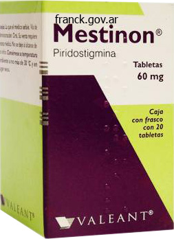
Pyridostigmine dosages: 60 mg
Pyridostigmine packs: 30 pills, 60 pills, 90 pills, 120 pills, 180 pills, 270 pills

Purchase generic pyridostigmine on line
Scarring may be a extreme sequela of this an infection, and it may be made worse by bacterial superinfection. The presence of any honey-colored crusting or increasing erythema outdoors the dermatome ought to counsel the chance of secondary impetigo or cellulitis. Prompt recognition and remedy are required to assist forestall severe, disfiguring scarring. The presence of multinucleated big cells on a Tzanck preparation taken from a vesicular rash in a dermatomal distribution confirms the prognosis. If one have been to biopsy a vesicle, ballooning degeneration of the keratinocytes would be present. Most cases occur with advancing age, as cell-mediated immunity tends to wane with time. Once the virus reactivates, it begins to replicate and to trigger necrosis of the affected nerve cells. Treatment: Treatment with antiviral medicines from the acyclovir household must be instituted instantly. The sooner therapy is started, the better is the prospect of lowering the size of illness. Painful vesicles, erosions with an erythematous base Dendritic keratitis (herpes simplex) demonstrated by fluorescein Technique of making use of fluorescein strip in previously anesthetized eye Acute keratitis (ciliary injection, irregular corneal surface) lower the danger of postherpetic neuralgia, but giant studies have up to now shown inconclusive information to assist this approach. The remedy has the best likelihood of changing the course of the disease if given within the first seventy two hours after the onset of illness signs. A stay attenuated zoster vaccine for the prevention of herpes zoster is being given to sufferers older than 60 years of age. As with all live vaccines, its use in immunosuppressed patients is contraindicated. Amitriptyline, gabapentin, lidocaine patches, pregabalin, anticonvulsants, and opioids are all used with various success. They are able to inflicting disease in any individual, but extreme infections seem to be extra probably in those who are immunocompromised. Warts can affect any cutaneous floor, and unique wart subtypes are extra susceptible to cause disease in different medical locations. Types sixteen and 18 are believed to have been responsible for up to 70% of cervical cancers. Clinical Findings: Verruca vulgaris, additionally known as the common wart, is the most prevalent wart that infects the human. These warts often appear as small papules with a rough surface studded with pinpoint, darkish purple to black dots. Most warts are between 5 mm and 1 cm in diameter, but some can become fairly massive and embody a lot larger areas of the skin. A good rule of thumb is that 50% of verrucae will disappear spontaneously in 2 years. The filiform wart is represented by a small verrucal papule with finger-like projections extruding from the bottom of the papule. The flat wart is incessantly encountered and manifests as a 3- to 5-mm, flat papule with a slight pink to pink to purple coloration. Flat warts are frequently seen on the legs of women and in the beard area of men, and they can be organized in a linear sample if the warts are unfold through the act of shaving. They are deep-seated papules and plaques that will coalesce in to massive mosaic warts. Uterine cervix Vagina Vaginal lumen Stratified cervical epithelium Basal layer of the cervical epithelium Virus Lamina propria the warts are well outlined and characteristically interrupt the skin strains. This is in contrast to a callus, by which the skin lines are retained, and this signal can be used to differentiate the two circumstances. Palmar warts are similar to plantar warts and have the identical medical look. Subungual and periungual warts, a subclassification of palmar/plantar warts, are discovered around and under the nail equipment.

Buy pyridostigmine 60mg line
However, many circumstances of medicationinduced pyogenic granulomas require some method of surgical elimination. In distinction to the other histiocytoses, patients with reticulohistiocytoma have regular lipid ranges. Reticulohistiocytomas can happen as a solitary development or as a quantity of growths in a condition known as multicentric reticulohistiocytosis. On histopathological examination, the two medical variants are identical in nature. It can typically be a marker of internal malignancy, and sufferers are afflicted with a extreme arthritis. Clinical Findings: Solitary lesions are sometimes small, agency dermal nodules ranging from 1 to 2 cm in diameter. They are found mostly on the pinnacle and neck area of the body however have been described in all places. Multicentric reticulohistiocytosis is unique in that it happens in an older inhabitants, with a higher percentage of females affected. The a number of reticulohistiocytomas discovered on this condition are most often localized to the dorsal facet of the arms and to the face. A distinctive finding is that of small papules along the lateral and proximal nail folds. These sufferers also have a extreme arthropathy, and this analysis ought to lead one to search for an underlying malignancy. The arthropathy almost all the time affects the interphalangeal joints, significantly the distal interphalangeal joints. Multicentric reticulohistiocytosis is believed to be a paraneoplastic situation in as a lot as 25% of the circumstances. The kind of malignancy is variable, with no predominant kind more prevalent than another. In about one third of patients with multicentric reticulohistiocytosis, the joint symptoms precede the growths; in one third, they appear on the same time; and one third of the sufferers develop only clinically minor or no arthropathy. Early recognition and treatment has helped lower the development in to severe mutilating arthritis. Many sufferers have cardiac involvement, and almost all organ techniques have been reported to be affected, some with deadly outcomes. Pathogenesis: Multicentric reticulohistiocytosis and solitary reticulohistiocytoma are believed to symbolize a uncommon dysfunction of histiocytes. Histology: the tumor exhibits a well-circumscribed dermal infiltrate and not using a capsule. The infiltrate is made up nearly totally of histiocytes with a "ground-glass" Multicentric reticulohistiocytomas. Organs Involved in Reticulohistiocytoma Inflammatory arthritis (hands, knees, shoulders) Lungs Bone marrow Eyes Heart Associated Autoimmune Diseases and Malignancy Systemic lupus erythematosus Breast cancer Colon most cancers Primary biliary cirrhosis Lymphoma Lung cancer Low energy. Treatment: Solitary reticulohistiocytomas are cured with a simple elliptical excision. Screening and fixed vigilance for an underlying malignancy is required in all instances. Corticosteroids, methotrexate, hydroxychloroquine, and cyclophosphamide have all been used. The objectives are to forestall or suppress the arthropathy and to display screen for malignancy. These growths are out there in all sizes and shapes and invariably could be found on any human older than forty years of age. They begin to manifest within the third to fifth decade of life and continue to improve in number thereafter. The basic description is that of a 1- to 2-cm plaque with a "stuck-on" appearance and small horn cysts. Stucco keratoses are small (1-5 mm), gray-tan papules with a stuck-on appearance or skinny patches on the decrease extremities. Dermatosis papulosis is a condition by which a number of seborrheic keratoses occur on the face and neck.

Generic pyridostigmine 60 mg without a prescription
This results in elevated manufacturing of thyroid hormones, both triiodothyronine (T3) and (T4), by the thyroid. The improve in metabolic functioning of the thyroid leads to diffuse enlargement and goiter. Histology: Biopsy specimens of the pretibial skin present large quantities of mucin deposits inside the center and decrease dermis, between collagen bundles. The mucin is so thick that it causes the dermal collagen bundles to be splayed apart. Biopsy specimens from clinically nonaffected skin may present a number of the same histological findings however on a lesser scale. Ablation of the thyroid may be achieved with radiation therapy or surgical removing. Abscess, sinus tract formation, and significant scarring lead to exquisitely tender areas of involvement. It may be an isolated medical discovering, or it may be associated with cystic acne, dissecting cellulitis of the scalp, and pilonidal cysts. Clinical Findings: Hidradenitis suppurativa is most commonly encountered in postpubertal girls. This situation preferentially affects areas which would possibly be rich in apocrine glands and terminal hairs. Hidradenitis suppurativa begins as tiny red papules or nodules that are probably to be folliculocentric. At this point, the differential analysis contains an early folliculitis or furunculosis. As the disease progresses, the onerous nodules become fluctuant and spontaneously drain to the surface of the skin. Eventually, sinus tracts develop that interconnect multiple subcutaneous nodules with a number of cutaneous openings. Clinically, pressing on one of many nodules might produce drainage from a distant sinus tract. Long-standing disease has been associated with the development of squamous cell carcinoma. The tumors are probably to be giant at prognosis the drainage from the cutaneous nodules typically requires extensive bandaging to hold clothing from getting dirty. The draining sinus tracts and nodules are sometimes colonized with varied micro organism, and cultures of the purulent drainage present development of a quantity of totally different organisms, together with Staphylococcus aureus and streptococcal species. The bacteria in these circumstances are present secondary to the underlying inflammatory condition and the lack of normal cutaneous pores and skin barrier operate. Pathogenesis: Hidradenitis suppurativa is an inflammatory illness with secondary bacterial superinfection and colonization. Hidradenitis is theorized to be attributable to rupture of the mature follicular epithelium along areas of apocrine glands; hence, its propensity to happen in areas with high densities of apocrine glands. Once the hair follicle ruptures, an inflammatory cascade is about off and causes the ensuing nodules, cysts, fistulas, and scarring. Histology: Chronic lesions show a dense, blended inflammatory infiltrate with abscess and sinus tract formation. Treatment: Therapy is usually geared toward decreasing irritation and bacterial superinfection. Topical clindamycin and other antibacterial products corresponding to benzyl peroxide are sometimes the first-line brokers employed for delicate illness. Other brokers that have had limited success include isotretinoin, etanercept, and infliximab. Surgical choices embrace broad native excisions to remove the affected tissue and restore with complicated flap closure. Liposuction has additionally been tried in an try and take away the affected apocrine gland hair follicle unit.

Purchase pyridostigmine 60mg with amex
There are extra extreme types of keratosis pilaris in which patients current to the dermatologist for therapy. Clinical Findings: Keratosis pilaris is one of the commonest dermatoses and is assumed by some to be a variant of normal. It is discovered in more than 40% of the adult population and in as many as 80% of youngsters. Most instances are asymptomatic and are of no concern to the patient or of only beauty concern. Small (1-2 mm), pink-to-red follicular hyperkeratotic papules are current to a various extent. Other circumstances are more widespread and can embody the higher thighs, shoulders, and cheeks. Widespread cases tend to be extra noticeable, and the small papules tend to be more inflammatory in nature. This inflammatory form of keratosis pilaris is also called keratosis pilaris rubra. It is often manifested by brilliant pink, small, hyperkeratotic papules that will resemble pustules. A small scraping of the inflammatory lesion ends in removal of a small keratin plug rather than the contents of an acneiform pustule. The location on the outer arms and upper thighs additionally helps to differentiate this situation from pimples. Ulerythema ophryogenes is a keratosis pilaris variant that manifests in early childhood. The lateral one third of the eyebrow is affected with minute, pink keratotic papules. Over time, alopecia could develop in the affected areas, especially the lateral eyebrows. It manifests as small, hyperkeratotic plugs on the cheeks that resolve and leave behind small, atrophic scars in a fantastic mesh-like sample. Erythromelanosis follicularis faciei et colli is similar in nature to atrophoderma vermiculata, nevertheless it lacks any proof of scarring. This situation has been reported to happen mostly in younger males during the second and third decades of life. Postinflammatory hyperpigmentation is another unusual feature not seen with the opposite variants. Keratosis follicularis spinulosa decalvans is probably the least frequent keratosis pilaris variant. It is manifested by areas of skin thickening and follicular plugging Ulerythema ophryogenes exhibiting loss of the lateral eyebrows Keratosis pilaris of upper thighs. Small 1- to 2-mm hyperkeratotic red papules Keratosis Pilaris Variants Keratosis pilaris rubra Ulerythema ophryogenes (keratosis pilaris atrophicans faciei) Atrophoderma vermiculata (folliculitis ulerythematosa reticulata) Erythromelanosis follicularis faciei et colli Keratosis follicularis spinulosa decalvans Keratosis pilaris atrophicans faciei. It is believed to be brought on by an abnormality in follicular keratinization of the infundibulum. The plug is often 1 to 2 mm in diameter and should lie on top of a meager lymphocytic infiltrate. After discontinuation, nonetheless, the rash of keratosis pilaris returns over a period of some weeks to months. The cream is utilized daily and has been successful in eradicating the redness and hyperkeratosis. Historically, the disease was categorized primarily based on the grouping of signs and organs affected, with names corresponding to Letterer-Siwe illness and HandSch�ller-Christian disease. These histiocytoses are a heterogeneous group of diseases which will affect each the skin and varied internal organs. The primary pathological discovering is the accumulation of pathological Langerhans cells throughout the affected tissue. The diagnosis is made on scientific, histological, laboratory, and radiographic findings. Prognosis and therapy depend on the organ methods concerned and the number of systems implicated. Usually, the situation is first noticed in childhood, but adult-onset illness does occur.

Order pyridostigmine amex
If the steroid is tapered too quickly, the patient could expertise a poststeroid flare of their dermatitis, which can be proof against further corticosteroid therapy. Without the utilization of patch testing, the allergen will stay unknown and the dermatitis will persist. Atopic dermatitis has been estimated to affect up to 10% of all kids and 1% of adults, and its prevalence has been steadily increasing. Patients frequently have a household history of atopic dermatitis, bronchial asthma, or skin sensitivity. Infants a couple of months old might initially current with pruritic, purple, eczematous patches on the cheeks and extremities in addition to the trunk. The itching is often severe and causes the child to excoriate the pores and skin, which might lead to secondary skin infections. These children have difficulty sleeping due to the severe pruritus associated with the rash. During flares of the dermatitis, sufferers could develop weeping patches and plaques that are extremely pruritic and occasionally painful. With time, the patches start to localize to flexural regions, significantly the antecubital and popliteal fossae. Patients with atopic dermatitis are more vulnerable to react to contact and systemic allergens. Sensitivity to contact allergens is likely a consequence of the frequent use of topical medicaments and the broken skin barrier. This mixture results in increased publicity to overseas antigens which might be capable of inducing allergic contact dermatitis. Laboratory testing generally shows an eosinophilia and an elevated immunoglobulin E (IgE) level. It might manifest with the appearance of honey-colored, crusted patches within the excoriated areas, which indicates impetigo. It can also manifest as a quantity of follicle-based pustules, representing folliculitis, or as deep pink, tender macules, indicating a deeper soft tissue an infection. The fee of colonization of atopic patients is much higher than in regular controls, most probably because of the disruption of the underlying dermis. Acquisition of a widespread herpesvirus an infection can have severe and doubtlessly lifethreatening consequences. The extensive areas of irregular, broken skin provide the proper setting for the event of this widespread viral infection. It is estimated that 10% of circumstances will resolve by the age of 1 year, 50% by 5 years, 70% by 7 years, and so forth. A small share of kids with atopic dermatitis continue on with the rash in to adulthood. They embody anything that irritates the pores and skin, similar to warmth, sweating, stress, many chemical compounds, and varied types of clothes. Atopic dermatitis is believed to be brought on by an aberrant T-cell (Th2) response within the skin with elevated levels of Th2 cytokines. These cytokines are responsible for eosinophil production and recruitment and for IgE production. Ultimately, the barrier of the epidermis is disrupted, and this is evident by the increase in transepidermal water loss, which could be measured. Histology: A nonspecific lymphocytic infiltrate is seen, with related exocytosis of lymphocytes in to the epidermis with widespread spongiosis. Treatment: Therapy consists of patient and family schooling in regards to the natural history of the illness and the episodic waxing and waning. Bathing regimens must be totally defined, and the use of cleaning soap must be discouraged. The patient ought to take shorter baths in lukewarm water, followed immediately by moisturization and utility of topical steroid medications as appropriate. The use of topical immunomodulators, alternating with topical corticosteroids or alone, decreases the atrophogenic side effects of the topical corticosteroids. On occasion, oral steroids could also be wanted to calm the inflammation and provides the patient some well-needed, albeit short-term, reduction. If any query exists as to whether or not a food is potentially exacerbating the dermatitis, an allergist may be consulted to perform particular food allergy testing.

Buy pyridostigmine online pills
Pruritic urticarial papules and plaques of pregnancy wholly abated with one week twice daily software of fluticasone propionate lotion: a case report and evaluate of the literature. Choroidal neovascularization in angioid streaks and pseudoxanthoma Integumentary System elasticum: 1 yr follow-up. Systemic therapies for pediatric atopic dermatitis: a evaluation for the first care doctor. Biofeedback, cognitive-behavioral methods, and hypnosis in dermatology: is it all in your thoughts Femoropopliteal occlusive disease: prognosis, indications for therapy, and outcomes of interventional therapy. Systemic and local management at the onset of Stevens-Johnson syndrome and poisonous epidermal necrolysis with ocular problems. Commentary: remedy of lichen planopilaris: some progress, however a protracted method to go. A case of neutrophilic eccrine hidradenitis associated with streptococcal infectious endocarditis. Sarcoidosis of the skin-a dermatological puzzle: important differential diagnostic elements and tips for scientific and histopathological recognition. New insights in to the administration of zits: an replace from the worldwide alliance to improve outcomes in zits group. The epidemiology of hand eczema within the common population-prevalence and major findings. Efficacy of 4 insect repellents against mosqui to bites: a double-blind randomized placebocontrolled subject research in Senegal. Long-term observe up, clinical features, and quality of life in a collection of 103 sufferers with hyperimmunoglobulinemia D syndrome. Expert consensus for the therapy of disseminated intravascular coagulation in Japan. Juvenile dermatomyositis: new developments in pathogenesis, assessment and remedy. Clinical, etiologic, and histopathologic features of Stevens-Johnson syndrome throughout an 8-year period at Mayo Clinic. Necrobiotic xanthogranuloma: a review of 17 cases with emphasis on scientific and pathologic correlation. Combination therapy of intravenous immunoglobulin and corticosteroid within the therapy of poisonous epidermal necrolysis and Stevens-Johnson syndrome: a retrospective comparative research in China. Kostman disease-infantile genetic agranulocytosis: historic views and new features. Paraneoplastic pemphigus- an autoimmune mucocutaneous disease associated with neoplasia. Therapy of paraneoplastic pemphigus with rituximab: a case report and evaluate of literature. Paraneoplastic autoimmune multiorgan syndrome: evaluation of the literature and help for a cytotoxic role in pathogenesis. Mucous membrane pemphigoid: medical aspects, immunopathological features and remedy. Advances in pemphigus and its endemic pemphigus foliaceus (Fogo Selvagem) phenotype: a paradigm of human autoimmunity. Structural foundation of the interplay between integrin alpha6beta4 and plectin on the hemidesmosomes. Cross-talk between hemidesmosomes and focal contacts: understanding subepidermal blistering illnesses. Drug-induced linear IgA bullous dermatosis demonstrating the isomorphic phenomenon. Epidermal basement membrane zone components: ultrastructural distribution and molecular interactions. Dynamic relationship of focal contacts and hemidesmosome protein complexes in reside cells. New insights in to the immunoultrastructural group of cutaneous basement membrane zone molecules.

Order pyridostigmine 60 mg with visa
The most superficial cells are bigger than the others and send projections down over the lateral surfaces of the cells beneath them, sometimes having an umbrella-like look. These "umbrella cells" have ample eosinophilic cytoplasm and may be binucleate. From the minor calyces onward, these cells rest on high of a skinny lamina propria, twin muscle layer, and adventitia. The outer of the two muscular layers consists of "typical" clean muscle cells, which enhance in quantity near the ureteropelvic junction and lengthen in to the ureter. The inner layer, in contrast, accommodates "atypical" easy muscle cells that terminate at the ureteropelvic junction. At present these atypical cells are thought to be the pacemaker cells answerable for the initiation of peristalsis. They are smaller than typical cells and their contractile filaments, as a substitute of working parallel, seem randomly scattered, as in cardiac pacemaker cells. Another population of cells, which resembles interstitial cells of Cajal, has just lately been identified in the renal calyces of some mammals, but its function remains to be being decided. These cells sit on a well-developed, free lamina propria that accommodates small vessels and nerves. Outside of the lamina propria is the muscularis propria, which is continuous with the layer of "typical" smooth muscle cells seen in the renal pelvis. Its contractile fibers are loosely organized and interspersed with connective tissue. In the decrease half of the ureter, an additional outer ring of longitudinal fibers tends to appear. Because of the urothelial folds and the well-developed longitudinal musculature, sizable calculi might cross by way of the ureter without harm to the mucosa. The outermost layer of the ureter incorporates a thick adventitia with longitudinally oriented small blood vessels. Urothelium in distended bladder the bladder incorporates five to eight irregularly folded layers of urothelial cells. An exception happens on the trigone, where there are usually fewer layers of urothelial cells with a smooth, unfolded arrangement. As the bladder is distended, the urothelial cells flatten out, with essentially the most superficial cells flattening out to such an extent that they turn into barely visible. During this course of, vesicles near the apical surfaces of the cells fuse with the plasma membrane to provide further floor space. Unlike in the ureter, the lamina propria often incorporates a muscularis mucosae, which appears discontinuous and accommodates a haphazard arrangement of wispy, skinny bundles of clean muscle cells. In some situations, the fibers can turn into hypertrophic and resemble these of the muscularis propria. The muscularis propria is named the detrusor muscle; as within the decrease ureter, it consists of inside and outer longitudinal fibers with an intervening layer of circumferential fibers. Except in the space of the bladder neck, these layers are typically indistinct, appearing as a meshwork of criss-crossed thick muscle bundles. Interspersed by way of the muscular layers are blood vessels, lymphatics, nerve fibers, and even adipose tissue. After the fourth week, during which the embryo undergoes a fancy folding course of, the intermediate mesoderm forms a lateral nephrogenic twine and a medial genital (gonadal) ridge. The nephrogenic wire offers rise to three successive kidney precursors, whereas the genital ridge offers rise to the gonads. The three kidney precursors-known as the pronephros, mesonephros, and metanephros, in order of appearance-develop in a cranial-to-caudal sequence alongside the nephrogenic cord. Many signaling pathways have been found to play roles in the improvement of the kidneys, with the already expansive list growing on a daily basis. A detailed dialogue of these pathways, however, is past the scope of this text, which is in a position to instead focus on the anatomic adjustments that occur throughout growth.
Cost of pyridostigmine
It is indicated for use as an anticoagulant in the treatment of a selection of situations, together with atrial fibrillation and deep venous thrombosis, and after coronary heart valve substitute surgery. Warfarin acts by inhibiting the enzymes that are liable for carboxylation of glutamate residues and epoxide reductase. This each decreases the out there clotting factors and induces vitamin K deficiency, resulting in added discount of available clotting factors. Clinical Findings: Vitamin K antagonists have been proven to trigger a selected sort of cutaneous eruption known as warfarin necrosis, which occurs in approximately zero. Warfarin necrosis affects the areas of the physique which have increased physique fat, such as the breasts, the stomach pannus, and the thighs. Inactive K Active K Purple toe syndrome associated with vitamin K antagonist therapy Circulatory system Intracranial hemorrhage, after trauma, within the occipital lobe in a affected person taking warfarin initially develops small, red to violaceous petechiae and macules preceded by paresthesias. These regions become erythematous and purple (ecchymoses) with intense edematous skin. Painful cutaneous ulcers might occur, with some extending in to the subcutaneous tissue, together with muscle. The affected areas proceed to endure necrosis unless the warfarin is withheld and the affected person is handled with a different class of anticoagulant. The feet and decrease extremities may have a reticulated, purplish discoloration referred to as "purple toe syndrome. Histology: Skin biopsies from areas of warfarin necrosis present an ulcer with a blended inflammatory infiltrate. Thrombosis is seen inside the small vessels (venules and capillaries) of the cutaneous vasculature. Findings of irritation, a neutrophilic infiltrate, arterial involvement, a powerful lymphocytic infiltrate, or the presence of micro organism in or around vessels mitigate in opposition to the diagnosis of warfarin necrosis. Bacteria will be present on the surface of the ulcer and are believed to be a secondary phenomenon. Preactivation of those clotting elements requires the action of vitamin K carboxylation on glutamate amino acid residues. Once preactivated, the clotting factors can be found for full activation and clot formation when exposed to calcium and phospholipids on the surface of platelets. Inhibition of these clotting factors by vitamin K antagonists leads to anticoagulation. On the other hand, protein C and protein S are answerable for turning off the clotting cascade and play a natural regulatory function in normal coagulation. When these proteins are inhibited, the clotting cascade may proceed unimpeded, permitting for extreme clotting. Therefore, when individuals are handled initially with warfarin, the degrees of protein C and protein S are depleted earlier than the other factors, leading to a prothrombic state. This initial prothrombic state is answerable for the medical signs and signs of microvasculature blood clotting and pores and skin necrosis. The clotting takes place in areas of elevated adipose tissue because of the sluggish move of blood via the fantastic vasculature in these regions. For this reason, most patients are given heparin or a similar anticoagulant until the total effect of warfarin on all clotting factors has occurred. Therapy: Treatment of warfarin necrosis requires discontinuation of warfarin and initiation of heparin anticoagulation and supportive care with fresh-frozen plasma and vitamin K replace the lost protein C and protein S. Surgical debridement could additionally be required, and one must be vigilant for any indicators or signs of secondary infection. Cross-linked fibrin (clot) Fibrinogen Element Ca++ Fe++ Site of absorption Mechanism Duodenum Active and jejunum Duodenum Facilitated diffusion and jejunum Mixed micelle Na+-coupled/2� active Na+-coupled/2� energetic Na+-coupled/2� energetic Na+-coupled/2� lively Facilitated diffusion Passive diffusion Bile acid Exocytosis Fat-soluble vitamin Chylomicron Passive diffusion Passive diffusion Passive diffusion Passive diffusion Water-soluble vitamins Ileum Vitamin C Jejunum Thiamin (B1) Jejunum Riboflavin (B2) Jejunum Biotin Ileum Vitamin B12 Jejunum Pyridoxine (B6) and ileum Fat-soluble nutritional vitamins Vitamin A Vitamin D Vitamin E Vitamin K Jejunum and ileum Jejunum and ileum Jejunum and ileum Jejunum and ileum In to lacteal alternative of vitamin K and supportive care. Vitamin K deficiency in neonates and infants is identified by an isolated elevation within the prothrombin time. The levels of the vitamin K�dependent clotting cofactors can each be measured, and vitamin K substitute ought to be administered to those who are poor. The best methodology for supplementation has yet to be decided, however it can be achieved with a onetime intramuscular injection or with oral substitute. The main scientific findings relate to nervous system involvement and liver illness.
Real Experiences: Customer Reviews on Pyridostigmine
Charles, 57 years: The rhomboideus major muscle arises from the spinous processes of T2 to T5 and inserts on the medial border of the scapula under its backbone.
Jose, 46 years: In females, the dorsal urethral defect might likewise be brief, leading to a patulous urethral orifice, or extra in depth, inflicting an open defect that reaches the neck of the bladder.
Kapotth, 63 years: Fever is common, as is the following growth of dry eyes and mouth as a result of the widespread, usually painless, inflammation of the affected glands.
8 of 10 - Review by A. Muntasir
Votes: 46 votes
Total customer reviews: 46
References
- Schwartz ML, Cox GF, Lin AE, et al. Clinical approach to genetic cardiomyopathy in children. Circulation. 1996;94:2021-38.
- Gum PA, Kottke-Marchant K, Welsh PA, et al. A prospective, blinded determination of the natural history of aspirin resistance among stable patients with cardiovascular disease. J Am Coll Cardiol 2003;41:961-67.
- Wen JG, Yang L, Xing L, et al: A study on voiding pattern of newborns with hypoxic ischemic encephalopathy, Urology 80(1):196n199, 2012.
- Garstang SV, Stitik TP. Osteoarthritis: epidemiology, risk factors, and pathophysiology. Am J Phys Med Rehabil 2006; 85(11 Suppl):S2-11.
- Hanevold CD, Kaiser BA, Palmer J, et al: Vesicoureteral reflux and urinary tract infections in renal transplant recipients, Am J Dis Child 141(9):982n984, 1987.

