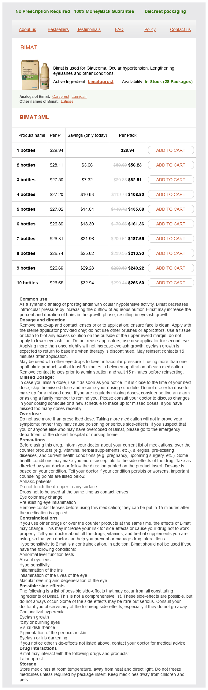
Bimatoprost dosages: 3 ml
Bimatoprost packs: 1 bottles, 2 bottles, 3 bottles, 4 bottles, 5 bottles, 6 bottles, 7 bottles, 8 bottles, 9 bottles, 10 bottles
Order bimatoprost 3 ml with amex
In this anatomical anomaly, the higher and decrease poles of the kidney-called moieties-are every drained by a separate ureter. Duplication is found in roughly 1 in 4000 pregnancies, is extra widespread in females, and is bilateral in 15 to 20 percent of circumstances (James, 1998; Vergani, 1998; Whitten, 2001). Development of hydronephrosis or ureteral dilatation may happen because of irregular implantation of 1 or each ureters inside the bladder-a relationship that reflects the anatomical Weigert-Meyer rule. The upper pole ureter could develop obstruction from a ureterocele within the bladder, whereas the lower pole ureter has a shortened intravesical segment that predisposes to vesicoureteral reflux. Thus, each moieties could become dilated from totally different etiologies, and both are at risk for lack of operate. Renal pelvis dilatation is visible in each the upper (U) and decrease (L) pole moieties, which are separated by an intervening band of renal tissue (arrowhead). The bladder, encircled by the highlighted umbilical arteries, contains a ureterocele (arrowhead). Renal Agenesis the prevalence of bilateral renal agenesis is roughly 1 in 8000 births, whereas that of unilateral renal agenesis is 1 in one thousand births (Cragan, 2009; Dolk, 2010; Sheih, 1989; Wiesel, 2005). When a kidney is absent, color Doppler imaging of the descending aorta demonstrates absence of the ipsilateral renal artery. In addition, the ipsilateral adrenal gland typically enlarges to fill the renal fossa, termed the lying down adrenal sign (Hoffman, 1992). As with other fetal anomalies, amniocentesis for chromosomal microarray analysis ought to be thought-about. In this coronal image of the fetal abdomen, colour Doppler reveals the course of the stomach aorta. The ultrasound beam is perpendicular to the aorta, demonstrating absence of the renal arteries bilaterally. This coronal image of a fetus with unilateral renal agenesis exhibits the adrenal gland (arrowheads) filling the renal fossa, termed the "lying-down" adrenal sign. If renal agenesis is bilateral, no urine is produced, and the resulting anhydramnios leads to pulmonary hypoplasia, limb contractures, and a distinctively compressed face. Multicystic Dysplastic Kidney this extreme type of renal dysplasia ends in a nonfunctioning kidney. Contralateral renal abnormalities are current in 30 to forty percent-most incessantly vesicoureteral reflux or ureteropelvic junction obstruction (Schreuder, 2009). Nonrenal anomalies have been reported in 25 % of cases, and cystic dysplasia may occur as a element of many genetic syndromes (Lazebnik, 1999; Schreuder, 2009). It is related to severely decreased amnionic fluid volume beginning early in gestation. The differential prognosis for these findings contains several genetic syndromes, aneuploidy, or normal variant. Bladder Outlet Obstruction Distal obstruction of the urinary tract is extra frequent in male fetuses, and the most typical etiology is posterior urethral valves. Characteristically, the bladder and proximal urethra are dilated, termed the "keyhole" signal, and the bladder wall is thick. Oligohydramnios, significantly earlier than midpregnancy, portends a poor prognosis because of pulmonary hypoplasia. Evaluation features a careful seek for related anomalies, which may occur in forty percent of instances, and for aneuploidy, which has been reported in 5 to 8 percent (Hayden, 1988; Hobbins, 1984; Mann, 2010). Evaluation and therapy of fetal bladder outlet obstruction is discussed in Chapter sixteen (p. In this 19-week fetus with extreme bladder outlet obstruction, the bladder is dilated and thick-walled, with dilatation of the proximal urethra that resembles a "keyhole. Skeletal Abnormalities the 2015 revision of the Nosology and Classification of Genetic Skeletal Disorders consists of a formidable 436 skeletal anomalies in forty two groups, characterized by genetic abnormalities, phenotypic features, or radiographic standards (Bonafe, 2015). The two forms of skeletal dysplasias are osteochondrodysplasias-the generalized irregular improvement of bone and/or cartilage, and dysostoses-which are abnormalities of particular person bones, for instance, polydactyly.

Cheap bimatoprost 3ml with amex
Recall that evolution of the renal system passes sequentially via the pronephric and mesonephric phases to attain the everlasting metanephric system. Between the 4th and fifth weeks, each mesonephric duct gives rise to a ureteric bud, which grows cephalad toward its respective mesonephros. As every bud lengthens, it induces differentiation of the metanephros, which can turn out to be the final kidney. Each mesonephros degenerates near the end of the primary trimester, and without testosterone, the mesonephric ducts regress as well. By the seventh week it turns into divided by the urorectal septum to create the rectum and the urogenital sinus. The urogenital sinus is taken into account in three components: (1) the cephalad or vesicle portion, which forms the urinary bladder; (2) the middle or pelvic portion, which creates the feminine urethra; and (3) the caudal or phallic part, which supplies rise to the distal vagina and to the larger vestibular (Bartholin) and paraurethral glands. Embryology of the Genital Tract the fallopian tubes, uterus, and upper vagina derive from the m�llerian ducts, additionally termed paramesonephric ducts, which kind adjoining to every mesonephros. These ducts lengthen downward after which turn medially to meet and fuse collectively within the midline. The uterus is fashioned by this union of the two m�llerian ducts at approximately the 10th week. Fusion to create the uterus begins in the center and then extends both caudally and cephalad. With mobile proliferation at the higher portion, a thick wedge of tissue creates the attribute piriform uterine form. At the same time, dissolution of cells at the decrease pole types the first uterine cavity. As the higher wedge-shaped septum is slowly reabsorbed, the ultimate uterine cavity is often formed by the twentieth week. In distinction, resorption failure of the frequent tissue between them ends in numerous levels of persistent uterine septum. As the distal end of the fused m�llerian ducts contacts the urogenital sinus, this induces endodermal outgrowths from the sinus termed the sinovaginal bulbs. These bulbs proliferate and fuse to type the vaginal plate, which later resorbs to form the vaginal lumen. However, the lumen stays separated from the urogenital sinus by the hymeneal membrane. The shut affiliation of the mesonephric (wolffian) and paramesonephric (m�llerian) ducts explains the simultaneous abnormalities in their finish organs. Kenney and colleagues (1984) confirmed that up to half of females with uterovaginal malformations have related urinary tract defects. Anomalies most frequently related to renal defects are unicornuate uterus, uterine didelphys, and agenesis syndromes, whereas arcuate and bicornuate are much less generally linked (Reichman, 2010). With m�llerian anomalies, ovaries are functionally normal but have a higher incidence of anatomical maldescent into the pelvis (Allen, 2012; Dabirashrafi, 1994). As discussed, the mesonephric ducts normally degenerate, nevertheless, persistent remnants may turn out to be clinically obvious. These are typically positioned within the proximal anterolateral vaginal wall however could also be found at different websites along the vaginal length. Intraabdominal wolffian remnants within the feminine embody a few blind tubules within the mesovarium-the epo�phoron-and related ones adjacent to the uterus- paro�phoron. The epo�phoron or paro�phoron could develop into clinically identifiable cysts in the grownup. Embryology of the Gonads At approximately four weeks, gonads derive from coelomic epithelium masking the medial and ventral surface of the nephrogenic wire at a website between the eighth thoracic and fourth lumbar segments. Because of this separate gonadal and m�llerian derivation, women with m�llerian defects usually have functionally normal ovaries and are phenotypic females. The coelomic epithelium thickens to form the genital ridge, also referred to as the gonadal ridge. Strands of these epithelial cells extend into the underlying mesenchyme as the first intercourse cords. By the sixth week, primordial germ cells have migrated from the yolk sac to enter the genital ridge mesenchyme.

Purchase bimatoprost 3ml line
Management Both medical and surgical remedy depend upon the precise anatomy and splenic status. Asplenic sufferers often have more severe coronary heart defects, usually precluding biventricular repair and necessitating staged single ventricle palliation. Biventricular circulation is extra frequent in polysplenia syndrome given much less severe lesions. The toddler with asplenia syndrome and ductaldependent pulmonary circulation will become progressively cyanotic with ductal constriction. Here, standard intensive care stabilization and quick surgical repair could additionally be needed. The infant with polysplenia syndrome hardly ever presents with cyanosis as pulmonary blood move obstruction is rare. Rather, they present with coronary heart failure and metabolic acidosis associated to left coronary heart obstruction with potential pulmonary overcirculation. Surgical treatment of patients with heterotaxy syndrome is complicated and first requires a decision whether biventricular repair is feasible. Most sufferers with asplenia syndrome initially require a secure supply of pulmonary blood flow. The staged palliation culminates in the Fontan circulation, though mortality and morbidity is higher with asplenia syndrome at this stage compared with different single ventricular lesions [14]. Improved outcomes in most up-to-date studies might mirror better surgical tehcniques in addition to better postoperative management [14]. Biventricular repair is more common in sufferers with polysplenia syndrome, though 50�70% require single ventricle palliation. Risk components for poor outcomes in sufferers with polysplenia syndrome embrace biliary atresia, low birth weight, complete heart block, coarctation, and single ventricle physiology [11]. Some patients will eventually need cardiac transplantation, occurring extra regularly after single ventricle palliation. Outcomes Patients with asplenia and polysplenia syndromes have variable prognosis relying on the severity of the precise lesion(s). The pure history may be dire, and asplenic sufferers normally have worse outcome than patients with polysplenia [15]. Spontaneous intrauterine demise happens 3�4 times more regularly in fetuses with polysplenia syndrome, probably due to rhythm disturbances and ventricular dysfunction [16]. Conclusions Heterotaxy syndrome represents a posh group of anomalies involving the thoracoabdominal viscera. Cardiac malformations are rarely singular and often occur as a constellation of abnormalities specifically associated with the disorders of laterality. However, sufferers with heterotaxy syndrome carry a worse prognosis than these with comparable isolated cardiac lesions. The name stems from the Greek word "ektopos" which suggests departure from the suitable hometown. The full spectrum of pentalogy of Cantrell consists of five anomalies: 1) 2) 3) 4) 5) Defect in the decrease sternum; Midline supraumbilical belly wall defect; Deficiency of the anterior diaphragm; Defect within the diaphragmatic pericardium; and Various congenital intracardiac abnormalities [2]. Note the abdominal wall defect presenting as a big omphalocele (green arrow) with the stomach contents outdoors the cavity and partially lined by a sac. There is protrusion of the apical portion of the heart (red arrow) exterior the chest cavity (blue arrow). The analysis, if not made by fetal ultrasound and fetal echocardiography, is clear on inspection of the infant at start. It is crucial to decide any intracardiac defects by sterile careful epicardial echocardiography. Surgical correction contains 4 necessary steps: coverage of the bare heart; palliation or complete restore of main intracardiac defects; placement of the heart into the thoracic cavity; and sternal or thoracic reconstruction. The heart protrudes through the sternal defect in real-time during respiratory movements. Intracardiac lesions embrace ventricular septal defect in twin A (not shown) and atrial septal defect in twin A and twin B (not shown). Complex cardiac fusion is essentially the most important determinant of surgical separation and survival [8, 9]. References 301 abnormalities are main determinants of feasibility of postnatal surgical separation of the twins and in counseling the mother and father regarding prognosis [10, 11]. McMahon and Spencer [12] have summarized the cardiac anomalies discovered in additional than 1200 pairs of conjoined twins reported within the literature.

Cheap bimatoprost online mastercard
Abnormalities of Chromosome Structure Structural chromosomal abnormalities embrace deletions, duplications, translocations, isochromosomes, inversions, ring chromosomes, and mosaicism (see Table 13-1). Identification of a structural chromosomal abnormality raises two major questions. First, what phenotypic abnormalities or later developmental abnormalities are related to this finding Second, is evaluation of parental karyotype indicated-specifically, are the mother and father at increased danger of carrying this abnormality Deletions and Duplications A chromosomal deletion signifies that a portion of a chromosome is missing, whereas a duplication means that a portion has been included twice. Most deletions and duplications happen during meiosis and result from malalignment or mismatching in the course of the pairing of homologous chromosomes. The misaligned segment might then be deleted, or if the mismatch remains when the two chromosomes recombine, it might end in a deletion in one chromosome and duplication within the other. When a deletion or duplication is recognized in a fetus or infant, parental karyotyping should be supplied, as a end result of if both mother or father carries a balanced translocation, the recurrence danger in subsequent pregnancies is significantly elevated. Common deletions could additionally be referred to by eponyms -for instance, del 5p known as cri du chat syndrome. These chromosomal deletions or duplications-smaller than three to 5 million base pairs-are too small to be detected with normal karyotyping. This syndrome is also referred to as DiGeorge syndrome, Shprintzen syndrome, and velocardiofacial syndrome. It is the most typical microdeletion, with a prevalence of 1 in 3000 to 6000 births. Although inherited in an autosomal dominant fashion, more than 90 % of cases come up from de novo mutations. The full deletion consists of three million base pairs, encompasses 40 genes, may embrace a hundred and eighty totally different features, and thus poses some counseling challenges (Shprintzen, 2008). In approximately seventy five percent of affected people, related abnormalities embrace conotruncal cardiac anomalies, corresponding to tetralogy of Fallot, truncus arteriosus, interrupted aortic arch, and ventricular septal defects (McDonald- McGinn, 2015). Immune deficiency, corresponding to T-cell lymphopenia, additionally develops in approximately seventy five %. Learning disabilities, autism spectrum disorder, and mental incapacity are additionally frequent. Other manifestations include hypocalcemia, renal anomalies, esophageal dysmotility, hearing loss, behavioral problems, and psychiatric illness-particularly schizophrenia. Short palpebral fissures, bulbous nasal tip, micrognathia, short philtrum, and small or posteriorly rotated ears are attribute facial features. A double-segment or reciprocal translocation begins when breaks occur in two different chromosomes. The broken fragments are then exchanged, so that every affected chromosome contains a fraction of the other. If no chromosomal material is gained or lost on this process, the translocation is taken into account balanced. The prevalence of reciprocal translocations approximates 1 in 600 births (Nussbaum, 2007). Although the balanced translocation provider is often normal phenotypically, repositioning of specific genes inside chromosomal segments can cause abnormalities. The danger of a significant structural or developmental abnormality in an obvious balanced translocation provider is approximately 6 %. Balanced translocation carriers are in danger to produce unbalanced gametes, resulting in abnormal offspring. The observed risk of a specific translocation can typically be estimated by a genetic counselor. In basic, translocation carriers identified after the start of an abnormal baby have a 5- to 30-percent risk of manufacturing liveborn offspring with an unbalanced translocation. Carriers recognized for other reasons, for instance, throughout an infertility analysis, have only a 5-percent risk. This is likely because the gametes are so irregular that conceptions are nonviable. These involve only acrocentric chromosomes, which are chromosomes 13, 14, 15, 21, and 22. In a robertsonian translocation, the q arms of two acrocentric chromosomes fuse at one centromere to type a derivative chromosome.
Discount bimatoprost line
Progressive aortic valve regurgitation is the most common late downside, highlighting the potential profit to patch closing each the aortic and ventricular orifices of the tunnel in an effort to provide added assist to the aortic valve leaflets. The morphology, origin, and exit factors are variable, permitting potential connections between any coronary artery department and any cardiac chamber, venous channel, or pulmonary artery [1]. The typical murmur is steady, peaking in diastole when the strain gradient is maximal. The medical features are the identical as another reason for left-to-right shunt inflicting coronary heart failure. This accounts for the extensive distribution of the anomalies throughout the coronary tree [3]. Approximately 60% arise from the right coronary artery; 20% from the left descending coronary artery; and 20% from the left major stem or circumflex artery. The key echocardiographic features embody a bigger than regular coronary artery caused by increased flow into the low strain exit chamber. This position was achieved by first forming a guide wire loop by way of the fistula from the native coronary artery. Occlusion here was achieved using coils which were deployed immediately from a catheter positioned via the length of the coronary artery. The course of the fistula can generally be traced from the origin to the exit by colour Doppler, allowing appreciation of the degree of tortuosity and relationship to normal coronary branches. Pulsed and steady wave Doppler interrogation ought to show a steady trace with more outstanding diastolic than systolic move. The diastolic predominance is because the aortic root (the origin of the coronary fistula flow) often has the best diastolic pressure within the circulation, leading to a stress gradient to the exit chamber. The actual nature of the flow pattern will rely upon the exit chamber involved and its strain profile. Following echocardiographic prognosis, a call must be made about additional diagnostic imaging. In some cases, it could be appropriate to proceed straight to cardiac catheterization with a view to occlusion of the defect on the similar procedure. Management Strategies the administration technique is decided by symptoms or evidence of deleterious effects on the guts and circulatory operate. Evidence of chamber dilation or an increase in pulmonary artery stress must be sought. In these cases, normal medical management to management heart failure should be instituted alongside contemplation of definitive remedy [10]. Interventional Catheterization Currently, interventional catheterization is the mainstay of definitive management. Such imaging may additionally be used to resolve whether an interventional approach is feasible or if a surgical strategy should be adopted. Radiation dosage, want for basic anesthesia, and the relative tachycardia of young sufferers have to be taken into consideration [8, 9]. The main objective in catheter therapy is full occlusion of the fistula at the most distal level, to restrict the danger of occlusion of an important coronary branch [13]. The preliminary method relies around angiographic delineation of the coronary circulation: the origin, course, and exit level of the fistula and the relationships of the fistula. Because of the individual variations corresponding to multiple feeding vessels, it is very important delineate the non-fistulous in addition to the fistulous coronary artery. Multiple angiographic injections and angulations may be required to achieve essentially the most useful views to plan an intervention. Three-dimensional rotational angiography in a contemporary catheterization laboratory is especially helpful in long tortuous vessels [14]. This is often achieved by passing a guidewire from the native coronary artery via the fistula and out of the exit point. Thus, an arteriovenous guidewire circuit is shaped, which allows a larger delivery catheter or sheath to be coaxially railroaded over the guidewire from the venous side into a position to allow supply of an acceptable system [6]. In either case, the objective is to occlude the vessel on the most distal web site possible, allowing upkeep of move in all the normal coronary side branches, which lie proximal to the fistulous connection.

Generic 3 ml bimatoprost free shipping
Pregnancy Outcomes Some outcomes extra widespread with hydramnios embody birthweight >4000 g, cesarean delivery, and importantly, perinatal mortality. Pregnancies with idiopathic hydramnios are associated with birthweights exceeding 4000 g in almost 25 % of instances, and the likelihood seems to be greater if the hydramnios is reasonable or severe (Luo, 2016; Odibo, 2016; Wiegand, 2016). A rationale for this association is that larger fetuses have higher urine output, by advantage of their increased quantity of distribution, and fetal urine is the biggest contributor to amnionic fluid quantity. Cesarean delivery rates are additionally greater in pregnancies with idiopathic hydramnios, with reported rates of 35 to fifty five percent (Dorleijn, 2009; Khan, 2017; Odibo, 2016). An unresolved question is whether hydramnios alone raises the chance for perinatal mortality. Some studies have discovered no enhance in stillbirth or neonatal dying charges with idiopathic hydramnios, whereas others show a higher threat (Khan, 2017; Pilliod, 2015; Wiegand, 2016). Using delivery certificates data from the state of California, Pilliod and coworkers (2015) identified hydramnios in zero. At 37 weeks, the stillbirth danger was sevenfold larger in pregnancies with hydramnios. By 40 weeks, this danger was greater than tenfold higher66 per 10,000 births in contrast with 6 per 10,000 without hydramnios. Risks appear to be compounded when a growth-restricted fetus is recognized with hydramnios (Erez, 2005). When an underlying cause is identified, degree of hydramnios has been associated with chance of preterm delivery, small-for-gestational age newborn, and perinatal mortality (Pri-Paz, 2012). However, idiopathic hydramnios is generally not related to preterm delivery (Magann, 2010; Many, 1995; Panting- Kemp, 1999). Occasionally, severe hydramnios may result in early preterm labor or the event of maternal respiratory compromise. The technique is just like that for genetic amniocentesis, described in Chapter 14 (p. Approximately a thousand to 2000 mL of fluid is slowly withdrawn over 20 to half-hour, relying on the severity of hydramnios and gestational age. Hydramnios severe sufficient to necessitate amnioreduction virtually invariably has an underlying trigger, and subsequent amnioreduction procedures could also be required as typically as weekly and even semiweekly. Complications inside forty eight hours of amnioreduction included delivery in 4 % and ruptured membranes in 1 p.c. There was no instance of chorioamnionitis, placental abruption, or bradycardia requiring supply (Dickinson, 2014). Oligohydramnios complicates approximately 1 to 2 p.c of pregnancies (Casey, 2000; Petrozella, 2011). When no measurable pocket of amnionic fluid is recognized, the term anhydramnios could additionally be used. Unlike hydramnios, which is often gentle and sometimes confers a benign prognosis within the absence of an underlying etiology, oligohydramnios is all the time a cause for concern, as mentioned on page 231. Etiology Pregnancies sophisticated by oligohydramnios embody those during which the amnionic fluid quantity has been severely diminished for the reason that early second trimester and people by which the fluid quantity was regular until near-term and even full-term. Whenever oligohydramnios is recognized, it turns into an essential consideration in clinical management. Early-Onset Oligohydramnios When amnionic fluid quantity is abnormally decreased from the early second trimester, it could replicate a fetal abnormality that precludes regular urination, or it may characterize a placental abnormality sufficiently severe to impair perfusion. Ruptured membranes must be excluded, and focused sonography is performed to assess for fetal and placental abnormalities. Oligohydramnios after Midpregnancy When amnionic fluid quantity turns into abnormally decreased in the late second or within the third trimester, it is very usually associated with fetal-growth restriction, with a placental abnormality, or with a maternal complication corresponding to preeclampsia or vascular illness (Table 11-4). The underlying trigger in such instances is frequently uteroplacental insufficiency, which may impair fetal development and scale back fetal urine output. Exposure to selected drugs has also been linked with oligohydramnios as mentioned subsequently.
Discount bimatoprost 3ml
Nasal packing causes vital discomfort to the patient with symptoms corresponding to headache, dryness of the mouth, watering of the eyes, and blood-stained discharge through the pack engorged with nasal secretions. In less critical cases, head finish elevation, nasal decongestant drops, antifibrinolytic agents corresponding to tranexamic acid, and medications to management elevated blood strain or medical situations if any, may be adopted. Embolization of the sphenopalatine artery on one or either side may be carried out for traumatic epistaxis without any important unwanted effects [10]. Embolization is carried out via the transfemoral route using the Seldinger technique. Although nasal packing might be done as an emergency procedure in circumstances selected for the above procedures, there exist different and fewer invasive ways to cut back the blood loss by way of the nose so as to facilitate the same. The different is by administering a higher palatine artery block via the larger palatine foramen situated over the palate utilizing an intraoral method. The presence of severe trismus as a end result of complicated facial fractures would preclude the use of these strategies. In cases the place nasal packing has already been done, additional nervousness awaits both doctor and patient at the time of removal of the pack. The pack could also be eliminated in 24�72 h relying on the case and the propensity to bleed once more. Pack elimination is as traumatic for the affected person as is its insertion and due care should be exercised throughout each the steps. A well-lubricated pack and proper method are important, and otolaryngologists are suitably educated in these principles. It is healthier to start instilling the liquid paraffin drops utilizing a dropper or syringe at least 2 h earlier than the elimination of the pack in ideal circumstances but additionally at the time of the particular procedure of removing utilizing more quantities of the solution. Bleeding throughout pack removing can be managed in the same means as has been described above within the methods to avoid nasal packing using easy measures. It is usually not essential to reinsert one other nasal pack, however somewhat endurance and perseverance are wanted. The completely different routes by which surgical entry can be gained are the Lynch-Howarth, subciliary, transconjunctival, gingivobuccal, lateral rhinotomy, and midfacial degloving approaches if open surgical procedure is being contemplated or the transnasal endoscopic approach. Fixation could also be done by wiring and plating, utilizing a titanium mesh, implant or prosthesis, or by a regional or free tissue switch. Cranialization, on the other hand, is undertaken for disruption of the posterior table of the frontal sinus bone and herniation of mind or meninges into the sinus. In this procedure, the posterior table and sinus mucosa are eliminated, the frontal outflow tract obliterated, and the inner lining of the frontal sinus changed with periosteum and allowed to be in contiguity with the intracranial cavity. Frontal sinus harm might manifest in long-term signs, and late problems within the type of cellulitis, meningitis, and osteomyelitis have been known to seem even up to 25 years. Frontal sinus fractures were traditionally repaired using open surgical approaches. Fractures of the posterior plate of the frontal bone and sinus had been mounted with the cavity of the sinus made continuous with the intracranial cavity after complete removal of any remnants of frontal sinus mucosa. Alternatively, fractures of the anterior table have been handled with complete exenteration of the frontal sinus contents and repair utilizing soft tissue and bone graft so as to restore cosmetic look of the face. In common, traumatic leaks if small tend to close spontaneously with minimal intervention and long-term sequelae. Fractures of the anterior cranial base on the region of the cribriform plate not often endure spontaneous closure and require surgical administration. The good news is that the majority of those may be approached nasally with an endoscope and thus keep away from the many sequelae seen within the case of open procedures similar to a frontal craniotomy or bicoronal approach/bifrontal craniofacial resection. Only conservative administration is adequate for many of the leaks attributable to trauma, and nearly 70�85% close this way within a few weeks. Controlled optimistic stress with using straws, incentive spirometers, and Valsalva maneuver can be beneficial within the closure of small leaks. This may be undertaken when a history of meningitis is current however no fluid is discovered on straining. Repair of such leaks must be carried out as they may have turn out to be true epithelialized fistulous tracts, and the repair should be accomplished in the same fashion as major repair.
Real Experiences: Customer Reviews on Bimatoprost
Bram, 42 years: These hyperechoic deposits can simply be seen sonographically, and a grading scale from 0 to three displays rising calcification with increasing numerical grade (Grannum, 1979).
Nafalem, 40 years: Small general measurement, indicating poor progress in utero, is a crucial clue to a genetic prognosis.
Koraz, 54 years: Activation of proteases probably performs a pivotal function in weakening the follicular basement membrane and ovulation (Curry, 2006; Ny, 2002).
Stejnar, 51 years: Though individual preferences vary, surgical correction by either the open or closed method could additionally be done, and both give related results.
Aila, 26 years: Live births, stillbirths, and terminated pregnancies have been included and totaled approximately 32,000 cases and nearly 12,000 controls.
Nemrok, 53 years: Close follow-up by a pediatric heart specialist is required through the childhood years, with compulsive transition to adult care by a specialist in adult congenital coronary heart disease.
Larson, 52 years: Large cystic hygromas are usually associated with hydrops fetalis, hardly ever resolve, and carry a poor prognosis.
Jesper, 33 years: In the present period the need for cardiac catheterization with angiography in cor triatriatum is uncommon, particularly for sufferers recognized in the neonatal interval.
10 of 10 - Review by Z. Deckard
Votes: 94 votes
Total customer reviews: 94
References
- National Collaborating Centre for Chronic Conditions. Hypertension: management of hypertension in adults in primary care: partial update. London: Royal College of Physicians, 2006.
- Murray MD, Deer MM, Ferguson JA, et al. Open-label randomized trial of torsemide compared with furosemide therapy for patients with heart failure. Am J Med 2001;111:513-520.
- Muysoms EE, Hauters PJ, Van Nieuwenhove Y, et al. Laparoscopic repair of parastomal hernias: a multi-centre retrospective review and shift in technique. Acta Chir Belg 2008;108(4):400-404.
- Berman B, Harrison-Balestra C, Perez OA, et al. Treatment of keloid scars post-shave excision with imiquimod 5% cream: A prospective, double-blind, placebo-controlled pilot study. J Drugs Dermatol 2009;8(5):455-8.
- Tang NL, Fan HP, Chang KC, et al. Genetic association between a chemokine gene CXCL-10 (IP-10, interferon gamma inducible protein 10) and susceptibility to tuberculosis. Clin Chim Acta 2009; 406: 98-102.
- Bush WH, Swanson DP: Acute reactions to intravascular contrast media: types, risk factors, recognition, and specific treatment, AJR Am J Roentgenol 157:1153-1161, 1991.