
Grisactin dosages: 250 mg
Grisactin packs: 30 pills, 60 pills, 90 pills, 120 pills, 180 pills, 360 pills

Cheapest generic grisactin uk
Multiple erythematous papules in a generalized distribution seem a month or extra after the illness. These lesions evolve to small papules and plaques, clinically and histologically resembling lupus vulgaris. Uniform hyperplasia of the ear pinna and lobe could closely mimic "turkey ear," as described in sarcoidosis. When the mucous membranes are concerned, the lesions turn into papillomatous or ulcerative. On the tongue, irregular, deep, painful fissures happen, typically related to microglossia to the degree that vitamin is compromised. The fee of development of lupus vulgaris is gradual, and a lesion might stay limited to a small space for a number of decades. In some sufferers, the lesions turn into papillomatous, vegetative, or thickly crusted, with a rupioid appearance. Colloid milia, pimples vulgaris, sarcoidosis, and rosacea might simulate lupus vulgaris. Differentiation from tertiary syphilis, continual discoid lupus erythematosus, Hansen illness, systemic mycoses, and leishmaniasis could also be tougher, and biopsy and tissue cultures could also be required. The disease is harmful, incessantly causes ulceration, and on involution leaves deforming scars because it slowly spreads peripherally over the years. Lupus vulgaris lesions of the head and neck can be associated with lymphangitis or lymphadenitis in some instances. If lesions contain the nostril or the earlobes, these buildings are shrunken and scarred, as if nibbled away. The tip of the nose could also be sharply pointed and beaklike, or the whole nostril could also be destroyed, with solely the orifices and the posterior parts of the septum and turbinates seen. The higher lip, a website of predilection, could turn into diffusely swollen and thickened, with fissures, adherent skinny crusts, and ulcers. On the trunk and extremities, lesions could also be annular or serpiginous or might kind gyrate patterns. On the palms and feet and across the genitals or buttocks, lesions might trigger mutilation by destruction, scar formation, warty thickenings, and elephantiasic enlargement. Scrofuloderma is tuberculous involvement of the pores and skin by direct extension from an underlying focus of infection. It occurs most incessantly over the cervical lymph nodes but in addition may happen over bone or around joints if these are involved. Clinically, the lesions start as subcutaneous masses, which enlarge to type nodules. Surgical procedures might incite lesions of scrofuloderma over joints or the belly cavity, apparently by releasing the loculated focus and contaminating the track along which instruments are inserted. Scrofuloderma heals with characteristic cordlike scars, frequently permitting the diagnosis to be made a few years later. Histologically, in scrofuloderma, the tuberculous course of begins in the underlying lymph node or bone and extends through the deep dermis. At the periphery, more typical okay sf re Cutaneous Tuberculosis From Endogenous Source by Direct Extension (Scrofuloderma and Periorificial Tuberculosis). Scrofuloderma ought to be differentiated from atypical mycobacterial infection, sporotrichosis, actinomycosis, coccidioidomycosis, and hidradenitis suppurativa. Lesions ulcerate from the start and prolong quickly, with no tendency to spontaneous healing. Classically, this begins with a distal lesion, and new lesions showing extra proximally. Less usually, a proximal lesion is present initially, and new lesions appear distally (retrograde lymphatic spread). Lesions are symmetrically distributed on the extensor extremities, especially on the ideas of the elbows and on the knees; dorsal surfaces of the hands and ft; buttocks; face and ears; and glans penis.
Purchase grisactin now
Tetanus toxoid ought to be given if the patient has not acquired the immunization inside 10 years Some data suggest a development towards higher outcomes with injection of intralesional triamcinolone, with anecdotal reviews of the injection site being spared necrosis however the areas above and under the positioning exhibiting necrosis. Severe ache normally develops inside a couple of minutes and spreads throughout the extremities and trunk Within a few hours, the victim might have chills, vomiting, violent cramps, delirium or partial paralysis, spasms, and abdominal rigidity. The abdominal pains are incessantly most severe and may be mistaken for appendicitis, colic, or meals poisoning. Colchicine has additionally been disappointing in animal models, however tetracyclines show some promise and deserve further study. Funnel Web Spiders Funnel internet spiders include Tegenaria agrestis (hobo spider or aggressive home spider of Pacific Northwest) and Atrax robustus (Sydney funnel web spider of Australia). In the United States the rattlesnake, water (cottonmouth) moccasin, copperhead, and coral snake are the venomous snakes most regularly encountered. Patients are normally young males, with 98% of bites on the extremities, most frequently the hands or arms. In Europe, 39% of envenomations from exotic pets are snakebites from rattlesnakes, cobras, mambas, or different venomous snakes. Snake venom has effects on the cardiovascular, hematologic, respiratory, and nervous methods. Local results on the chunk web site embrace the speedy onset of swelling, erythema, and ecchymosis. Fang marks are often seen and ache is frequent, except with Mojave rattlesnake bites. In the japanese United States, copperheads inflict most snakebites, adopted by rattlesnakes and cottonmouths. Successful remedy is usually attained by immersing the injured part in sizzling water for 30�60 minutes. The water should be as sizzling as could be tolerated, as a result of the venom is detoxified by warmth. Meperidine hydrochloride administered intravenously or intramuscularly may be necessary. Systemic toxicity normally resolves spontaneously with supportive care within 1 or 2 days. The desmoglein compensation hypothesis, based mostly on the differential expression of desmoglein 1 and 3 at totally different amounts at totally different ranges of the epidermis and mucosa, for example, perfectly explains the observed variations within the phenotypes of pemphigus foliaceus and vulgaris. High-frequency ultrasound has been anecdotally reported as a possible tool to decide blister location. Usually, antibodies are circulating and could be discovered sure in perilesional and nonbullous lesional skin, whereas blistered skin often fails to reveal deposits. Biopsies of decrease extremity skin should be averted if possible, because it might be vulnerable to false-negative results. Salt-split-skin preparations are useful in determining the site of deposition of the autoantibodies. A 1-M solution of sodium chloride (NaCl) predictably splits pores and skin at the degree of the lamina lucida. Localization of immune deposits to the roof or ground of this split is diagnostically useful. An n-serrated sample corresponds to a break up above the basal lamina, whereas a u-serrated sample corresponds to a sub�lamina densa cut up (see photographs on ExpertConsult). Newer immunohistochemical stains corresponding to C3d stain can also aid in the prognosis in some cases. Data range in regards to the sensitivity and specificity of these tests, and not every test is universally available. Specific dermatoses of pregnancy are mentioned beneath the differential diagnosis of herpes gestationis. The outlook for immunobullous illnesses has improved because the introduction of rituximab, intravenous immunoglobulins, and extra targeted, autoantibody-directed therapies serving to shift away from broadly poisonous immunosuppressive regimens.
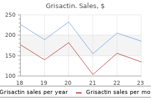
Order grisactin 250 mg free shipping
Cocks N, et al: Community seroprevalence survey for yaws and trachoma in the western division of Fiji. N Engl J Med 2016; 375: 1093 Engelman D, et al: Opportunities for built-in control of uncared for tropical ailments that have an result on the pores and skin. Kwakye-Maclean C, et al: A single dose oral azithromycin versus intramuscular benzathine penicillin for the remedy of yaws. Marks M, et al: Metaanalysis of the performance of a combined treponemal and nontreponemal speedy diagnostic test for syphilis and yaws. Clin Infect Dis 2016; sixty three: 627 Marks M, et al: Prevalence of active and latent yaws in the Solomon Islands 18 months after azithromycin mass drug administration for trachoma. They are characterised by the power to produce latent however lifelong infection by infecting immunologically protected cells (immune cells and nerves). The structural parts of a viral particle (virion) consist of a central core of nucleic acid a protective protein coat (capsid), and (in certain teams of viruses only) an outermost membrane or envelope. The capsid of the only viruses consists of many identical polypeptides (struc ural units) that fold and interact with each other to form morphologic models (capsomeres). The number of capsomeres is believed to be fixed for every virus with cubic symmetry, and it is a crucial criterion in the classification of viruses. The protein coat determines serologic specificity, protects the nucleic acid from enzymatic degradation in biologic environments, controls host specificity, and increases the effectivity of an infection. The outermost membrane of the enveloped viruses is essential for the attachment to , and penetration of, host cells. Some viruses are distinguished by their mode of transmission: arthropod-borne viruses, respiratory viruses, fecal-oral or intestinal viruses, venereal viruses, and penetrating-wound viruses. Serologic data present that many extra people are infected than give a history of medical disease. Instead, the ini ial clinical presentation known as a primary episode and may characterize a real main an infection or a recurrence. Persons with continual or acute immunosuppression might have extended and atypical medical courses. Although the technique is fast, its success depends closely on the skill of the interpreter. A positive serologic check indicates solely that the individual is infected with that virus, not that the viral infection is the reason for the current lesion. In addition to determining the infection price in various populations, serologic checks are most useful in evaluating couples in which only one partner gives a historical past of genital herpes (discordant couples), in t. The onset is commonly accompanied by high fever, regional lymphadenopathy, and malaise. The herpetic lesions in the mouth are normally damaged vesicles that appear as erosions or ulcers coated with a white membrane. The erosions might turn into widespread on the oral mucosa, tongue, and tonsils, and the gingival margin is usually eroded. It might trigger pharyngitis, with ulcerative or exudative lesions of the posterior pharynx. Oral therapeutic options embrace acyclovir suspension, 15 mg/kg five times day by day for 7 days; valacyclovir, 1 g twice daily for 7 days; or famciclovir, 500 mg twice daily for 7 days. The most frequent clinical manifestation of orolabial herpes is the "chilly sore" or "fever blister. Outbreaks are variable in severity, partly associated to the trigger of the outbreak Some outbreaks are small and resolve quickly, whereas others could additionally be severe, involving each the upper and the lower lip. A prodrome of up to 24 hours of tingling, itching, or burning may precede the outbreak. Local discomfort, in addition to headache, nasal congestion, or mild flulike symptoms, could occur. All topical therapies for the acute therapy of recurrent orolabial herpes have restricted efficacy, reducing disease length and ache by 1 day or less. Tetracaine cream, penciclovir cream, and acyclovir cream (not ointment) have some limited efficacy. Topical acyclovir ointment and docosanol cream provide minimal to no reduction in therapeutic time or discomfort. Intermittent therapy with valacyclovir, 2 g twice day by day for 1 day, or famciclovir, 1.
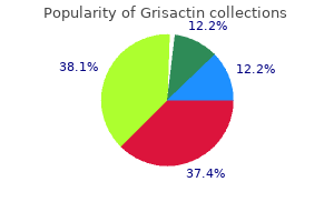
Discount 250mg grisactin with mastercard
The fungus is probably acquired from water, soil, or vegetation in forested areas the place the illness is prevalent. Agricultural laborers have been most frequently affected, with 90% of instances occurring in males. The organisms are usually quite a few and seem in chains of spheres related by short, narrow tubes. Surgical excision of the affected areas may be healing when the lesions are small, but recurrence is frequent. Complete decision of keloidal blastomycosis has been reported in a patient treated for 1 year with a mix of itraconazole, a hundred mg/day, and clofazimine, a hundred mg/day. Mestre T, et al: Mycetoma of the foo -diagnosis of the etiologic agent and surgical treatment. Nenoff P, et al: Eumycetoma and actinomycetoma-an replace on causative agents, epidemiology, pathogenesis, diagnostics and remedy. Conjunctival, lacrimal, oral, and urethral tissues may be involved, and genital lesions could resemble condylomata. The lesions begin as small papillomas and become pedunculated tumors with fissured and warty surfaces. Grayish white flecks could also be famous on the tissue, corresponding to transepithelial elimination of huge sporangia. Conjunctival lesions begin as small, pinkish papillary nodules, which later become bigger, dark, and lobulated. The illness is endemic in Sri Lanka and India but also occurs in parts of East Asia, Latin America, the southern United States, the United Kingdom, and Italy. Rhinosporidium seeberi, a decrease aquatic fungus present in stagnant water is the causative organism. The organisms appear as spherules 7�10 �m in diameter, that are contained within giant, cystic sporangia that might be as massive as 300 �m in diameter. Culture on Sabouraud dextrose agar is made of nasal discharge, abscess fluid, or biopsy specimens. Biopsy specimens will present fibroblastic proliferation and an inflammatory reaction with lymphocytes, plasma cells, histiocytes, eosinophils, and large cells. The organisms appear as broad hyphae which might be usually aseptate and may be branched at right angles. The SplendoreHoeppli phenomenon is common and seems as eosinophilic sleeves across the hyphae. Pythiosis, caused by Pythium insidiosum, a primitive aquatic hyphal organism that acts as a zoonotic pathogen, might have an effect on people and has an identical look. Infection happens often in healthy people, and in contrast to mucormycosis, usually runs an indolent course. The infections may be categorised as cutaneous, subcutaneous, visceral, or disseminated. Subcutaneous lesions happen in two fundamental sorts, each involving different anatomic websites, both as well-circumscribed subcutaneous plenty involving the nostril, paranasal tissue, and upper lip, or as nodular, subcutaneous lesions located on the extremities, buttocks, and trunk. The pathogenic genera include Rhizopus, Absidia, Mucor, Cunninghamella, Apophysomyces, Rhizomucor, Saksenaea, Mortierella, and Cokeromyces. Histologically, the organism generally seems as eosinophilic, thick-walled hyphae that look hole in cross part. The organisms are extremely vasculotropic and dissect alongside the media of muscular vessels, leading to infarction of tissue. The two orders inside this class that cause cutaneous an infection most often are the Entomophthorales and Mucorales. A granulomatous response is seen in about 50% of patients, and gigantic overseas body big cells can rarely be noted filled with organisms. Destruction of the concerned space by excision or electrosurgery is the most typical methodology of treatment. Mucormycosis Mucormycosis refers to infections brought on by the order Mucorales of the class Zygomycetes. When invasive, infections charac eristi cally are acute, rapidly developing, and sometimes deadly. Infection has additionally been related to methotrexate, prednisone, and infliximab remedy.
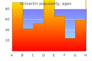
Buy generic grisactin canada
Half the mothers are asymptomatic at delivery, though many will subsequently develop arthralgia, Sj�gren syndrome, or different delicate systemic findings. Although the pores and skin lesions are transient, half the sufferers have an associated isolated congenital heart block, usually third diploma, which is permanent. In youngsters with cutaneous involvement, thrombocytopenia and hepatic illness might happen as incessantly as cardiac disease. Malar rash Discoid rash Photosensitivity Oral ulcers (21%) Arthritis Proteinuria >0. Skin involvement occurs in 80% of circumstances and is commonly useful in arriving at a prognosis. Heterozygous deficiency of both complement part C4A or C4B has a frequency of approximately 20% in white populations. Chemotherapeutic agents implicated embrace fluorouracil, capecitabine, cemcitabine, docetaxel, paclitaxel, and doxorubicin, with emerging stories of nivolumab, other checkpoint inhibitors, and small molecule kinase inhibitors corresponding to masitinib. Biopsies at all websites show interface dermatitis and a scant perivascular lymphoid infiltrate. Neutrophils are present in or beneath the lamina densa on immunofluorescent electron microscopy. The recognition of this subset as distinct is made clear by its often dramatic therapeutic response to dapsone. Capillary loops within the Osler-Weber-Rendu syndrome reveal ectasia of half the capillary loop. The palms, soles, elbows, knees, or buttocks may turn out to be persistently erythematous or purplish, typically with overlying scale. Leg ulcers, usually deeply punched out and with very little inflammation, could additionally be seen on the pretibial or malleolar areas. Many of those patients current with a livedoid pattern, and heaps of have an antiphospholipid antibody. Sneddon syndrome is composed of livedo reticularis and strokes associated to a hyalinizing vasculopathy. These skin-colored to erythematous lesions with a smooth, ulcerated or umbilicated surface could show vasculitis or, in older lesions, a palisaded granulomato s inflammation. The earliest modifications famous could additionally be transitory or migratory arthralgia, usually with periarticular inflammation. Arthralgia is usually the earlies abnormality and will remain the only real symptom for a while. It could also be of either nephritic or nephrotic type, main in both case to chronic renal insufficiency with proteinuria and azotemia. Pericarditis, the most frequent cardiac manifestation, and endocarditis also happen. Coombs-positive hemolytic anemia, neutropenia, and lymphopenia are different hematologic findings. The severity of thrombocytopenia may be a prognostic marker, however may additionally be followed as a biomarker for response to remedy. Overlap with any of the connective tissue illnesses could additionally be seen, occurring in approximately 25% of sufferers. Myopathy of the vacuolar sort could produce muscular weak point, myocardial disease, dysphagia, and achalasia of the esophagus. A historical past of exposure to excessive daylight earlier than the onset of the disease or earlier than an exacerbation is usually obtained. Some patients may have solely mild constitutional symptoms for weeks or months, but instantly after exposure to strong sunlight, they could develop the facial eruption and extreme illness complications. Risk of fetal death is increased in ladies with a previous history of fetal loss and anticardiolipin or anti-Ro antibodies. For the patient with these antibodies but without a history of previous fetal loss, the danger of fetal loss or neonatal lupus is low. One femaleto-male transgender affected person skilled vital enchancment after testosterone therapy. IgG levels could additionally be excessive, the albumin/globulin ratio is reversed, and serum globulin is increased, especially the -globulin or 2 fraction.
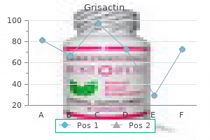
Buy 250 mg grisactin overnight delivery
I n contrast, endometrial cells are more probably to have rounded nuclei on the periphery of cellular clusters. Endometrial clusters are sometimes closely associated with neutrophils (including intracytoplasmic neutrophils) and may comprise stromal parts within the heart of the cluster ("double-contour" appearance). Other related modifications may be seen, such as elevated inflammation, histiocytes, parakeratosis, degenerated cells, and granular particles. I n atrophy, the basal cells may have an elevated N /C ratio secondary to focal nuclear enlargement. I n addition, features such as hyperchromasia and nuclear membrane irregularities can be seen in benign atrophic samples, which may trigger concern for squamous dysplasia. Atrophic squamous cells have dense cytoplasm F and enlarged nuclei (2-3 times the size of an intermediate cell nucleus) with high N /C ratios. The round uniform nuclei, retained polarity, and association with background single parabasal cells are a clue to the benign nature of a cluster of basal cells. This fragment has a syncytial look because the borders between cells are tough to determine. The nuclei are small and uniform in dimension but are darkish and have gentle nuclear border irregularities. However, the nuclei on this fragment are well organized, and a few cells have a reassuring quantity of cytoplasm (Pap stain). The numerous squamous cells in this large fragment have vague mobile borders, making assessment of the nuclear to cytoplasmic (N/C) ratio troublesome. The nuclei are somewhat dark but are uniform in measurement and have only delicate nuclear contour irregularities; the nuclear to cytoplasmic (N/C) ratios are low. A n extra difficult problem in atrophic smears is the occasional presence of granular particles, acute irritation, and apoptotic bodies. This cell and its pink cytoplasm, along with the presence of degenerated cells ("blue blobs") and granular material within the background, might cause initial concern for a keratinizing squamous cell carcinoma (Pap stain). Note the necrotic particles connected to viable tumor cells ("clinging diathesis") as nicely as current in the background. A single keratinizing cell with pink cytoplasm and an irregularly shaped, dark nucleus may be seen within the heart of the sector (Pap stain). A nswer: D istinguishing dysplasia within atrophic smears may be one of the challenging diagnostic problems on the Pap take a look at. The cells have high nuclear to cytoplasmic ratios, which is regarding regardless of the small nuclear sizes. Tubal Metaplasia O ccasionally, endocervical cells bear tubal metaplasia and undertake the traits of the glandular cells lining the fallopian tubes. Most notably, these cells are columnar and characteristically comprise a terminal bar with cilia at the luminal finish. This fragment accommodates crowded, dark cells with enlarged nuclei and high nuclear to cytoplasmic (N/C) ratios. However, at the fragment edges, the columnar nature of the cells may be seen together with terminal bars and cilia. Once tubal metaplasia is identified in a specimen, one must consider whether or not other hyperchromatic crowded groups in a the specimen simply represent tubal metaplasia lacking identifiable cilia versus a second cell sort. A strip of endocervical cells with basally oriented nuclei and ciliated terminal bars. Finding a subject such as this might be reassuring if different fields contain metaplastic cells that appear extra hyperchromatic and lack definitive cilia or terminal bars (Pap stain). I nconspicuous nucleoli could additionally be seen on some preparations, though sometimes the chromatin has a powdery or coarse look. The chromatin is even but coarsely granular, giving a hyperchromatic look (Pap stain). The neoplastic cells on this small cluster have high nuclear to cytoplasmic ratios and overlapping nuclei. Note the marked variation in measurement and nuclear overlap seen in these cells (Pap stain). While the cells seem properly organized within the tissue fragment, the projection of elongated nuclei past the fragment edge (creating a ragged look i.
Diseases
- Microcephaly brain defect spasticity hypernatremia
- Chromosome 4 ring
- Nakamura Osame syndrome
- Posterior tibial tendon rupture
- Cephalopolysyndactyly
- Stein Leventhal syndrome
- Right ventricle hypoplasia
- Hypogonadotropic hypogonadism without anosmia, X linked
- Trimethylaminuria
- Genuphobia
250mg grisactin
Leishmania is demonstrated in the cutaneous and mucous membrane lesions by direct smears or cultures. In Wright-stained biopsy materials, intracellular and extracellular organisms are seen with typical morphology of two chromatic structures: nucleus and parabasal body. In later mucosal lesions, the shortage of parasites makes identification tough. In ulcerous leishmaniasis, marked irregular acanthosis and sometimes pseudoepitheliomatous hyperplasia may be discovered. The dermis shows a dense infiltration of histiocytes, lymphocytes, and plasma cells. Numerous organisms are current (mostly in histiocytes), that are nonencapsulated and comprise a nucleus and a paranucleus. Wright, Giemsa, and monoclonal antibody staining may be helpful in figuring out the organisms. In patients with granuloma ous infiltrates containing intracellular parasites within histiocy es, leishmaniasis is considered one of several ailments to be thought of, together with rhinoscleroma, histoplasmosis, granuloma inguinale, Chagas illness, Penicillium marneffei infection, and toxoplasmosis. Touch smears stained with Giemsa are useful in many circumstances of cutaneous and mucocutaneous leishmaniasis. Two perpendicular grooves on the union of the osseous palate and gentle tissues, within the midst of the vegetative infiltration of the whole pharynx, are referred to as the palate cross of espundia. Only in exceptional instances does American leishmaniasis invade the genital or ocular mucous membranes. Mucocutaneous leishmaniasis is mainly attributable to Leishmania (Viannia) braziliensis braziliensis and L. The typical morphology of leishmania, as present in vertebrates, is round or oval, usually with one extremity extra rounded than the other, measuring 2�4 �m � 1. The earliest lesion is the cutaneous nodule or leishmanioma, which happens on the website of the preliminary sandfly inoculation. Kala-azar, which means "black fever," acquired its name due to the patchy macular darkening of the pores and skin brought on by deposits of melanin that develop in the later course of the illness. These patches are most marked over the forehead and temples, periorally, and on the midabdomen. The main goal for the parasites is the reticuloendothelial system; the spleen, liver, bone marrow, and lymph nodes are attacked. An intermittent fever, with temperatures ranging from 39�C�40�C (102�F�104�F), ushers within the disease Hepatosplenomegaly, agranulocytosis, anemia, and thrombocytopenia occur. Chills, fever, emaciation, weight reduction, weak point, epistaxis, and purpura develop because the illness progresses. They are nonflagellate oval organisms about three mm in diameter, generally identified as Leishman-Donovan our bodies. Leishmania donovani donovani causes visceral leishmaniasis in India, with the major reservoir being people and the vector being Phlebotomus argentipes. American visceral leishmaniasis principally impacts domestic dogs, though explosive outbreaks of the human infection occur sporadically, when the number of Lutzomyia longipalpis builds as much as a excessive level in the presence of contaminated canines. Canine visceral infections with Leishmania infantum have been reported in foxhounds in numerous elements of the United States and Canada. Miltefosine, an oral alkyl-phosphocholine analog, has proved as efficient as amphotericin B in some trials. Mixed infections involving each Leishmania and Trypanosoma cruzi are becoming increasingly frequent in Central and South America due to overlapping endemic areas. Amiodarone has been used as an unconventional antiparasitic drug on this setting along with commonplace therapy. During and after recovery from the disease, a particular type of dermal illness often known as post�kala-azar dermal leishmaniasis seems. This situation appears throughout or shortly after treatment in the African type, but its appearance may be delayed as a lot as 10 years after therapy within the Indian type. It follows the treatment of visceral leishmaniasis in 50% of Sudanese sufferers and 5%�10% of those seen in India. There are two constituents of the eruption: a macular, depigmented eruption discovered mainly on the face, arms, and upper a part of the trunk and a warty, papular eruption by which amastigotes could be discovered. Because it may persist for as much as 20 years, these sufferers could act as a persistent reservoir of infection. Leishman-Donovan our bodies may be present in the blood in people with kala-azar of India.

Best buy for grisactin
Determining whether or not p53 is diffuse in a population of singly dispersed carcinoma cells is difficult, as unfavorable staining cells may be missed on the counterstain (p53 immunostain). Several overtly malignant cells may be seen in this cell block preparation, some with a "signet ring" morphology. I f the cells are overtly malignant and morphologically consistent with a historical past of a given malignancy, the specimen can be thought of diagnostic. Cells of mesothelial origin are doubtless reactive mesothelial cells once mesothelioma is excluded (see "Mesothelioma" under the "Tissue Fragments Pa ern"). Cells of epithelial origin might represent benign and subclinical proliferations (such as endosalpingiosis), low-grade neoplasms implanted on a cavity floor (such as a borderline serous tumor), or a metastatic carcinoma (see "Metastatic A denocarcinoma" under the "Tissue Fragments Pa ern"). However, in most situations patients could have a identified history of illness, which can help help the workup. Even at excessive magnification, the cells could additionally be troublesome to determine as a "fourth cell" inhabitants. The malignancies that fall into this class often exist as monotonous dispersed cells with spherical, regular nuclei, only mild pleomorphism, and ample cytoplasm. Common examples include melanoma, lobular breast carcinoma, and adenocarcinomas with signet ring morphology. Upon immunostaining with particular markers, the "fourth inhabitants" turns into obvious. For this cause, it could be very important create cell block material and carry out immunostains in patients with a history of any of those malignancies. However, the cell has an irregular nuclear membrane and was found in a patient with a historical past of an invasive gastric signet ring carcinoma (Pap stain). In this separate specimen of metastatic lobular breast carcinoma, nearly all of cells present are malignant. Some have irregular nuclear borders and high nuclear to cytoplasmic (N/C) ratios (Pap stain). Cursory examination at low magnification may not be enough in such a specimen (Pap stain). Some of the options present (multinucleation, prominent nucleoli, and mitotic activity) may also be seen in a reactive mesothelial cell population, which is why the creation of cell block materials for ancillary studies is important in serous fluid specimens (Pap stain). The specimen seems to include two populations of cells-lymphocytes and bigger cells with plentiful cytoplasm. However, the cells are very massive and have nuclear membrane irregularities and coarse chromatin, findings which collectively are convincing for malignancy (Pap stain). These implants may be invasive or noninvasive and arise from a borderline or malignant neoplasm (discussed further within the "Tissue Fragments Pattern"). Benign processes in addition to processes of uncertain malignant potential also can current as a "fourth" population of single cells in a fluid specimen. In this case, the affected person had noninvasive implants from a borderline serous tumor. The tumor cells have vacuolated cytoplasm and small nucleoli, but their nuclear contours are regular. To exclude the potential of a reactive mesothelial cell proliferation, it is recommended to observe the workup for mesothelioma (discussed in the "Tissue Fragments Pattern"). This affected person had an increased number of giant mesothelial cells in a pleural fluid specimen, raising suspicion for a mesothelioma, which was later confirmed. A separate field with three atypical mesothelial cells with nucleolar prominence (Pap stain). It is troublesome to exclude a reactive mesothelial process using cytomorphology alone (Pap stain). A cell block preparation of the identical case shows a mixture of mesothelioma and background cells. A calretinin stain highlights the mesothelial cells within the specimen, that are increased in number which suggests a minimal of a reactive population of mesothelial cells and possibly a mesothelioma (calretinin immunostain). While its expression is lost in a few of the atypical cells, expression in a few of these cells precludes a definitive prognosis of mesothelioma. The specimen incorporates a massive number of mesothelioma cells with elevated cell measurement and outstanding nucleoli. Despite being markedly atypical, the cells preserve mesothelial features: abundant cytoplasm, two-tone cytoplasm, and "lacy skirt" morphology (Pap stain).

Buy 250 mg grisactin with visa
The uppermost portion of the follicle, which extends from its surface opening to the entrance of the sebaceous duct, known as the infundibular section. It resembles the floor dermis and its keratinocytes could additionally be of epidermal origin. The portion of the follicle between the sebaceous duct and the insertion of the arrector pili muscle is the isthmus. The inferior portion includes the lowermost part of the follicle and the hair bulb. Throughout life, the inferior portion undergoes cycles of involution and regeneration. Primary follicles are surrounded by the looks of two secondary follicles; different secondary follicles subsequently develop around the principal Apocrine Units Apocrine items develop as outgrowths not of the floor epidermis, however of the infundibular or higher portion of the hair follicle. Although immature apocrine models are found masking the complete pores and skin floor of the human fetus, these regress and are absent by the point the fetus reaches time period. The straight excretory portion of the duct, which opens into the infundibular portion of the hair follicle, consists of a double layer of cuboidal epithelial cells. Apocrine coils seem extra extensively dilated than eccrine coils, and apocrine sweat stains more deeply purple in H&E sections, contrasting with the pale pink of eccrine sweat. Controversy surrounds the mode of secretion in apocrine secretory cells, whether merocrine, apocrine, holocrine, or all three. Pro ein, carbohydrate, ammonia, lipid, and iron are all found in apocrine secretion. It appears milky white, though lipofuscin pigment might rarely produce dark shades of brown and gray blue (apocrine chromhidrosis). Apocrine secretion is mediated by adrenergic innervation and by circulating catecholamines of adrenomedullary origin. Vasoactive intestinal polypeptide can also play a task in stimulating apocrine secretion. Although sometimes present in an ectopic location, apocrine models of the human physique are generally confined to the following sites: axillae, areolae, anogenital region, exterior auditory canal (ceruminous glands), and eyelids (glands of Moll). The density of pilosebaceous units decreases all through life, possibly due to dropout of the secondary follicles. In mouse models, signaling by molecules designated as ectodysplasin A and noggin is essential for the development of main hair follicles and induction of secondary follicles. Arrector pili muscles contained inside the follicular unit interconnect on the stage of the isthmus. The hair shaft and internal root sheath transfer collectively because the hair grows upward until the fully keratinized, inner root sheath sheds on the stage of the isthmus. The epidermis of the higher part of the follicular canal is contiguous with the outer root sheath. The upper two portions of the follicle (infundibulum and isthmus) are everlasting; the inferior segment is completely changed with each new cycle of hair progress. Normally, about 85%�90% of all scalp hairs are in the anagen part, a figure that decreases with age and reduces sooner in individuals with male-pattern baldness (as length of anagen decreases dramatically). Most websites on the body have a much shorter anagen and for a lot longer telogen, resulting in shor hairs that stay in place for lengthy intervals without rising longer. Most commonly, telogen effluvium is the end result of early release from anagen, similar to that induced by a febrile illness, surgery, or weight reduction. Pregnancy is typically accompanied by retention of an elevated variety of scalp hairs in anagen, as nicely as a prolongation of telogen. Soon after delivery, telogen loss may be detected as abnormally extended telogen hairs are launched. At the identical time, abnormally extended anagen hairs are transformed synchronously to telogen. Patients receiving chemotherapy typically have hair loss as a outcome of the drugs intrude with the mitotic activity of the hair matrix, leading to the formation of a tapered fracture.
Buy grisactin canada
In the southern states, blacks have the next incidence than whites, and the mortality fee is also greater among African Americans. Amphotericin B given for 1�2 weeks adopted by itraconazole 6�12 months for extra severe illness is indicated. Organisms are usually few in number and are most regularly discovered inside large cells or intraepidermal abscesses. The organism is a thick-walled yeast, often 5�7 �m in diameter, although large types have been reported in tissue. Rarely, acute pores and skin lesions could lack pseudoepitheliomatous hyperplasia and show a diffuse neutrophilic dermal infiltrate. Primary cutaneous blastomycosis demonstrates a neutrophilic infiltrate with many budding cells of blastomycetes. The lymph nodes might show marked inflammatory changes, big cells containing the organisms, lymphocytes, and plasma cells. Lung involvement could show many changes which may be suggestive of tuberculosis with tubercle formation. There is a male/female ratio of roughly 6:1, and most sufferers are over age 60. Often, the cutaneous type happens in patients without a known history of pulmonary lesions. Outdoor exercise after durations of heavy rain is a risk factor for acute pulmonary disease. Beaver lodges are a typical website for the fungus, and some stories have linked outbreaks of disease with outings close to a beaver lodge. Blastomycosis has additionally been reported from the bite of a dog with pulmonary blastomycosis. The mucocutaneous sort usually begins in the mouth, where small papules and ulcerations appear. With time, adjacent tissues are affected, and intensive ulcerations ultimately destroy the nostril, lips, and face. The lymphangitic type manifests itself by enlargement of the regional lymph nodes quickly after the appearance of the preliminary lesions in regards to the mouth. Nodes could turn into tremendously enlarged and break down with ulcerations that secondarily contain the skin, causing severe ache and dysphagia with progressive cachexia and dying. The infection could carefully simulate Hodgkin illness, particularly when the suprahyoid preauricular, or retroauricular teams of lymph nodes are involved. There is a visceral kind, brought on by hematogenous unfold of the illness to the liver, adrenal glands, spleen, intestines, and other organs. There is also a mixed type that has the mixed symptomatology of the mucocutaneous, lymphangitic, and visceral sorts. For extreme illness preliminary amphotericin adopted by itraconazole as in North American blastomycosis should be given. Immunodiffusion exams are often used for prognosis and posttherapy follow-up the take a look at is extremely specific but solely about 90% sensitive. Biopsies could reveal pseudoepitheliomatous hyperplasia, abscess formation, or ulceration. A granulomatous inflammatory infiltrate is incessantly current, consisting of lymphocytes, epithelioid cells, and Langerhans giant cells. This persistent granulomatous illness is endemic in Brazil and also occurs in Argentina and Venezuela. Occasional circumstances have been reported within the United States, Mexico, Central America, Europe, and Asia. The disease is generally discovered amongst laborers, largely in males Although the initial an infection is usually respiratory, some individuals could turn out to be infected by selecting their tooth with twigs or from chewing leaves. South American blastomycosis is 15 instances extra common in men, which is of specific interest because it has been shown that 17-estradiol inhibits transition from the mycelial to the issue-invasive yeast type. The earliest manifestation may be a small nodule which will heal and disappear earlier than the onset of other lesions. When the lesions occur on the face, the lymphatic drainage is radial, quite than linear, and secondary nodules occur as rosettes around the primary lesion. Regional lymphangitic sporotrichosis is the widespread kind, accounting for 75% of circumstances.
Real Experiences: Customer Reviews on Grisactin
Marik, 62 years: It is an acute febrile disease characterized by the looks of an preliminary lesion at the website of the mite bite about a week earlier than the onset of the fever.
Lukar, 48 years: Recurrent respiratory papillomatosis has a bimodal distribution: in youngsters underneath age 5 years and after age 15.
Brenton, 49 years: Pramoxine, a nonsteroidal topical anesthetic, is also typically effective, especially in a lotion form combined with hydrocortisone.
Kapotth, 37 years: Fever polyarthralgia and polymyalgia, damaging arthritis, erythema nodosum, and myopathy have been reported.
9 of 10 - Review by W. Ballock
Votes: 91 votes
Total customer reviews: 91
References
- Denys MA, Decalf V, Kumps C, et al: Pathophysiology of nocturnal lower urinary tract symptoms in older patients with urinary incontinence, Int J Urol 24:808n815, 2017.
- Hautvast RW, Brouwer J, DeJongste MJ, Lie KI: Effect of spinal cord stimulation on heart rate variability and myocardial ischemia in patients with chronic intractable angina pectoris: A prospective ambulatory electrocardiographic study. Clin Cardiol 1998;21:33-38.
- Szumla-ski CL, Wei-shilboum RM. Sulphasalazi-e i-hibitio- of thiopuri-e methyltra-sferase: possible mecha-ism for i-teractio- with 6-mercaptopuri-e a-d azathiopri-e. Br J Cli- Pharmacol. 1995;39:456-459.
- Rogers WJ, Canto JG, Lambrew CT, et al. Temporal trends in the treatment of over 1.
- Mamisch TC, Menzel MI, Welsch GH, et al. Steady- state diffusion imaging for MR in- vivo evaluation of reparative cartilage after matrix- associated autologous chondrocyte transplantation at 3 Tesla- preliminary results. Eur J Radiol 2008; 65(1):72-9.