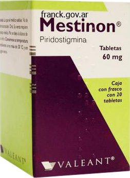
Mestinon dosages: 60 mg
Mestinon packs: 30 pills, 60 pills, 90 pills, 120 pills, 180 pills, 270 pills

Cheap mestinon 60 mg with mastercard
Let border, terminal part o the arch o aorta, pulmonary trunk, let auricle, and let ventricle. The typical anatomical apex, i current, is oten inerior to the shadow o the diaphragm. Three primary sorts o cardiovascular shadows happen, relying primarily on body sort or habitus. Because breast cancer cells have an uncommon anity or iodide, they turn out to be recognizable. It is especially useul or analyzing the viscera and lymph nodes o the mediastinum and roots o the lungs, by means o each planar and reconstructed. The arch o the aorta (20) is obliquely placed (more sagittal than transverse), with the ascending finish anteriorly within the midline, and the descending finish posteriorly and to the let o the vertebral our bodies (17). The pulmonary trunk (27) orms the stem o an inverted Y, with the arms ormed by the best (28) and let (29) pulmonary arteries. The proper pulmonary artery (28) passes beneath the arch o the aorta [between ascending (24) and descending (25) aortae]. A scan on the degree o the maximum diameter o the guts demonstrates all our chambers (32�35) and the diagonal slant o the interventricular septum (between 33 and 35)-see inset. Reconstructed rom information generated and accrued by spiral magnetic resonance imaging. The remainder o the structures within the superior mediastinum pass through the superior thoracic aperture to the basis o the neck or cross between the neck and stomach. Within the superior mediastinum, constructions happen in systemic layers, proceeding rom anterior to posterior: (1) lymphoid system (thymus), (2) blood vascular system (veins frst, then arteries), (3) respiratory system (trachea), (4) alimentary system (esophagus), and (5) lymph vascular system. The pattern o the branches o the arch o the aorta is atypical in roughly 35% o people. Contents embrace the esophagus and esophageal nerve plexus, thoracic aorta, thoracic duct and lymphatic trunks, posterior mediastinal lymph nodes, and azygos and hemi-azygos veins. The azygos/hemi-azygos venous system constitutes the venous counterpart to the thoracic aorta and its posterior mediastinal branches. The thoracic portion o the sympathetic trunks and thoracic splanchnic nerves may or will not be thought of components o the posterior mediastinum. Anterior mediastinum: the smallest subdivision o the mediastinum, between sternum and transversus thoracis muscles, signifcant primarily as a surgical aircraft, accommodates primarily free connective tissue and, in inants and children, the inerior extend o the thymus. Surace anatomy o thoracic viscera: the heart and nice vessels are within the central thorax, surrounded laterally and posteriorly by the lungs, and are overlapped anteriorly by the strains o pleural reection and anterior borders o the lungs, sternum, and the central half o the thoracic cage. The place o the mediastinal viscera is decided by position relative to gravity, part o respiration, and the construct and physical situation o the particular person. The transverse thoracic plane intersects the sternal angle and demarcates the great vessels superiorly rom the pericardium/ heart. The xiphisternal junction provides an indication o the central tendon o the diaphragm. It is a fexible, dynamic container, housing most o the organs o the alimentary system and half o the urogenital system. Containment o the belly organs and their contents is provided by musculo-aponeurotic partitions anterolaterally, the diaphragm superiorly, and the muscular tissues o the pelvis ineriorly. Interposed between the extra inflexible thorax and pelvis, this association enables the abdomen to enclose and defend its contents while providing the fexibility required by respiration, posture, and locomotion. Through voluntary or refexive contraction, its muscular roo, anterolateral walls, and foor can raise inner (intraabdominal) stress to aid expulsion o air rom the thoracic cavity (lungs and bronchi) or o fuid. The anterolateral stomach wall and various other organs mendacity against the posterior wall are lined on their inside aspects with a serous membrane or peritoneum (serosa) that refects (turns sharply and continues) onto the belly viscera (L. Thus, a bursal sac or lined potential space (peritoneal cavity) is ormed between the partitions and the viscera that normally contains only sufficient extracellular (parietal) fuid to lubricate the membrane masking most o the suraces o the constructions orming or occupying the stomach cavity. Visceral movement related to digestion happens reely, and the double-layered refections o peritoneum passing between the walls and the viscera provide passage or the blood vessels, lymphatics, and nerves. Variable quantities o at may occur between the walls and viscera and the peritoneum lining them. The abdominal cavity orms the superior and major part o the abdominopelvic cavity. The airplane o the pelvic inlet (superior pelvic aperture) arbitrarily, however not bodily, separates the abdominal and the pelvic cavities. Consequently, the extra superiorly positioned stomach organs (spleen, liver, part o the kidneys, and stomach) are protected by the thoracic cage.
Generic 60 mg mestinon with amex
Medium muscular arteries (distributing arteries) have partitions that consist chiefy o circularly disposed easy muscle bers. Their capacity to decrease their diameter (vasoconstrict) regulates the fow o blood to dierent parts o the physique as required by circumstance. Pulsatile contractions o their muscular partitions (regardless o lumen caliber) quickly and rhythmically constrict their lumina in progressive sequence, propelling and distributing blood to various components o the physique. Most o the named arteries, including these observed in the physique wall and limbs during dissection such because the brachial or emoral arteries, are medium muscular arteries. Small arteries and arterioles have relatively slender lumina and thick muscular partitions. The arteries (A) and veins (B) proven right here carry oxygen-rich blood rom the center to the systemic capillary beds and return low-oxygen blood rom the capillary beds to the guts, respectively, constituting the systemic circulation. Although commonly depicted and considered as single vessels, as shown here, the deep veins o the limbs often happen as pairs o accompanying veins. Small arteries are often not named or specically identied during dissection, and arterioles could be noticed solely underneath magnication. I a major channel is occluded, the smaller alternate channels can usually increase in dimension over a interval o time, providing a collateral circulation or alternate pathway that ensures the blood provide to buildings distal to the blockage. Occlusion o an finish artery interrupts the blood supply to the construction or segment o an organ it supplies. True terminal arteries supply the retina, or example, where occlusion will result in blindness. While not true terminal arteries, unctional terminal arteries (arteries with ineectual anastomoses) provide segments o the brain, liver, kidneys, spleen, and intestines; they could additionally exist in the coronary heart. Both eects make it easier or the musculovenous pump to overcome the orce o gravity to return blood to the center. Examples o medium veins embody the named supercial veins (cephalic and basilic veins o the upper limbs and nice and small saphenous veins o the lower limbs) and the accompanying veins that are named according to the artery they accompany. Large veins are characterized by broad bundles o longitudinal easy muscle and a well-developed tunica adventitia. Although their partitions are thinner, their diameters are often bigger than those o the corresponding artery. The skinny walls permit veins to have a big capability or growth and do so when blood return to the center is impeded by compression or internal pressures. Since the arteries and veins make up a circuit, it could be expected that hal the blood volume would be in the arteries and hal in the veins. Although oten depicted as single vessels in illustrations or simplicity, veins are inclined to be double or a quantity of. This association serves as a countercurrent warmth exchanger, the nice and cozy arterial blood warming the cooler venous blood because it returns to the heart rom a chilly limb. The accompanying veins occupy a relatively unyielding ascial vascular sheath with the artery they accompany. Systemic veins are more variable than arteries, and venous anastomoses-natural communications, direct or oblique, between two veins-occur extra oten between them. The outward enlargement o the bellies o contracting skeletal Veins usually return low-oxygen blood rom the capillary beds to the heart, which gives the veins a darkish blue look. The giant pulmonary veins are atypical in that they carry oxygen-rich blood rom the lungs to the center. Because o the decrease blood stress within the venous system, the partitions (specically, the tunica media) o veins are thinner than those o their companion arteries. Small veins are the tributaries o bigger veins that unite to orm venous plexuses (networks o veins), such because the dorsal venous arch o the oot. In the limbs, and in another places where the fow o blood is opposed by the pull o gravity, the medium veins have valves. Venous valves are cusps (passive faps) o endothelium with cup-like valvular sinuses that ll rom above. The valvular mechanism also breaks columns o blood within the veins into shorter segments, Accompanying veins (L. Although most veins o the trunk happen as large single vessels, veins in the limbs occur as two or extra smaller vessels that accompany an artery in a common vascular sheath. Cardiovascular System forty one muscular tissues in the limbs, restricted by the deep ascia, compresses the veins, "milking" the blood superiorly towards the guts; another (musculovenous) type o venous pump.

Buy mestinon 60 mg lowest price
The developmental complex of "parachute mitral valve," supravalvular ring of left atrium, subaortic stenosis and coarctation of aorta. They concluded that youthful age on the time of pulmonary valve substitute and valve oversizing in sufferers less than 20 years of age on the time of pulmonary valve replacement were significant predictors of structural valve deterioration. They suggested that these findings might probably have an effect on the timing of pulmonary valve substitute and the extent of valve oversizing in babies. No statistically significant distinction in valve performance was seen between bioprosthetic valve types at short-term follow-up. On the contrary, they discovered that they perform well and lead to a much lower reoperation rate than has been reported after allograft utilization. Congenitally bicuspid aortic valve inflicting extreme, pure aortic regurgitation without superimposed infective endocarditis. Quadricuspid aortic valve related to fibromuscular subaortic stenosis and aortic regurgitation handled by conservative surgery. Pathogenetic mechanisms of prolapsing aortic valve and aortic regurgitation associated with ventricular septal defect. Mechanisms of aortic valve incompetence: finite-element modeling of Marfan syndrome. Current management of severe congenital mitral stenosis: outcomes of transcatheter and surgical remedy in 108 infants and youngsters. Left ventricular apical approach for the surgical remedy of congenital mitral stenosis. Aortic valve-preserving process for enlargement of the left ventricular outflow tract and mitral annulus. Left atrialleft ventricular conduit for reduction of congenital mitral stenosis in infancy. Systemic atrioventricular conduit for extracardiac bypass of hypoplastic systemic atrioventricular valve. Mitral valve restore for congenital mitral valve stenosis within the pediatric inhabitants. Surgical repair of congenital mitral valve malformations in infancy and childhood: a single-center 36-year experience. Surgical therapy of congenital mitral valve disease: midterm outcomes of a repairoriented coverage. Clinical course and hemodynamic observations after supraannular mitral valve alternative in infants and youngsters. Surgical reconstruction techniques for mitral valve insufficiency from lesions with restricted leaflet movement in infants and youngsters. Toward comprehensive evaluation of mitral regurgitation using cardiovascular magnetic resonance. Repair versus replacement for degenerative mitral valve illness with coexisting ischemic coronary heart illness. The long-term outcomes of commissure plication annuloplasty for congenital mitral insufficiency. Uber einen sehr seltenen Fall von lnsufficienz der Valvular tricuspidalis, bedingt durch eine angeborene hochgradige Missbildung deselben. Pulmonary valve substitute in tetralogy of Fallot: influence on survival and ventricular tachycardia. Younger age and valve oversizing are predictors of structural valve deterioration after pulmonary valve replacement in sufferers with tetralogy of Fallot. A new method for alternative of the mitral valve by a semilunar valve homograft. It lies centrally deep within the coronary heart immediately adjacent to the two atrioventricular valves. In many instances obstruction happens at a number of levels and though the degree of obstruction in anybody area may not be severe, the combined impact of obstruction at a number of levels is clinically essential. At one end of the spectrum are sufferers with isolated aortic valve stenosis, normal-sized left heart buildings and a traditional aortic arch and isthmus.


Buy genuine mestinon line
The median sacral artery, an unpaired parietal branch, may be mentioned to occupy a ourth (posterior) airplane as a outcome of it arises rom the posterior side o the aorta just proximal to its biurcation. Although markedly smaller, it may be thought-about a midline "continuation" o the aorta, by which case its lateral branches, the small lumbar arteries and lateral sacral branches, would also be included as half o the paired parietal branches. Right Intermediate Hepatic veins (middle) Left Hemi-azygos vein Left inferior phrenic vein Posterior intercostal veins Left suprarenal vein Left renal vein Left gonadal vein (testicular or ovarian) Right gonadal vein (testicular or ovarian) Ascending lumbar vein Left frequent iliac vein Left external iliac vein Left inner iliac vein Median sacral vein Right common iliac vein Azygos vein Right inferior phrenic vein Inferior vena cava Right suprarenal vein Right renal vein 1st 2nd Lumbar 3rd veins 4th the stomach aorta descends anterior to the our bodies o the T12�L4 vertebrae. On the best, the aorta is said to the azygos vein, cisterna chyli, thoracic duct, proper crus o the diaphragm, and right celiac ganglion. On the let, the aorta is related to the let crus o the diaphragm and the let celiac ganglion. The veins that correspond to the unpaired visceral branches o the aorta are instead tributaries o the hepatic portal vein. The branches corresponding to the paired visceral branches o the belly aorta include the right suprarenal vein, the right and let renal veins, and the best gonadal (testicular or ovarian) vein. Lymphatic vessels and lymph nodes o the posterior belly wall and lymphatic trunks o the stomach. All lymphatic drainage rom the lower hal o the body converges in the stomach to enter the start o the thoracic duct. Lymph rom the frequent iliac lymph nodes passes to the right and let lumbar lymph nodes. Lymph rom the alimentary tract, liver, spleen, and pancreas passes alongside the celiac and superior and inerior mesenteric arteries to the pre-aortic lymph nodes (celiac and superior and inerior mesenteric nodes) scattered around the origins o these arteries rom the aorta. Eerent vessels rom these nodes orm the intestinal lymphatic trunks, which may be single or multiple, and participate within the confuence o lymphatic trunks that gives rise to the thoracic duct. These nodes obtain lymph directly rom the posterior belly wall, kidneys, ureters, testes or ovaries, uterus, and uterine tubes. They also receive lymph rom the descending colon, pelvis, and lower limbs by way of the inerior mesenteric and common iliac lymph nodes. Eerent lymphatic vessels rom the massive lumbar lymph nodes orm the best and let lumbar lymphatic trunks. The inerior end o the thoracic duct lies anterior to the our bodies o the L1 and L2 vertebrae between the proper crus o the diaphragm and the aorta. The thoracic duct begins with the convergence o the primary lymphatic ducts o the abdomen, which in only a small proportion o people takes the orm o the generally depicted, thin-walled sac or dilation, the cisterna chyli (chyle cistern). Consequently, basically all of the lymphatic drainage rom the lower hal o the body (deep lymphatic drainage inerior to the extent o the diaphragm and all supercial drainage inerior to the extent o the umbilicus) converges within the abdomen to enter the start o the thoracic duct. The thoracic duct ascends via the aortic hiatus in the diaphragm into the posterior mediastinum, where it collects extra parietal and visceral drainage, notably rom the let upper quadrant o the body. The duct finally ends by getting into the venous system at the junction o the let subclavian and internal jugular veins (the let venous angle). Hiccups result rom irritation o aerent or eerent nerve endings, or o medullary centers within the brainstem that control the muscles o respiration, significantly the diaphragm. Hiccups have many causes, similar to indigestion, diaphragm irritation, alcoholism, cerebral lesions, and thoracic and stomach lesions, all which disturb the phrenic nerves. Reerred Pain rom Diaphragm Pain rom the diaphragm radiates to two dierent areas as a result of o the dierence within the sensory nerve provide o the diaphragm (Table 5. Pain ensuing rom irritation o the diaphragmatic pleura or the diaphragmatic peritoneum is reerred to the shoulder region, the world o skin equipped by the C3�C5 segments o the spinal wire (see the Clinical Box "Visceral Reerred Pain" earlier in this chapter). Irritation o peripheral areas o the diaphragm, innervated by the inerior intercostal nerves, is extra localized, being reerred to the skin over the costal margins o the anterolateral abdominal wall. Rupture o Diaphragm and Herniation o Viscera Rupture o the diaphragm and herniation o viscera can result rom a sudden massive increase in either the intrathoracic or intra-abdominal stress. The common trigger o this damage is severe trauma to the thorax or abdomen during a motorized vehicle accident. Most diaphragmatic ruptures are on the let side (95%) as a outcome of the substantial mass o the liver, intimately related to the diaphragm on the right facet, provides a bodily barrier. Posterior Abdominal Wall 545 A nonmuscular space o variable size called the lumbocostal triangle normally occurs between the costal and lumbar parts o the diaphragm. This half o the diaphragm is normally ormed solely by usion o the superior and inerior ascias o the diaphragm.

Purchase mestinon without a prescription
Capillaries consist only o this tunic, with blood capillaries additionally having a supporting basement membrane. Arteries, veins, and lymphatic ducts are distinguished by the thickness o this layer relative to the dimensions o the lumen, its organization, and, within the case o arteries, the presence o variable amounts o elastic bers. Blood beneath excessive pressure leaves the guts and is distributed to the physique by a branching system o thick-walled arteries. The nal distributing vessels, arterioles, ship oxygenrich blood to capillaries. Capillaries orm a capillary mattress, the place the interchange o oxygen, vitamins, waste products, and different substances with the extracellular fuid occurs. Blood rom the capillary bed passes into thin-walled venules, which resemble wide capillaries. The largest veins, the superior and inerior venae cavae, return low-oxygen blood to the center. The dierent types o arteries are distinguished rom each other on the basis o total dimension, relative amounts o elastic tissue or muscle in the tunica media. There are three types o arteries: Large elastic arteries (conducting arteries) have many elastic layers (sheets o elastic bers) in their partitions. Their elasticity allows them to expand once they obtain the cardiac output rom the ventricles, minimizing the strain change, and return to regular size between ventricular contractions, as they proceed to push the blood into the medium arteries downstream. This maintains the blood stress in the arterial system between cardiac contractions (at a time when ventricular stress alls to zero). Overall, this minimizes the ebb in blood pressure as the heart contracts and relaxes. The partitions o most blood vessels have three concentric layers o tissue, called tunics (L. With less muscle, veins are thinner walled than their companion arteries and have wide lumens (L. Cardiovascular System 39 Examples o giant elastic arteries are the aorta, the arteries that originate rom the arch o the aorta (brachiocephalic trunk, subclavian and carotid arteries), and the pulmonary trunk and arteries. The valves o the veins break up the columns o blood, thus relieving the more dependent elements o excessive stress, permitting venous blood to fow only toward the center. Capillaries are typically organized in capillary beds, networks that join the arterioles and venules. The blood enters the capillary beds by way of arterioles that control the fow and is drained rom them by venules. As the hydrostatic pressure within the arterioles orces blood into and through the capillary bed, it also orces fuid containing oxygen, nutrients, and other cellular materials out o the blood at the arterial finish o the capillary mattress (upstream) into the extracellular areas, permitting change with cells o the surrounding tissue. Muscular contractions within the limbs unction with the venous valves to transfer blood towards the heart. The outward growth o the bellies o contracting muscular tissues is proscribed by deep ascia and turns into a compressive orce, propelling the blood against gravity. In some situations, blood passes via two capillary beds beore returning to the center; a venous system linking two capillary beds constitutes a portal venous system. The venous system by which nutrient-rich blood passes rom the capillary beds o the alimentary tract to the capillary beds or sinusoids o the liver-the hepatic portal system-is the most important instance. A widespread orm, atherosclerosis, is related to the buildup o at (mainly cholesterol) in the arterial partitions. A calcium deposit orms an atheromatous plaque (atheroma)-well-demarcated, hardened yellow areas or swellings on the intimal suraces o arteries. The consequences o atherosclerosis include ischemia (reduction forty two Chapter 1 Overview and Basic Concepts o blood supply to an organ or region) and inarction (local dying, or necrosis, o an area o tissue or an organ ensuing rom reduced blood supply). Varicose veins have incompetent valves; thus, the column o blood ascending toward the guts is unbroken, placing increased strain on the weakened partitions, urther exacerbating the varicosity drawback. Incompetent ascia is incapable o containing the enlargement o contracting muscles; thus, the (musculoascial) musculovenous pump is ineective. A weakened vein dilates beneath the pressure o supporting a column o blood in opposition to gravity. The Starling hypothesis (see "Blood Capillaries" in this chapter) explains how most o the fuid and electrolytes coming into the extracellular spaces rom the blood capillaries can additionally be reabsorbed by them. I this materials had been to accumulate within the extracellular areas, a reverse osmosis would happen, bringing even more fuid and leading to edema (an extra o interstitial fuid, maniest as swelling). The lymphoid system thus constitutes a sort o "overfow" system that gives or the drainage o surplus tissue fuid and leaked plasma proteins to the bloodstream, in addition to or the removal o debris rom cellular decomposition and inection.
Purchase 60mg mestinon fast delivery
At irregular intervals, gastric peristalsis pushes the chyme via the pyloric canal and orice into the small gut or urther mixing, digestion, and absorption. In the supine place, the pyloric half o the abdomen lies at the level o the transpyloric aircraft, halfway between the jugular notch superiorly and the pubic crest ineriorly. The angular incisure (notch), probably the most inerior part o the curvature, indicates the junction o the physique and pyloric half o the stomach. Because o the unequal lengths o the lesser curvature on the proper and the higher curvature on the let, in most individuals the shape o the stomach resembles the letter J. Surace anatomy and eect o body type (bodily habitus) on disposition and form o abdomen. The most typical place o the abdomen in a person o medium build in the supine or susceptible position. Pyloric half: the unnel-shaped outfow area o the abdomen; its wider half, the pyloric antrum, leads into the pyloric canal, its narrower half. It is a marked thickening o the round layer o smooth muscle that controls discharge o the abdomen contents through the pyloric orifce (inerior opening or outlet o the stomach) into the duodenum. Intermittent emptying o the abdomen occurs when intragastric stress overcomes the resistance o the pylorus. When contracted, the gastric mucosa is thrown into longitudinal ridges or wrinkles known as gastric olds (gastric rugae). During swallowing, a brief lived groove or urrow-like gastric canal orms between the longitudinal gastric olds along the lesser curvature. Saliva and small quantities o masticated ood and different fuids drain along the gastric canal to the pyloric canal when the stomach is usually empty. The two layers o the lesser omentum prolong across the stomach and go away its higher curvature because the greater omentum. Anteriorly, the stomach is expounded to the diaphragm, let lobe o the liver, and anterior abdominal wall. The let half o the liver is reduce away in order that the lesser omentum and omental oramen could be seen. Circular peristaltic waves start in the body o the abdomen and sweep towards the pyloric canal, as proven in (E) (arrowheads), where they stop. Radiograph demonstrating the pyloric area o the abdomen and the superior half o the duodenum. The transverse colon is said ineriorly and laterally to the stomach as it programs along the greater curvature o the abdomen to the let colic fexure. The mattress o the stomach, on which the abdomen rests within the supine place, is ormed by the constructions orming the posterior wall o the omental bursa. From superior to inerior, the mattress o the abdomen is ormed by the let dome o the diaphragm, spleen, let kidney and suprarenal gland, splenic artery, pancreas, and transverse mesocolon. Because this vein is obvious in dwelling individuals, surgeons use it or identiying the pylorus. They drain lymph rom its anterior and posterior suraces toward its curvatures, the place the gastric and gastro-omental lymph nodes are positioned. The eerent vessels rom these nodes accompany the large arteries to the celiac lymph nodes. The ollowing is a summary o the lymphatic drainage o the stomach: Lymph rom the superior two thirds o the stomach drains along the best and let gastric vessels to the gastric lymph nodes; lymph rom the undus and superior half o the body o the stomach additionally drains along the brief gastric arteries and let gastro-omental vessels to the pancreaticosplenic lymph nodes. Lymph rom the best two thirds o the inerior third o the abdomen drains alongside the best gastro-omental vessels to the pyloric lymph nodes. Lymph rom the let one third o the higher curvature drains to the pancreaticoduodenal lymph nodes, that are situated along the quick gastric and splenic vessels. It runs toward the lesser curvature o the stomach, where it offers o hepatic and duodenal branches, which leave the abdomen within the hepatoduodenal ligament. The relaxation o the anterior vagal trunk continues along the lesser curvature, giving rise to anterior gastric branches. The larger posterior vagal trunk, derived mainly rom the proper vagus nerve, enters the abdomen on the posterior surace o the esophagus and passes toward the lesser curvature o the abdomen. The posterior vagal trunk provides branches to the anterior and posterior suraces o the stomach.
Cheap mestinon on line
A is between the submucosal esophageal veins draining into both the azygos vein (systemic) or the let gastric vein (portal); when dilated, these are esophageal varices. B is between the inerior and center rectal veins draining into the inerior vena cava (systemic) and the superior rectal vein, persevering with as the inerior mesenteric vein (portal). The submucosal veins concerned are normally dilated (varicose in appearance), even in newborns. D is on the posterior aspects (bare areas) o secondarily retroperitoneal viscera, or the liver, the place twigs o visceral veins-or instance, the colic vein, splenic veins, or the portal vein itsel (portal system)-anastomose with retroperitoneal veins o the posterior belly wall or diaphragm (systemic system). As it approaches the porta hepatis, the hepatic portal vein divides into right and let branches. The hepatic portal vein collects blood with reduced oxygenation but wealthy in nutrients rom the abdominal half o the alimentary system, together with the gallbladder and pancreas, as nicely as the spleen, and carries it to the liver. Within the liver, its branches are distributed in a segmental pattern (see "Blood Vessels o Liver") and finish in expanded capillaries, the venous sinusoids o the liver. Portal�systemic anastomoses, in which the portal venous system communicates with the systemic venous system, are ormed in the submucosa o the inerior esophagus, in the submucosa o the anal canal, within the peri-umbilical area, and on the posterior features (bare areas) o secondarily retroperitoneal viscera, or the liver. However, the quantity o blood orced by way of the collateral routes may be extreme, resulting in doubtlessly atal varices (abnormally dilated veins) (see the Clinical Box "Portal Hypertension," p. Blunt trauma to the let side or to other areas o the abdomen that trigger a sudden, marked enhance in intra-abdominal stress. The close relationship o the spleen to the ribs that normally protect it could be detrimental when there are rib ractures. Severe blows on the let side may racture a quantity of o these ribs, and rupture the underlying spleen, or sharp bone ragments might lacerate the spleen. When the spleen is diseased, resulting rom, or instance, granulocytic leukemia (high leukocyte and white blood cell count), it could enlarge to 10 or extra times its regular measurement and weight (splenomegaly). Generally, i its lower edge could be detected when palpating under the let costal margin on the finish o inspiration. Accessory Spleen(s) and Splenosis One or more small accent spleens may develop prenatally near the splenic hilum. They could additionally be e embedded partly or wholly within the tail o the pancreas, between the layers o the gastrosplenic ligament, in n the inracolic compartment, in the mesentery, or in close proximity to an ovary or testis. Accessory spleens are relatively frequent, are normally small (approximately 1 cm in diameter, and range rom zero. Awareness o the potential presence o an accessory spleen is essential as a outcome of i not eliminated during a splenectomy, the signs that indicated removing o the spleen. Splenosis-generalized autoimplantation o ectopic splenic tissue into the peritoneum, omentum, or mesenteries- sometimes ollows splenic rupture. This potential space descends to the extent o the tenth rib within the midaxillary line. Its existence should be kept in thoughts when doing a splenic needle biopsy, or when injecting radiopaque materials into the spleen or visualization o the hepatic portal vein (splenoportography). Blockage o Hepatopancreatic Ampulla and Pancreatitis Because the principle pancreatic duct joins the bile duct to orm the hepatopancreatic ampulla and pierces the duodenal wall, a gallstone passing alongside the extrahepatic bile passages could lodge in the constricted distal finish o the ampulla, the place it opens at the summit o the major duodenal papilla. In this case, each the biliary and pancreatic duct methods are blocked and neither bile nor pancreatic juice can enter the duodenum. However, bile might back up and enter the pancreatic duct, usually leading to pancreatitis (infammation o the pancreas). A similar refux o bile generally results rom spasms o the hepatopancreatic sphincter. Normally, the sphincter o the pancreatic duct prevents refux o bile into the pancreatic duct; however, i the hepatopancreatic ampulla is obstructed, the weak pancreatic duct sphincter may be unable to stand up to the excessive pressure o the bile in the hepatopancreatic ampulla. I an accessory pancreatic duct connects with the primary pancreatic duct and opens into the duodenum, it may compensate or an obstructed main pancreatic duct or spasm o the hepatopancreatic sphincter. Utilizing the fuoroscopic visualization provided by the contrast medium, devices operated through the endoscope are then utilized or the intervention.

Buy 60 mg mestinon with visa
The inerior end o the radius moves around the relatively fxed finish o the ulna throughout supination and pronation o the hand. The two bones are frmly united distally by the articular disc, reerred to clinically as the triangular ligament o the distal radio-ulnar joint. It has a broad attachment to the radius however a narrow attachment to the styloid course of o the ulna, which serves as the pivot point or the rotary motion. During pronation, the inerior finish o the radius strikes anteriorly and medially across the inerior end o the ulna, carrying the hand with it. In radiographs o the wrist and hand, the "joint house" at the distal end o the ulna appears wide as a outcome of o the radiolucent articular disc. This coronal part o the best hand demonstrates the distal radio-ulnar, wrist, intercarpal, carpometacarpal, and intermetacarpal joints. Although they appear to be steady when considered radiographically in parts A and C, the articular cavities o the distal radio-ulnar and wrist joints are separated by the articular disc o the distal radio-ulnar joint. Kucharczyk, Proessor and Neuroradiologist Senior Scientist, Department o Medical Resonance Imaging, University Health Network Toronto, Ontario, Canada. The proximal surace o this disc articulates with the distal aspect o the head o the ulna. Hence, the joint cavity is L-shaped in a coronal part; the vertical bar o the L is between the radius and ulna, and the horizontal bar is between the ulna and the articular disc. The articular disc separates the cavity o the distal radio-ulnar joint rom the cavity o the wrist joint. During supination, the radius uncrosses rom the ulna, its distal end moving (rotating) laterally and posteriorly so the bones become parallel. The synovial membrane extends superiorly between the radius and ulna to orm the sacciorm recess o the distal radio-ulnar joint. This redundancy o the synovial capsule accommodates the twisting o the capsule that happens when the distal finish o the radius travels around the comparatively xed distal end o the ulna throughout pronation o the orearm. These the anterior and posterior interosseous nerves provide the distal radio-ulnar joint. The place o the joint is indicated approximately by a line joining the styloid processes o the radius and ulna or by the proximal wrist crease. The wrist (carpus), the proximal segment o the hand, is a fancy o eight carpal bones, articulating proximally with the orearm via the wrist joint and distally with the ve metacarpals. Circumduction o the hand consists o successive fexion, adduction, extension, and abduction. The distal finish o the radius and the articular disc o the distal radio-ulnar joint articulate with the proximal row o carpal bones, besides or the pisiorm. The pisiorm lies in a plane anterior to the other carpal bones, articulating with only the triquetrum. Most activities require a small quantity o wrist fexion; nonetheless, tight grip (clenching o the st) requires extension on the wrist. The synovial membrane lines the interior surace o the brous layer o the joint capsule and is attached to the margins o the articular suraces. They are strong and directed in order that the hand ollows the radius during supination o the orearm. The dorsal radiocarpal ligaments take the same direction so that the hand ollows the radius throughout pronation o the orearm. The joint capsule can also be strengthened medially by the ulnar collateral ligament, which is connected to the ulnar styloid process and triquetrum. The joint capsule can be strengthened laterally by the radial collateral ligament, which is hooked up to the radial styloid course of and scaphoid. The midcarpal joint, a fancy joint between the proximal and distal rows o carpal bones. The pisotriquetral joint, ormed rom the articulation o the pisiorm with the palmar surace o the triquetrum. The actions at the wrist joint could also be augmented by additional smaller actions at the intercarpal and midcarpal joints. The actions are fexion�extension, abduction�adduction (radial deviation�ulnar deviation), and circumduction. Observe the palmar radiocarpal ligaments, passing rom the radius to the two rows o carpal bones. These strong ligaments are directed so that the hand ollows the radius throughout supination.
Real Experiences: Customer Reviews on Mestinon
Stan, 60 years: Tracheo- and bronchomalacia could persist for a quantity of weeks to months and can continue to compromise ventilation. The genital organs are demonstrated: testis, epididymis, ductus deerens, ejaculatory duct, and penis, with the accessory glandular constructions (seminal gland, prostate, and bulbo-urethral gland). The veins o the plexus normally merge to orm a singular ovarian vein, which leaves the lesser pelvis with the ovarian artery.
Arokkh, 28 years: With the exception o the subcostal nerve (T12) and lumbosacral trunk (L4�L5), the somatic nerves o the posterior stomach wall are products o the lumbar plexus, ormed by the anterior rami o L1�L4 deep to the psoas. It divides into these terminal branches because it seems within the cubital ossa, anterior to the lateral epicondyle o the humerus, between the brachialis and brachioradialis. A comparable experience has been described by the group from Toronto in a report published in 2000 by Shukla et al.
Masil, 46 years: Viscera o Thoracic Cavity 341 I a penetrating wound opens through the thoracic wall or the surace o the lungs, air might be sucked into the pleural cavity as a outcome of o the adverse stress. The smaller anterior circumex humeral artery passes laterally, deep to the coracobrachialis and biceps brachii. The anterior facet o the scrotum is equipped by derivatives o the lumbar plexus: anterior scrotal nerves, derived rom the ilio-inguinal nerve, and the genital department o the genitoemoral nerve (Table 6.
Nafalem, 65 years: This will scale back the quantity of button that can be harvested from the sinus below the coronary ostium and necessitates more careful and finer suturing with a higher threat of subsequent hemorrhage. The umbilical (let sagittal) fssure is the continuous groove ormed Caudate lobe Right sagittal fissure * Left lobe anteriorly by the fssure or the spherical ligament and posteriorly by the fssure or the ligamentum venosum. They pass through the paravertebral ganglia o the trunks without synapsing to enter the abdominopelvic splanchnic nerves, which convey them to the prevertebral ganglia o the abdominal cavity.
9 of 10 - Review by M. Konrad
Votes: 192 votes
Total customer reviews: 192
References
- Dickneite G, Pragst I. Prothrombin complex concentrate vs fresh frozen plasma for reversal of dilutional coagulopathy in a porcine trauma model. Br J Anaesth. 2009;102:345-354.
- El-Serag HB. Hepatocellular carcinoma. New Engl J Med 2011;365(12):1118-1127.
- Labauge P, Laberge S, Brunereau L, et al. Hereditary cerebral cavernous angiomas: clinical and genetic features in 57 French families. Societe Francaise de Neurochirurgie. Lancet 1998;352: 1892-7.
- Paden ML, Conrad SA, Rycus PT, Thiagarajan RR, Registry E. Extracorporeal Life Support Organization Registry Report 2012.
- Revel A, Ariel L, Rein AJ, et al: Fetal endocardial fibroelastosis. J Clin Ultrasound 1994; 22:355-356.
- Durkin M, Witt J, Lemonte A, Wheat B, Connolly P. Antigen assay with the potential to aid in diagnosis of blastomycosis. J Clin Microbiol 2004;42 (10):4873-5.
- Liu GJ, Luo J, Zhang L. Meta-analysis of the effectiveness and safety of prophylactic use of nimodipine in patients with an aneurysmal subarachnoid haemorrhage. CNS Neurol Disord Drug Targets. 2011;10(7):834-844.

