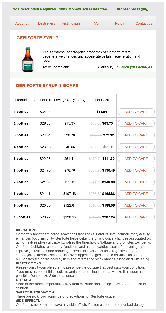
Geriforte Syrup dosages: 100 caps
Geriforte Syrup packs: 1 bottles, 2 bottles, 3 bottles, 4 bottles, 5 bottles, 6 bottles, 7 bottles, 8 bottles, 9 bottles, 10 bottles

Order geriforte syrup online from canada
Coronal computed tomography (A), endoscopic (B), and endoscopic ultrasound (C) photographs present a well-defined, submucosal mass (arrows) in the second portion of the duodenum. Coronal T2-weighted (A) and contrast-enhanced axial T1-weighted (B) magnetic resonance photographs show a quantity of giant, peritoneal-based lots (arrows) and ascites. Nuclear Medicine A tagged purple blood cell scan is useful in cases of acute gastrointestinal bleeding as a manifesting symptom of small bowel adenocarcinoma. Localization of the first tumor to facilitate surgery is healthier carried out utilizing octreoscan in patients with carcinoid tumors. It is necessary to think about small bowel malignancies as a possible trigger of those signs and to have a excessive degree of suspicion for catching them at an early stage. The scientific differential prognosis is huge and contains ischemic, infectious, and inflammatory causes and other major malignancies. Small bowel lymphoma and adenocarcinoma can manifest as small bowel obstruction or intussusception. The differential diagnosis of lymphoma can embrace tuberculosis and inflammatory small bowel disease. The differential diagnosis includes lymphoma, desmoid tumor, metastases, and mesenteric lymphadenopathy. Five-year survival rates vary from 10% to 60%, with a median of approximately 30%. This check is helpful in treatment planning and might predict the response to somatostatin analogs. Patients with constructive octreoscan findings are handled with somatostatin analogs, which leads to control of signs (75%), stabilization of tumor growth (71%), or tumor shrinkage (9%). Transarterial chemoembolization is the procedure of alternative for administration of inoperable carcinoid liver metastases and shows promising results with partial response in a minimum of 50% of sufferers and a mortality fee of 5%. The treatment could vary from resection adopted by chemotherapy for local tumor to only chemotherapy in advanced tumors. Radiation therapy is the treatment of selection for initially cumbersome tumor sites, remedy of residual illness after chemotherapy, or serious local issues. The most necessary factors that determine mode of remedy are small bowel tumor measurement and cell division rate. Adenocarcinoma of the small bowel: review of the National Cancer Data Base, 1985-1995. Diagnostic highquality double-contrast barium enema examination is an art, requiring skillful maneuvering of the affected person and barium pool while optimally utilizing fluoroscopy. Barium Enema Examination Single-contrast examination is most well-liked in motionless, elderly, or incontinent patients. Contraindications for the standard bowel preparation include bedridden patients, postoperative patients, sufferers with diabetes and hypothyroidism, and sufferers taking opiates. Before starting the study, compliance with the bowel preparation should be confirmed. Lubricant is applied on the exterior anal surface and the rectal catheter tip,9 and the catheter is gently inserted into the anal canal. The distal rectum is then drained by dropping the barium bag to the ground to keep away from bubbles when air is insufflated. The affected person is turned proper facet down to fill the proximal transverse colon and then was the supine place to fill the posteriorly situated hepatic flexure. The enema tip can then be eliminated, providing bodily aid to the affected person and permitting better evaluation of the distal rectum. The enema tip could need to be left in place in patients expelling gas and who may need extra air to visualize the terminal ileum. Technique Tips � Perform air insufflation only after barium has passed the splenic flexure. Technique Tips � Sigmoid colon spot films are obtained earlier than barium reaches the ascending colon. If barium refluxes via the ileocecal valve, the sigmoid colon may be partly obscured. Some technical challenges during a double-contrast barium enema examination and plausible options are summarized in Box 29-2.
Purchase geriforte syrup with visa
They appear as polypoid buildings that project into the gallbladder lumen and may be sessile or pedunculated and generally lower than 2 cm. Adenomas obstructing the cystic duct could current with signs of acute cholecystitis (see Chapter 56). Gallstones are differentiated based mostly on mobility and adherence to the gallbladder wall. Gallbladder carcinoma has a heterogeneous internal architecture with mucosal irregularity, adjacent parenchymal liver invasion, biliary duct dilatation, metastases, and lymphadenopathy. Transverse ultrasound image reveals an echogenic, rounded polyp hooked up to the gallbladder wall. Gallstones adherent to the gallbladder wall are echogenic and show posterior acoustic shadowing. Tumefactive sludge types one other differential prognosis but could be discerned by the distinction in morphology as proven with a change in position of the patient. Adenomas are smooth, lobulated or rounded masses with a homogeneous echotexture and an identifiable stalk in Document t�l�charg� de ClinicalKey. Longitudinal (A) and transverse (B) ultrasound images of the gallbladder in a 48-yearold woman with right higher quadrant pain show multiple small echogenic ldl cholesterol polyps (arrows) adherent to the gallbladder wall. Gallbladder carcinoma is clear by its heterogeneous inside structure with mucosal irregularity, adjoining parenchymal liver invasion, biliary duct dilatation, metastases, and lymphadenopathy. Large polyps mimicking gallbladder carcinoma may require cholecystectomy to rule out malignancy. Low-attenuation bands or nodules are visualized within the thickened gallbladder wall, which characterize abscesses of foci of xanthogranulomatous irritation. The wall margin with the liver may be indistinct, and extension of inflammatory course of into the liver might give a masslike appearance. Biliary dilatation when current is normally secondary to intraductal stones, hepatoduodenal ligament adenopathy, or coexistent malignancy of the gallbladder or bile duct. Ultrasonography Focal wall thickening with hypoechoic bands or nodules within the thickened gallbladder wall is seen. Extension of the inflammatory course of into the adjacent liver leads to loss of well-defined fats plane between the gallbladder and liver. Other ultrasonographic findings embody disruption of the mucosal line, pericholecystic fluid, stones, and intrahepatic biliary dilatation. Axial contrast-enhanced computed tomography scan of the abdomen exhibits irregular gallbladder wall thickening on the fundus with mucosal irregularity and extension into the surrounding hepatic parenchyma (arrow). When visualized, the Rokitansky-Aschoff sinus appears as a small cystic construction with water density inside the thickened wall. Multiple intramural cystic spaces are seen inside a focal mass that seem hypointense on T1-weighted photographs and hyperintense on T2-weighted pictures. Signal void may be seen inside the cystic spaces when intracystic calculi are present. On gadolinium administration, the focal mass exhibits enhancement while the cystic spaces are nonenhancing. A "diamond ring" appearance may be seen on transverse sections because of the ringlike distribution of the hyperintense cystic constructions across the gallbladder wall on T2-weighted pictures. Occasionally, this artifact could additionally be mistaken for air within the gallbladder lumen or wall (emphysematous cholecystitis), which may have a similar look. Whereas the dirty shadow of air is extra linear in configuration, the reverberation artifacts of adenomyoma are V shaped. The three variants of adenomyomatous hyperplasia are localized (or fundal), segmental, and diffuse. A to C, Axial contrast-enhanced computed tomography pictures of the stomach present focal wall thickening of the gallbladder at the fundus (arrows). The small cystic areas of water attenuation are best seen in B and C within the focal wall thickening and symbolize the Rokitansky-Aschoff sinuses crammed with bile.
Discount geriforte syrup 100 caps with mastercard
Measure full blood rely, liver and renal perform before starting treatment, then 1�2 weekly till remedy is established and 2�3 month-to-month thereafter. Treatment must be stopped immediately if abnormalities develop or if the affected person turns into breathless. Administration Communication Monitoring Cost Clinical tip-There are significant restrictions related to the prescription of methotrexate in order to reduce medicine errors and the chance of toxicity. They might have an essential role in reviewing or continuing prescriptions, for instance on the time of hospital admission. If in any doubt about the appropriateness of a methotrexate prescription, all the time search senior recommendation. Oral infections (such as dental abscess) or aspiration pneumonia attributable to Gram-negative anaerobes from the mouth. Surgical and gynaecological infections brought on by Gram-negative anaerobes from the colon, for example Bacteroides fragilis. In anaerobic bacteria, reduction of metronidazole generates a nitroso free radical. Bacterial resistance to metronidazole is usually low but is rising in prevalence. Mechanisms include decreased uptake of metronidazole and reduced era of nitroso free radicals. As with many antibiotics, metronidazole could cause gastrointestinal upset (such as nausea and vomiting) and quick and delayed hypersensitivity reactions (see Penicillins, broad-spectrum). When used at excessive doses or for a chronic course, metronidazole may cause neurological antagonistic results together with peripheral and optic neuropathy, seizures and encephalopathy. Metronidazole is metabolised by hepatic cytochrome P450 enzymes, so the dose should be lowered in folks with extreme liver disease. Metronidazole inhibits the enzyme acetaldehyde dehydrogenase, which is answerable for clearing the intermediate alcohol metabolite acetaldehyde from the body. Metronidazole has some inhibitory effect on cytochrome P450 enzymes, reducing metabolism of warfarin (increasing the danger of bleeding) and phenytoin (increasing the danger of toxicity, together with impaired cerebellar function). Metronidazole could be prescribed as a gel for topical administration to treat vaginal an infection such as bacterial vaginosis or to scale back the odour from an contaminated pores and skin ulcer. Explain that the purpose of treatment is to do away with an infection and enhance signs. Warn the affected person to not take alcohol during or for forty eight hours after treatment, explaining that if they do they may feel very unwell with nausea, vomiting, flushing and headache. If an allergy develops throughout remedy, give the patient written and verbal advice to not take this antibiotic in the future and be positive that the allergy is clearly documented in their medical records. Check that infection resolves by review of symptoms, signs and blood tests (improvement in inflammatory markers) if acceptable. For treatment exceeding 10 days, measure full blood count and liver operate tests to monitor for adverse effects. A 7-day course of non-proprietary oral metronidazole tablets taken one 8-hrly currently costs around �1. You ought to due to this fact select the decrease dose until there are overwhelming scientific indications for the upper dose. Administration Communication Monitoring Cost Clinical tip-Anaerobic micro organism are often resistant to penicillins because of production of -lactamases. However, co-amoxiclav (amoxicillin with the -lactamase inhibitor clavulanic acid) does have good efficacy against anaerobes. Naloxone binds to opioid receptors (particularly the pharmacologicallyimportant opioid �-receptors), the place it acts as a competitive antagonist. However, if an opioid is present, naloxone displaces it from its receptors and, in so doing, it reverses its effects. In opioid toxicity, that is used to restore an sufficient degree of consciousness and respiratory fee. Where naloxone is administered to reverse opioid toxicity in an opioid-dependent particular person, an opioid withdrawal reaction could also be precipitated. However, caution must be exercised in patients who might have developed opioid dependence (whether from therapeutic or recreational use) due to the danger of precipitating opioid withdrawal. Lower doses should be used within the palliative care setting to cut back the risk of complete reversal of analgesia.
Purchase geriforte syrup 100 caps fast delivery
Radiologic exams additionally can be useful to exclude stenosis before the capsule endoscopy and supply extra accurate localization of the pathologic course of after the capsule endoscopy. The selection of medication varies based on the severity of the disease, which relies on clinical, biochemical, endoscopic, and histologic findings. Irritable bowel syndrome and lactose intolerance can have comparable scientific presentation. In sufferers with out small bowel involvement, differentiation from ulcerative colitis could additionally be tough. Other differential diagnoses embody ischemia, neoplasm (lymphoma and, hardly ever, adenocarcinoma), ulcerative colitis, radiation remedy, vasculitis, and, in youngsters, lymphoid hyperplasia. Infectious Causes Infectious ailments of the small bowel may be brought on by numerous organisms, including bacteria, viruses, parasites, and fungi. Radiologic indicators are sometimes nonspecific, and bowel wall thickening is a typical finding. Clinical info similar to stool culture, the immune status, and geographic location is useful for a particular prognosis. Giardia lamblia is the most frequent cause of parasitic enteritis within the United States. Immune standing, medical setting, and geographic location are important elements in disease expression and remedy. Campylobacter and Salmonella may cause persistent diarrhea in sufferers with human immunodeficiency virus infection. In growing international locations, infectious enteritis may be endemic; and in most components of the world, seasonality is recognized within the incidence of acute diarrhea. If the host is a child, elderly, or immunocompromised, dehydration could additionally be frequently encountered. Enteropathogens could involve the complete small bowel, although certain pathogens usually tend to colonize at sure segments. However, ingestion of the toxin alone may cause infection (Staphylococcus aureus, Clostridium botulinum). Whereas most infections elicit an inflammatory response, parasites similar to Giardia or Cryptosporidium trigger minimal mucosal response, and it might be troublesome to localize these organisms within the villi. Acid-fast bacilli inside the histiocytes or in the stool samples could be recognized. Endoscopic findings in infectious enteritis range from normal gut (mostly viral infections) to inflammation, atrophic or blunted villi, erosions, and ulcers. If the course is prolonged, it is very important differentiate an infectious cause from inflammatory, neoplastic, and vascular causes. The terminal ileum is most severely affected in infections with Campylobacter and Yersinia, and demonstrates wall thickening with nodular folds and generally aphthous ulcers. In salmonellosis, barium studies are not often indicated, and findings are nonspecific with aphthous ulcers and wall thickening most commonly in the region of the terminal ileum. Strictures are normally short and have an hourglass configuration and typically cause small bowel obstruction. The cecum and ileocecal valve may be unrecognizable, with cephalad retraction of the cecum and straightening of the ileocecal angle. The barium study findings of cryptosporidiosis are nonspecific fold thickening and enhance in intraluminal fluid. Nonspecific wall thickening, gentle ileus secondary to Document t�l�charg� de ClinicalKey. The extent of the colonic involvement is more substantial in typhlitis, and the presence of known risk components favors the analysis of typhlitis (neutropenic colitis). Ultrasonography Acute infectious ileitis could show thickening of the ileal wall and mesenteric adenopathy. Demonstration of the normal appendix on ultrasonography can rule out appendicitis. Axial computed tomography image reveals peritoneal delicate tissue nodules (arrows) with small amount of ascites. Axial (A) and coronal (B) computed tomography images demonstrate wall thickening of the terminal ileum (arrow, A), cecum (C), and ascending colon (arrowheads, B). Specific radiologic findings, location, and extent of the illness can help in the correct prognosis when evaluated along with the clinical and laboratory info.
Buy cheap geriforte syrup on line
The inferior mesenteric vein, which drains the left colic, sigmoid, and superior hemorrhoidal veins, often terminates in the splenic vein or the superior mesenteric vein. Portal-to-portal collaterals could develop within the setting of persistent superior mesenteric vein occlusion; these are sometimes submucosal and are vulnerable to bleeding. B, Selective distinction injection of the celiac artery demonstrates the splenic artery (short arrow), widespread hepatic artery (single long arrow), right hepatic artery (double long arrows), gastroduodenal artery (single arrowhead), and superior pancreaticoduodenal arcade (double arrowheads). B, Selective distinction injection of the superior mesenteric artery (long arrow) demonstrates multiple jejunal and ileal branches (short arrows) in addition to the ileocolic artery (arrowhead). Prolonged ischemia might lead to tissue damage secondary to reperfusion injury, leading to elevated microvascular permeability. The scientific presentation is acute, with insufficient time to develop a collateral perfusion. Acute hemodynamic compromise, dehydration, or hypercoagulability within the setting of visceral artery stenosis could prompt the thrombotic occasion. In nonocclusive mesenteric ischemia, diminished mesenteric arterial circulate results from lowered perfusion pressure or vasoconstriction somewhat than from a physical impediment to blood move. This lowered perfusion pressure ensuing from varied Document t�l�charg� de ClinicalKey. B, Selective distinction injection of the inferior mesenteric artery (single arrowhead) demonstrates the ascending (single brief arrow) and descending (double brief arrows) branches of the left colic artery. The ascending branch anastomoses with the left branch of the center colic artery (single lengthy arrow), which is derived from the superior mesenteric artery. The marginal artery of Drummond (double lengthy arrows) provides off arborizing vasa rectae (double arrowheads) to colon. Mesenteric vein thrombosis, which is often associated with current surgical procedure, hypercoagulable state, or inflammatory issues, normally begins in the venous arcades and can propagate to the superior mesenteric vein and portal vein. Venous obstruction ends in hypovolemia and hemoconcentration, arteriolar vasoconstriction, and lowered arterial influx, in the end leading to hemorrhagic bowel infarction. Infarcted bowel is segmental, and the transition between normal and ischemic bowel is usually more gradual compared with other causes of acute mesenteric ischemia. Plain radiography is nonspecific in all causes of acute mesenteric ischemia and could also be regular in one fourth of circumstances. Plain radiographs might reveal dilated fluid-filled bowel loops, suggesting a nonspecific ileus, thumbprinting from focal submucosal hemorrhage, or separation of bowel loops because of mesenteric thickening. Plain radiography could help exclude some other causes of stomach pain corresponding to bowel obstruction. The arc of Riolan bridges the inferior and superior mesenteric arteries (long arrow). The celiac artery (1) gives off the frequent hepatic artery (3), splenic artery (4), and ultimately the gastroduodenal artery (5); the left (6) and proper (7) hepatic arteries; and superior pancreaticoduodenal arteries (8). The superior mesenteric artery (9) offers off the inferior pancreaticoduodenal arteries (10) as properly as the center colic (11) artery, which provides off left (12) and right (13) branches. Collateral circulation between the celiac axis and superior mesenteric artery could occur via a persistent direct fetal communication often identified as the arc of Buehler (2) or by way of the pancreaticoduodenal arcades, formed by communication of the superior (8) and inferior (10) pancreaticoduodenal arteries. The inferior mesenteric artery (14) gives off the left colic artery (15), which divides into ascending (16) and descending (18) branches. Collateral circulation between the superior and inferior mesenteric arteries could happen through the arc of Riolan (17) or the marginal artery of Drummond (19). An occluded vessel will seem dilated and may include echogenic particles with absent Doppler flow. Linear echogenic foci of intramural or portal venous gas could also be detected and are indicators of bowel infarction. A goal sign (arrows) is characterised by an outer and internal hyperattenuating and middle hypoattenuating layer. Focal nonenhancement in the left kidney (arrowheads) is consistent with infarct and is additional proof of embolic phenomena. B, A wedge-shaped space of hypoattenuation in the spleen (arrow) is in keeping with infarct from splenic artery embolism (arrowhead). Anteroposterior aortography is beneficial to assess the aorta, renal arteries, and distal mesenteric vessels. The angiographic prognosis of acute embolus is made when a filling defect is demonstrated that at least partially obstructs the artery, with absence of collateral vessels.
Buy generic geriforte syrup 100caps on line
In acute coronary syndrome, prescribe aspirin initially as a once-only loading dose of 300 mg followed by a regular dose of seventy five mg day by day. For acute ischaemic stroke, prescribe aspirin 300 mg every day for 2 weeks earlier than switching to 75 mg daily. For long-term prevention of thrombosis after an acute occasion or in individuals with atrial fibrillation, prescribe low-dose aspirin 75 mg day by day. Much larger doses of aspirin are required for the remedy of pain, with a most day by day dose of four g, taken in divided doses. Enteric-coated tablets might help additional, but are related to slower absorption and therefore not appropriate to be used in medical emergencies or for rapid pain relief. Advise patients that the aim of low-dose aspirin remedy is to stop heart attacks or strokes and to delay life. Warn them to be careful for indigestion or bleeding symptoms and report these to their physician if they happen. The purpose is that large-scale randomised-controlled trials and meta-analyses have discovered that absolutely the risk of great vascular events on this group is low (around 1/500), and any potential advantages of low-dose aspirin are offset by the increased risk of significant bleeding (around 1/1000). As a common alternative for sedation for interventional procedures, if basic anaesthesia is pointless or undesirable. Opening the channel permits chloride to circulate into the cell, making the cell more immune to depolarisation. The scientific manifestations of this embrace lowered anxiety, sleepiness, sedation and anticonvulsive results. This can be handled by introducing a benzodiazepine, which can then be withdrawn in a gradual and more controlled means. There is comparatively little cardiorespiratory depression in benzodiazepine overdose (in distinction to opioid overdose), but lack of airway reflexes can lead to airway obstruction and death. Abrupt cessation then produces a withdrawal reaction similar to that seen with alcohol. The aged are extra prone to the results of benzodiazepines so ought to receive a decrease dose. Benzodiazepines are greatest averted in sufferers with important respiratory impairment or neuromuscular illness. They also wants to be prevented in liver failure as they may precipitate hepatic encephalopathy; if their use is important. The effects of benzodiazepines are additive to those of different sedating drugs, including alcohol and opioids. Most rely upon cytochrome P450 enzymes for elimination, so concurrent use with cytochrome P450 inhibitors. Diazepam can be given rectally for seizures, but you should use the rectal resolution (rather than suppositories) to ensure rapid absorption. In sedation for interventional procedures, a short-acting drug is best, as this allows fast restoration after completion of the process or inadvertent over-sedation. Midazolam is most appropriate here, nevertheless it ought to only be utilized by individuals skilled in secure sedation practice. Intravenous administration of benzodiazepines, whether for seizures or sedation, ought to be undertaken only the place amenities and experience exist to cope with over-sedation (including capabilities for airway management). When treating insomnia and anxiety, advise your patient that pharmacological therapy is simply a short-term measure. Discuss the dangers of dependence, advising that this could be minimised by avoiding every day use if potential and taking them for now not than 4 weeks. Advise sufferers that they want to not drive or operate complex or heavy equipment after taking the drug, and warning them that sometimes sleepiness may persist the next day. In insomnia and nervousness, enquiry about symptoms and side effects is one of the best type of monitoring. Administration Communication Monitoring Cost Clinical tip-Flumazenil is a selected antagonist of benzodiazepines. In this context, flumazenil might precipitate seizures which � having now blocked the benzodiazepine receptor � might be difficult to treat. Hyperkalaemia: nebulised salbutamol may be used as an extra therapy (alongside insulin, glucose and calcium gluconate) for the pressing therapy of a excessive serum potassium focus.
Order 100caps geriforte syrup with visa
Axial unenhanced computed tomography scan reveals diffuse low attenuation of the liver in contrast with that of the spleen and the intrahepatic vessels. The second and third criteria try and overcome this limitation by expressing hepatic steatosis in relation to different organs known to be free of fats, such because the spleen. Perfusion alterations, timing of acquisitions, and contrast kind, dosage, and injection fee may affect hepatic and splenic attenuation. Sensitivity and specificity of these attenuation differences range from 54% to 93% and 87% to � 93%, respectively. Axial unenhanced computed tomography scan reveals diffuse low attenuation of the liver. Axial unenhanced computed tomography scan reveals a geographically formed area of excessive attenuation (arrow) within the subcapsular area of the proper lobe of a fatty liver, in keeping with focal fatty sparing. Axial unenhanced computed tomography scan reveals a focal small high-attenuation area (arrow) in a diffuse low-attenuation liver, according to an space of focal sparing in a fatty liver. Although areas of focal fat deposition and focal fat sparing are often geographic in form and occur at these particular locations, they are often nodular or occur in an atypical region, elevating concern for a real hepatic mass. This allows for processing of material decomposition photographs or multimaterial decomposition images, based on the attenuation differences of tissues at various power ranges. Material separation permits for the era of iodine, water, and fat photographs due to variations in chemical composition. In addition, digital monochromatic pictures may be processed to optimize the contrast-to-noise ratio. However, atypical fats distribution, like that seen in focal and multifocal fatty illness, may simulate other lesions and presents a diagnostic problem. Key distinguishing imaging features of fatty liver disease, apart from the fat content, embrace lack of increased gadolinium enhancement (isoenhancing to hypoenhancing compared with regular liver tissue), geographic distribution, ill-defined margins, attribute areas of focal fat deposition or sparing, and absence of mass impact on surrounding constructions. Because of the chemical shift, the fats and water protons resonate and precess at totally different frequencies. Frequency-selective imaging applies a saturation (or excitation) radiofrequency pulse to the fat or water frequency range to selectively suppress (or excite) fats or water alerts. In specific, fat saturation is a standard choice for many scientific imaging sequences, including most spin echo and gradient echo�based sequences at 1. With fats saturation, the pictures coincide with the water sign alone; with out fats saturation, they symbolize the sum of fat and water signals. In regular liver, fats saturation has no impact and the two sets of images have related signal intensities. Phase-interference imaging takes advantage of the echo time�dependent phase-interference impact between fat and water gradient echo indicators. Because fats and water indicators precess at totally different frequencies, they endure phase interference at predictable periodicity. Consequently, the fat and water alerts cancel at out-of-phase and add at in-phase echo occasions. A, Findings in a 49-year-old man with severe fatty liver present a large fats signal peak at 1. B, Findings in a healthy 14-year-old boy with solely hepatic water peak; fat fraction <1%. When attainable, the out-of-phase pulse sequence should be obtained before the in-phase sequence to ensure that perceived sign loss is due to fat deposition and not T2* decay. The fats fraction can then be calculated as a ratio of fat proton density to complete (fat and water) proton density. In spectroscopy, the proton density fat fraction has been validated using biochemical assay of tissue samples. Top, Findings in a 45-year-old man with 26% fats fraction by spectroscopy show marked signal loss on the out-of-phase picture compared with the in-phase image. Bottom, Findings in a 61-year-old wholesome man with 1% fats fraction by spectroscopy present no significant sign distinction between out-ofphase and in-phase images; fat fraction map demonstrates no fat within the liver.
Cheap 100caps geriforte syrup fast delivery
Pathophysiology Adenomas are small, well-differentiated tumors of the renal cortex. Most of these lesions are identified at post-mortem and are subsequently of little medical significance. Less commonly, patients current with signs of metastatic disease, corresponding to bone ache. Patients also might current with systemic symptoms of malignancy corresponding to fatigue, weight reduction, and fever. A well-described presentation in males is new unilateral (usually left) varicocele secondary to compression or obstruction of the ipsilateral renal vein by tumor or thrombus. The 5-year survival of papillary and chromophobe subtypes (80% to 90%) is significantly larger than for clear cell subtype (50% to 60%). Renal medullary carcinoma is extraordinarily aggressive, and metastases, especially to regional lymph nodes, are commonly present at diagnosis. Environmental danger components embody smoking; unopposed estrogen exposure; weight problems (particularly in women); occupational exposure to petroleum merchandise, heavy metals, and asbestos; and hypertension and its remedy. Bilateral or multifocal tumors are seen in approximately 5% of sporadic cases, sometimes with similar histologic subtype in the a quantity of tumors. A, Scout tomogram from a computed tomography scan exhibits calcification in the left higher quadrant (arrow). Axial contrast-enhanced (B) and coronal unenhanced (C) images present the irregular pattern of calcification associated with this tumor arising from the anterior facet of the decrease pole of the left kidney. Precontrast photographs are required to detect renal mass calcification and provide a baseline density measurement to evaluate the diploma and sample of lesion enhancement. Corticomedullary phase pictures are greatest for evaluation of lesion vascularity, assessment of vascular invasion, and depiction of renal vascular anatomy for surgical planning. Some lesions, particularly smaller lesions or papillary subtype, may be difficult to differentiate from normal renal medulla on corticomedullary phase photographs. Excretory part pictures provide data relating to involvement of the renal collecting system. Collecting duct carcinoma and renal medullary carcinoma seem infiltrative and heterogeneous as a result of intratumoral hemorrhage and necrosis. Axial unenhanced (A), corticomedullary phase (B), and nephrographic section (C) images present a ball-shaped mass arising from the best kidney. The presence of enhancement is required to confidently diagnose a stable renal tumor. Both quantitative and qualitative assessments of enhancement on subtraction pictures have been shown to be correct. Arterial section computed tomography images show enhancement of thrombus in the right renal vein (A) and inferior vena cava (B). Nephron-sparing surgical procedure was reserved for patients with bilateral renal tumors, solitary kidney, or renal insufficiency. However, partial nephrectomy is being carried out with increasing frequency to preserve renal operate. Other danger components embrace Balkan nephropathy, ureteral pseudodiverticulosis, and the hereditary nonpolyposis colon most cancers syndromes. In these patients, higher-grade, incompletely excised, or multifocal tumors improve the chance for growth of upper tract lesions. Upper tract lesions that develop after cystectomy are inclined to be more aggressive with worse prognosis; thus, routine postoperative surveillance is performed. Excretory urography has classically been used for the detection and characterization of upper urinary tract anomalies, although sensitivity is limited, starting from 43% to 64%. The surface of the filling defect could also be irregular, stippled, or frondlike, depending on the shape of the tumor. The lesions are isointense to barely hypointense on T1-weighted pictures and isointense to barely hyperintense on T2-weighted photographs. Infiltrating central tumors show obliteration of the conventional echogenic renal sinus fats. On computed tomography, transitional cell carcinoma and other infiltrating renal neoplasms may substitute the normal low-attenuation renal sinus fats with delicate tissue attenuation, as seen in the left kidney on these unenhanced (A) and contrast-enhanced (B) photographs. Current therapy regimens embody use of bacille Calmette-Gu�rin, mitomycin-C, thiotepa, or doxorubicin instilled through the bladder with a ureteral stent in place. The general response fee is reported to be 54% with sturdy response rates as low as 5% to 10%.
Real Experiences: Customer Reviews on Geriforte Syrup
Aidan, 49 years: Peristaltic activity of the patent ureter creates ureteral jets within the urinary bladder, and these may be readily recognized with colour Doppler imaging. The complexity of bile duct confluence and the variability of the left intrahepatic bile ducts account for the upper incidence of biliary problems after leftsided hepatectomy. They both induce weight loss of 2�3 kg and are minimally related to hypoglycemia. Normal nodes in the hepatoduodenal ligament are routinely seen clustered across the frequent hepatic artery and have been referred to as "daisy chain" nodes.
Kaffu, 34 years: Typical antipsychotics are relatively old medicine and can be found in inexpensive non-proprietary forms. Ototoxicity is extra doubtless if aminoglycosides are co-prescribed with loop diuretics. Transverse diffusion weighted photographs show two small metastases (arrow), which present hyperintensity at b 0 sec/mm2, and remain hyperintense at b a hundred and fifty sec/ mm2 (B) and b 600 sec/mm2 (C), indicating restricted diffusion. In the context of short-term remedy for nausea and vomiting, this is most likely to take the type of an acute dystonic response such as oculogyric crisis.
8 of 10 - Review by M. Varek
Votes: 316 votes
Total customer reviews: 316
References
- Schelenz S., Ross C.N. Limitations of caspofungin in the treatment of obstructive pyonephrosis due to Candida glabrata infection. BMC Infect Dis 2006;6:126.
- Handke M, Harloff A, Olschewski M, et al. Patent foramen ovale and cryptogenic stroke in older patients. N Engl J Med 2007;357:2262-8.
- Kanazawa H, Okamoto T, Hirata K, et al. Deletion polymorphisms in the angiotensin converting enzyme gene are associated with pulmonary hypertension evoked by exercise challenge in patients with chronic obstructive pulmonary disease. Am J Respir Crit Care Med 2000; 162: 1235-1238.
- Schiff E, Ben-Baruch G, Peleg E, et al. Immunoreactive circulating endothelin-1 in normal and hypertensive pregnancies. Am J Obstet Gynecol 1992;166(2):624-628.
- Niemann PS, Pinho L, Balbach T, et al. Anatomically oriented right ventricular volume measurements with dynamic threedimensional echocardiography validated by 3-Tesla magnetic resonance imaging. J Am Coll Cardiol. 2007;50:1668-76.
- Fein G, Feinberg I, Insell T, et al. Sleep mentation in the elderly. Psychophysiology 1985;22:218-25.