Selegiline dosages: 5 mg
Selegiline packs: 60 pills, 90 pills, 120 pills, 180 pills, 270 pills, 360 pills
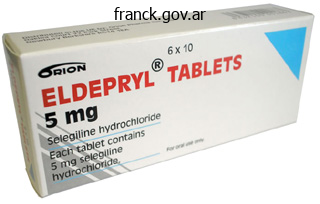
Effective 5mg selegiline
When the muscle shortens, it pulls the proximal phalanx of the second digit medially. The plantar interosseus muscle that originates on the medial side of the fourth metatarsal inserts on the medial aspect of the proximal phalanx of the fourth digit. Thus, when it shortens, it pulls the proximal phalanx of the fourth digit medially. The plantar interosseus muscle that originates on the medial facet of the fifth metacarpal inserts on the medial aspect of the proximal phalanx of the fifth digit, and thus pulls the fifth digit medially when it shortens. The combined movements of the three interossei muscular tissues is to deliver digits three, four, and 5 nearer to digit 2, which is identical as adducting the toes. Antagonists Dorsal interossei (abducts the toes) Implications of Shortened and/or Lengthened/ Weak Muscle Shortened: Limited capacity to abduct the toes is famous. The word brevis informs us that this muscle is shorter than flexor hallucis longus. Location Flexor hallucis brevis is a third-layer intrinsic foot muscle, positioned on the plantar surface of the foot and masking the first metatarsal. Palpation and Massage this muscle could be palpated on the plantar facet of the primary metatarsal. Extensor hallucis longus and brevis (extend the massive toe) Innervation and Arterial Supply Innervation: medial plantar nerve Arterial supply: medial plantar artery Notable Muscle Facts There are two tendons of insertion of flexor hallucis brevis, each of which contains a sesamoid bone. Notable Muscle Facts Adductor hallucis helps to help the transverse arch of the foot. Adductor hallucis is much like adductor pollicis in that each muscles have a transverse head and an indirect head. Location Adductor hallucis is a third-layer intrinsic foot muscle, positioned on the plantar floor of the foot. Implications of Shortened and/or Lengthened/ Weak Muscle Shortened: Inability to abduct the good toe is noted. Because the origin is proximal to the insertion, and the muscle crosses the plantar surface of the large toe, adductor hallucis additionally flexes the massive toe. Brevis indicates that the digiti minimi of the foot is smaller than that of the hand. Location Flexor digiti minimi brevis is a third-layer intrinsic foot muscle, situated on the plantar surface of the foot. Palpation and Massage Flexor digiti minimi brevis could be palpated and massaged by making use of direct pressure or friction to the muscle on the plantar floor of the fifth digit. Origin and Insertion Origin: base of the fifth metatarsal Insertion: base of the proximal phalanx of the fifth digit How to Stretch this Muscle Extend the fifth digit of the foot. Thus, the plantar surface of the proximal phalanx is pulled towards the fifth metatarsal. Location Lumbricals are situated fairly centrally on the plantar surface of the foot. Origin and Insertion Origin: tendon of origin of flexor digitorum longus Insertion: plantar side of the proximal phalanges of digits 2�5 and the extensor expansion, which covers the dorsal floor of the toes Notable Muscle Facts Lumbrical muscles in the hand have the identical actions as the lumbricals of the foot. Plantae refers to the truth that this muscle is positioned on the plantar surface of the foot. Location Quadratus plantae is situated on the proximal or posterior third of the plantar floor of the foot. Implications of Shortened and/or Lengthened/ Weak Muscle Shortened: Tension is felt in the heel area. Lengthened: Reduced capacity to flex the 4 lateral toes is famous, notably when the ankle is dorsiflexed. Origin and Insertion Origin: calcaneus Insertion: tendon of insertion of flexor digitorum longus Palpation and Massage Quadratus plantae could be palpated and massaged by applying friction and direct strain to the plantar surface of the calcaneus. Explanation of Actions By anchoring on the calcaneus and by pulling the tendon of flexor digitorum longus instantly towards the calcaneus, quadratus plantae helps to flex the toes.

Trusted selegiline 5mg
Small osseous remnants of the earlier episternum occurring within the ligaments of the sternoclavicular joint. Either of two massive, vertical grooves on both side of the vertebral column that are occupied by the lungs. Externally and internally visible process that projects laterally from the jugular foramen. It sometimes divides the jugular foramen into a lateral portion for the interior jugular vein and a medial section for nerves. Anthropometric landmark indicating essentially the most prominent point on the external occipital protuberance. Bony ridge occasionally present between the external occipital protuberance and the foramen magnum. Line arching externally from the higher margin of the external occipital protuberance. Outer surface of the occipital bone positioned superior to the exterior occipital protuberance. Cross-shaped bony prominence with the interior occipital protuberance at its middle. Thick bony ridge that occasionally extends from the internal occipital protuberance to the foramen magnum. Prominence that sometimes initiatives from the jugular course of within the direction of the transverse strategy of the atlas. Large opening in the occipital bone for passage of the medulla oblongata, vessels and nerves. Portion of occipital bone that projects superiorly from foramen magnum to sphenoid bone. Part of the basioccipital bone that slopes upwardly from the foramen magnum to the dorsum sellae. Prominence on the inferior floor of the basioccipital bone, for attachment of the pharyngeal raphe. Anatomic variant that types when the higher half of the squama occipitalis is separated by a transverse suture. Passage situated posterior to the occipital condyle, for transmission of a vein from the sigmoid sinus. Passage that originates from the lateral a half of the occipital bone anterior to the foramen magnum and ends exterior, anterior to the occipital condyle. Either of two small protuberances sometimes present, one on both side of the floor of the hypophysial fossa. Longitudinal groove lateral to the body of the sphenoid bone that lodges the internal carotid artery. Pointed process lateral to the doorway of the internal carotid artery into the cranial fossa. Median bony ridge on the anterior surface of the physique of the sphenoid bone that articulates with the perpendicular plate of the ethmoid. Originally paired, concave bony plate which fuses with the body of the sphenoid and varieties a part of the anterior and inferior wall of the sphenoidal sinus and different buildings. Cleft between the greater and lesser wings of the sphenoid for the passage of nerves and veins. Bony ridge between the vertical temporal surface and the horizontally-oriented inferior floor of the larger wing of the sphenoid. Round opening in the nice wing that extends anteriorly into the pterygopalatine fossa. Opening for passage of the mandibular nerve in the medial part of the great wing, positioned in front of the foramen spinosum. Opening often current medial to the foramen ovale for passage of an emissary vein from the cavernous sinus.
Selegiline 5 mg without a prescription
Neurons of the inferior colliculus project to the thalamus, which then sends auditory data to the cerebrum for the acutely aware notion of sound. The superior colliculus is the superior pair and combines sensory details about visual area, auditory area, and somatosensory area. Activity in the superior colliculus is said to orienting the eyes to a sound or touch stimulus. The mind stem includes three areas: the midbrain, the pons, and the medulla Cerebellum the cerebellum, as the name suggests, is the "little brain" and accounts for roughly 10 % of the mass of the mind. The cerebellum is largely answerable for comparing information from the cerebrum with sensory suggestions from the periphery by way of the spinal cord. Descending fibers from the cerebrum have branches that connect to neurons within the pons. Sensory data from the periphery, which enters via spinal or cranial nerves, also tasks to a nucleus within the medulla known as the inferior olive. Fibers from this nucleus enter the cerebellum and are in contrast with the descending instructions from the cerebrum. For instance, if the cerebrum sends a command right down to the spinal cord to provoke strolling, a duplicate of that motor command is shipped to the cerebellum. Sensory suggestions from the muscle tissue and joints, proprioceptive details about the actions of walking, and sensations of balance are despatched to the cerebellum via the inferior olive and then the cerebellum integrates all of that data. The output of the cerebellum is into the midbrain, which then sends a descending input to the spinal wire to correct motor data going to skeletal muscle tissue. It is also answerable for lifting the higher eyelid when the eyes level up, and for pupillary constriction. The trigeminal nerve (V) is answerable for cutaneous sensations of the face and controlling the muscular tissues of mastication. The vagus (X) nerve is answerable for contributing to homeostatic management of the organs of the thoracic and higher belly cavities by way of autonomic neurons. The cranial nerves can be categorised as sensory nerves, motor nerves, or a mixture of each, which means that the axons in these nerves can originate out of sensory ganglia exterior to the cranium or motor nuclei throughout the brain stem. Three of the nerves are solely composed of sensory fibers; 5 are strictly motor; and the remaining 4 are combined nerves that contain each sensory and motor fibers. The trigeminal and facial nerves both concern the face; one is primarily related the sensations and the other primarily associated with the muscle movements. The facial and glossopharyngeal nerves are both liable for conveying gustatory, or taste, sensations in addition to controlling salivary glands. The vagus nerve is concerned in visceral responses to taste, specifically the gag reflex. An important studying outcome for this lesson is to perceive and describe the capabilities of cranial nerves. There are many mnemonics others have created that can quickly be discovered via an internet search. However, one of the simplest ways to bear in mind a mnemonic, is to make your own with personally-relatable information. The anatomical arrangement of the roots of the cranial nerves observed from an inferior view of the mind. Describe the composition of gray and white mater and supply examples of mind buildings made of every. Required materials � None Procedure this activity shall be completed individually or in small groups. Describe and establish the mind meninges: dura mater, arachnoid mater, & pia mater three. Define the following structural options of the brain: gyrus, sulcus, fissure Required materials � None Procedure this activity might be accomplished individually or in small groups. Check Your Understanding Categorize the next terms and supply a one line definition for every of them. For the meninges, additionally rank them from the most superficial layer to the deepest layer.
Order discount selegiline online
Colchicine-There is one small managed trial of colchicine in comparison with placebo for acute gout; everyone taking colchicine developed diarrhoea and vomiting, regularly earlier than pain relief. Severe diarrhoea when immobilized with acute gout may be an unpleasant experience. Traditionally high doses of colchicine are recommended; however, a lower dose of zero. One trial, in a Hong Kong emergency department, in contrast indometacin one hundred fifty mg/day with prednisolone 30 mg per day for 5 days. Among the patients that received indometacin, 5% had a gastrointestinal haemorrhage and 11% had been admitted to hospital for remedy of a critical opposed impact. A second trial, in Dutch primary care, compared naproxen 1 g/day with prednisolone 35 mg per day and located effectiveness to be equivalent and an analogous incidence of opposed results. Clinical expertise helps the utilization of intra-articular steroids, but septic arthritis have to be positively excluded. Intra-articular injections in acute gout may be tough and very painful, particularly in smaller joints. Other treatments-Experience and some controlled trial proof counsel that some non-drug pain-relief modalities corresponding to using ice packs might give additional pain reduction. Desensitizing regimens of allopurinol may be tried in milder circumstances of hypersensitivity. Uricosuric drugs-Uricosuric medication lower serum urate by inhibiting its tubular reabsorption. Uricase drugs-Uricase drugs work by oxidizing uric acid to the extra soluble allantoin. Other drugs-Several other medicine have, coincidentally, been discovered to have urate-lowering results. Intercritical and persistent gout the mainstay of treatment for prevention of recurrent acute gout and persistent gout is decreasing serum urate enough to allow crystals to clear. Patients with two or extra assaults of gout per yr ought to be offered urate-lowering medicine. Starting urate-lowering medicine throughout an assault might delay decision and ought to be prevented as lowering serum urate can trigger acute gout. Medication review-Consider stopping diuretics or some other drugs recognized to enhance serum urate. Lifestyle interventions-Patients ought to be advised to: shed pounds; scale back the amount of meat and fish eaten; cut back alcohol intake and avoid beer; improve intake of low-fat dairy products. Xanthine oxidase inhibitors-These prevent the purine breakdown products xanthine and hypoxanthine being transformed into urate. Allopurinol-Allopurinol reduces serum urate, however its impact on recurrent gout is unclear. Only a minority of sufferers taking the standard dose of 300 mg/day achieve a goal urate of 0. Allopurinol hypersensitivity might occur in up to 2% of sufferers; this Pseudogout Pseudogout, which can be easily confused with gout, is caused by deposition of calcium pyrophosphate crystals. Typically it produces an episodic monoarthritis, but it can even have a scientific image much like osteoarthritis or rheumatoid arthritis. Its prevalence will increase from 3% in folks in their 60s to half of those in their 90s. It may be associated with hypothyroidism, hypercalcaemia, haemochromatosis or hypomagnesaemia. Diagnosis is predicated on figuring out pyrophosphate crystals or chondrocalcinosis seen on X-ray. Other crystal diseases A number of different crystals can produce acute musculoskeletal inflammation. Most widespread are hydroxyapatite crystals, which usually deposit in tendons, periarticular delicate tissue and synovium. Hydroxyapatite deposition may be asymptomatic however can every so often result in important joint destruction. Identifying and correcting an underlying reason for hypophosphataemia or hypercalcaemia might reduce the risk of future attacks. Its identification in joint fluid requires special staining with Alizarin Red dye. Serum uric acid and cardiovascular disease: current developments, and where do they go away us Use of oral prednisolone or naproxen for the treatment of gout arthritis: a doubleblind, randomised equivalence trial.
Discount 5 mg selegiline free shipping
The circulatory system is made up of the cardiovascular system and the lymphatic system. Heart the center is an organ in the chest that pumps blood through the veins and arteries. The coronary arteries carry oxygenatedandnutrient-filledbloodtothemyocardium (heart muscle). An aortogram is an invasive procedure by which a catheter is positioned in the aorta and a distinction material is injected. Arteriosclerosis refers to the thickening, hardening, and lack of elasticity of the arterial walls. Study the following desk for extra terms referring to pathological terms and diagnoses associated to the lymphatics. In more severe issues of the lymphatic system corresponding to most cancers, excision of the affected lymphatic construction may be needed. The name of the record produced by recording the electrical currents of the heart muscle is a. The following diagram illustrates the motion of air into the respiratory tract with the associated constructions. Combining Form alveol/o bronch/o, bronchi/o Meaning alveolus/alveoli bronchus/bronchi Word Association Alveolar ventilation refers to the quantity of gasoline expired from the alveoli. Laryngospasm is the uncontrolled and involuntary muscular contraction of the vocal folds. The medical specialty that offers with diseases involving the respiratory tract is named pulmonology. Methods used to address this problem might embrace the use of the Heimlich maneuver or, in extreme cases, endotracheal intubation. The following table lists some of the most common surgical procedures related to the respiratory system. Tiny air sacs by way of which the trade of oxygen and carbon dioxide takes place are called a. A 75-year-old girl with a left cerebrovascular accident (stroke) is now unable tospeak. An instructor says that this disease has been practically eradicated in developed nations. A 29-year-old girl is making an attempt to break up sputum by using which type of over-thecounter treatment This section will concentrate on medical vocabularies and jargons related to digestion, micturition or urination, and replica. Detailed discussion on these methods could be present in Chapters 9, 10, and eleven of your textbook. Alimentation (alimentum = to nourish) is the term used for the method of giving or receiving diet, whereas metabolism is used to describe all the body processes concerned in sustaining life. Nutrient Classification carbohydrates Associated Enzyme/s lactase (breaks down lactose) amylase (breaks down starch) proteins protease proteinase fat lipase Word Parts lact + ase amyl + ase prote + ase protein + ase lip + ase Study the following word parts related to digestion and vitamin: Word Part -ation bil/i, chol/e cirrh/o deglycos/o -orexia -pepsia vag/o Meaning action or process bile orange-yellow down, from, reversing, or eradicating sugar urge for food digestion vagus nerve Word Association Defecation is the process of passing out stool or feces via the anus. The vasovagal syncope is the sudden loss of consciousness caused by affectation of the vagus nerve. Alimentary Tract the alimentary tract, otherwise often recognized as the digestive tract, begins from the mouth and continues right down to the anus. Upper Gastrointestinal Tract Digestive Organs lips tooth gums tongue mouth esophagus abdomen Lower Gastrointestinal Tract Word Part cheil/o dent/i, dent/o, odont/o gingiv/o gloss/o, lingu/o or/o, stomat/o esophag/o gastr/o Word Association cheilosis dentistry gingivitis glossitis oropharynx esophagitis gastroenterologist intestines duodenum jejunum ileum colon or massive gut appendix cecum sigmoid colon anus or rectum rectum anus intestin/o, enter/o duoden/o jejun/o ile/o col/o, colon/o append/o, appendic/o cec/o sigmoid/o proct/o rect/o an/o intestinal, enteritis duodenal jejunostomy ileostomy colonoscopy appendectomy ileocecal sigmoidectomy proctologist rectal anal Accessory Organs of Digestion Proper digestion and absorption of vitamins is aided by the secretion of substances by the accessory organs of digestion. The following desk lists the word components related to the accessory organs of digestion. Word Part cholecyst/o choledocho/o hepat/o pancreat/o sial/o Meaning gallbladder widespread bile duct liver pancreas salivary gland Word Association Cholecystectomy is the surgical removal of the gallbladder. The presence of gallstones within the widespread bile duct is referred to as choledocholithiasis. Lack of insulin or insulin resistance ends in hyper + glycemia (hyper = elevated, glyc/o = sugar, emia = blood).
Generic 5 mg selegiline visa
Identify typical organelles present in an animal cell and summarize major capabilities of every Required Materials � None Procedure this exercise will be completed individually or in small groups. Name one major structural similarity and one main structural distinction between cells and organelles. Complete the table under to summarize the most important operate of each given organelle in one sentence or phrase. Summarize the main events for each a part of the cell cycle (interphase and mitosis) Required Materials � None Procedure this activity might be accomplished individually or in small groups. Match the cell cycle section to the major mobile occasions by completing desk under with the provided cell cycle phases. Label every of the next drawings of cells in numerous stages of mitosis and cytokinesis. Define histology Describe a tissue as part of the anatomical organization of the physique List and define the four main tissue varieties List and describe all the sub-types for epithelial and connective tissues Identify each of the epithelial and connective tissue varieties and sub-types via a picture or diagram List an example of a location for each tissue sub-type Summarize the function for every tissue sub-type Describe the structural modifications that contribute to particular perform for each tissue sort & sub-type Identify the most important structures of the cells in the tissues noticed � nuclei, cell membrane/cilia, extracellular material (fibers and matrix), specialised arrangements of cells the time period tissue is used to describe a gaggle of cells found collectively in the physique. Each of these tissue varieties is characterized by specific capabilities that contribute to the overall health and maintenance of the human body. Epithelial tissue, also referred to as epithelium, refers to the sheets of cells that cover exterior surfaces of the physique, strains internal cavities and passageways, and types certain glands. Connective tissue, as its name implies, binds the cells and organs of the physique collectively and capabilities within the protection, support, and integration of all parts of the body. Muscle tissue is excitable, responding to stimulation and contracting to provide motion. Nervous tissue is also excitable, allowing the propagation of electrochemical alerts in the form of nerve impulses that permit communication between completely different areas of the physique. Just as knowing the structure and performance of cells helps you in your study of tissues, data of tissues will allow you to perceive how organs function. Epithelial and connective tissues shall be covered on this lesson whereas muscle and nervous tissues shall be lined within the next lesson. Epithelial Tissue Most epithelial tissues are essentially large sheets of cells covering all of the surfaces of the physique exposed to the surface world and lining the surface of organs. Other areas embody the airways, the digestive tract, in addition to the urinary and reproductive techniques, all of that are lined by an epithelium. Epithelial tissue is very mobile, with little or no extracellular material current between cells. All epithelia share some important structural and practical options that outline epithelial tissue: � Polarity - Epithelial cells exhibit polarity with differences in structure and function between the exposed or apical-facing floor of the cell and the basal floor near the underlying body structures. Certain organelles are segregated to the basal sides, whereas different organelles and extensions, such as cilia, when current, are on the apical surface. Supported by connective tissue � the basement membrane, a combination of the basal and reticular laminas, connects epithelial tissues underlying connective tissues to provide structural and practical assist. The epithelial layer secretes the basal lamina, a combination of glycoproteins and collagen, which connects to a reticular lamina secreted by the underlying connective tissue. All supplies that enter or depart the epithelial layer should come by diffusion or absorption from underlying tissues or the floor. Innervated � Most epithelial tissues are provided by nervous tissue to enable interaction with the exterior surroundings. Regeneration - Many epithelial tissues are capable of quickly replacing damaged and lifeless cells. Sloughing off of broken or useless cells is a attribute of surface epithelium and allows our airways and digestive tracts to quickly replace broken cells with new cells. The cells of an epithelium act as gatekeepers of the physique controlling permeability and allowing selective transfer of supplies throughout a physical barrier. Some epithelia typically embrace structural features that enable the selective transport of molecules and ions across their cell membranes. Many epithelial cells are additionally capable of secretion and release mucous and specific chemical compounds onto their apical surfaces. Cells lining the respiratory tract secrete mucous that traps incoming microorganisms and particles. Similarly, the variety of cell layers in the tissue could be one-where every cell rests on the basal lamina-which is a straightforward epithelium, or more than one, which is a stratified epithelium and only the basal layer of cells rests on the basal lamina. Pseudostratified (pseudo- = "false") describes tissue with a single layer of irregularly formed cells that give the looks of multiple layer.
Buy selegiline 5mg
Examples embrace the cranial (skull) bones, the scapulae (shoulder blades), the sternum (breastbone), and the ribs. Flat bones function factors of attachment for muscular tissues and infrequently shield inside organs. These bones tend to have more advanced shapes, just like the vertebrae that assist the spinal wire and protect it from compressive forces. Many facial bones, particularly the ones containing sinuses, are categorised as irregular bones. Sesamoid Bones A sesamoid bone is a small, round bone that, as the name suggests, is shaped like a sesame seed. These bones type in tendons (the sheaths of tissue that join bones to muscles) the place a nice deal of strain is generated in a joint. Sesamoid bones vary in quantity and placement from individual to particular person but are typically present in tendons related to the ft, arms, and knees. The patellae (singular = patella) are the only sesamoid bones found in widespread with each individual. The diaphysis is the tubular shaft that runs between the proximal and distal ends of the bone which are known as epiphyses. The hole region in the diaphysis is recognized as the medullary cavity, which is crammed with yellow marrow in adults. The medullary cavity has a fragile membranous lining known as the endosteum (end- = "inside"; oste- = "bone"), the place bone development, restore, and remodeling happen. The outer floor of the bone is roofed with a fibrous membrane called the periosteum (peri- = "around" or "surrounding"). The periosteum accommodates blood vessels, nerves, and lymphatic vessels that nourish compact bone. In this region, the epiphyses are covered with articular cartilage, a skinny layer of cartilage that reduces friction and acts as a shock absorber. The two layers of compact bone and the inside spongy bone work collectively to defend the internal organs. If the outer layer of a cranial bone fractures, the brain is still protected by the intact inside layer. This cross-section of a flat bone reveals the spongy bone (diplo�) lined on both facet by a layer of compact bone. Bone Features the surface features of bones differ considerably, depending on the function and site in the body. There are three common classes of bone markings: (1) articulations, (2) projections, and (3) holes. These surfaces tend to conform to one another, corresponding to one being rounded and the other cupped, to facilitate the function of the articulation. In basic, their size and form is an indication of the forces exerted by way of the attachment to the bone. A hole is a gap or groove within the bone that permits blood vessels and nerves to enter the bone. As with the other markings, their size and shape mirror the scale of the vessels and nerves that penetrate the bone at these factors. These salt crystals form when calcium phosphate and calcium carbonate mix to create hydroxyapatite, which contains different inorganic salts like magnesium hydroxide, fluoride, and sulfate as it crystallizes, or calcifies, on the collagen fibers. Immature osteogenic cells are found in the deep layers of the periosteum and the marrow. Osteoblasts are liable for forming new bone and are discovered within the growing parts of bone, together with the periosteum and endosteum. As the secreted matrix surrounding the osteoblast calcifies, the osteoblast become trapped within it; in consequence, it adjustments in structure and turns into an osteocyte, the first cell of mature bone and the most typical type of bone cell. Each osteocyte is located in a space known as a lacuna and is surrounded by bone tissue. Osteocytes preserve the mineral focus of the matrix through the secretion of enzymes. They can communicate with each other and receive nutrients via lengthy cytoplasmic processes that reach through canaliculi (singular = canaliculus), channels within the bone matrix. The dynamic nature of bone means that new tissue is constantly shaped, and old, injured, or unnecessary bone is dissolved for restore or for calcium launch.
References
- Wolff JA, Kulovich S, Yu A, et al. The effectiveness of benzoate in the management of seizures in nonketotic hyperglycinemia. Am J Dis Child 1986;140:596.
- Hall MJ, Forman AD, Pilarski R, et al: Gene panel testing for inherited cancer risk, J Natl Compr Canc Netw 12:1339n1346, 2014.
- Matern, U., Koneczny, S. Safety, hazards and ergonomics in the operating room. Surg Endosc 2007;21:1965-1969.
- Perkes IE, Menon DK, Nott MT, Baguley IJ. Paroxysmal sympathetic hyperactivity after acquired brain injury: a review of diagnostic criteria. Brain Inj. 2011;25(10):925-932.
- Edlund C, Peeker R, Fall M: Clam ileocystoplasty: successful treatment of severe bladder overactivity, Scand J Urol Nephrol 35:190n195, 2001.
- Dalakas MC, Houff SA, Engel WK, Madden DL, Sever JL. CSF 'monoclonal' bands in chronic relapsing polyneuropathy. Neurology. 1980;30:864-867.

