Coreg dosages: 25 mg, 12.5 mg, 6.25 mg
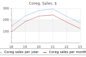
Coreg packs: 10 pills, 20 pills, 30 pills, 60 pills, 90 pills, 120 pills, 180 pills, 270 pills, 360 pills
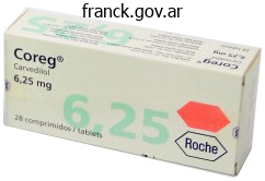
Coreg 25mg generic
The end result of sphincter restore is affected by factors similar to irritable bowel syndrome, rectal hypersensitivity, inflammatory bowel disease, diabetes mellitus, bowel transit, and bowel consistency. The patient should be made aware of attainable problems such as infection, wound breakdown, fistula formation, and failure of surgical procedure. Intraoperative the operation is often performed beneath common anesthesia with the affected person in lithotomy, though some surgeons choose to use the inclined jackknife place. The incision is then deepened into the ischioanal fossa and utilizing Metzenbaum scissors the sphincter muscle tissue are identified and mobilized. In the acute state of affairs, for instance, immediately after childbirth, the freshly torn muscle ends can be recognized and approximated by an skilled obstetrician. However, current publications have reported removed from perfect results with the overlapping technique as some sufferers developed new evacuation disorders. For extra support some surgeons carry out a levatorplasty by inserting two interrupted sutures using 2-0 delayed suture material. Finally a easy wound closure of the pores and skin is carried out with the midportion left open for drainage. Laxatives should be prescribed to maintain the stools gentle as passage of stool will be painful. Box 35-2 Caution Points Time restore of anal sphincter lacerations when wholesome granulation tissue is present. Overlapping anal sphincteroplasty procedures are challenging when the exterior anal sphincter defect is bigger than 90�. Sutures have been positioned by way of the 2 dissected flaps of the exterior anal sphincter. In patients with a extreme trauma to the perineum aside from after supply, a proximal colostomy is commonly constructed to avoid septic complications and to facilitate nursing administration. However, stoma-related problems are reported in additional than 50% of these patients. Depending on the extent of injury and the associated trauma, reconstructive surgical procedure could also be deferred. A third- or fourth-degree perineal tear have to be repaired immediately, though defects after repair are reported in as a lot as 85% and about 40% of those women ultimately develop incontinence. They discovered equal results with acceptable long-term useful consequence in each groups. In about 62%, a repeat restore can be anticipated to achieve success, although patients who had undergone more than two previous repairs appear to have poorer medical results. In the dorsal lithotomy place, a U-shaped incision gives sufficient access to the exterior anal sphincter defect. Use fine absorbable suture on the anal mucosa and inside anal sphincter; use delayed absorbable suture on the exterior anal sphincter. Perineal hygiene using a peri-bottle, sitz baths, and/or a handheld bathe head is necessary during the postoperative period to decrease the chance of wound infection. No affected person was fully continent (stool and gas), with 52% of patients nonetheless wearing a pad and 66% of sufferers reporting lifestyle restriction. General questionnaires have a long history of use with established reliability, validity, and population norms. It is of utmost significance that after successful repair, sufferers proceed to perform pelvic flooring exercises and biofeedback. Box 35-4 Complications and Morbidity Long-term full fecal continence following anal sphincteroplasty is uncommon, with most sufferers nonetheless requiring pad use. Preoperative counseling should stress that though most sufferers will enhance after sphincteroplasty surgical procedure, many have residual symptoms, and a few might develop de novo evacuation problems. With scalpel or scissors an higher pores and skin flap is dissected free towards the anal canal and lifted to obtain adequate exposure of, and access to , the intersphincteric aircraft. The dissection should begin laterally as this is the best place to develop the intersphincteric aircraft at an early stage. It is important that the surgeon follows the natural slightly posterior course of the intersphincteric groove as it nears the level of the pelvic ground. The dissection should proceed above the puborectalis muscle sling and above the levators to open up the postrectal area and in addition deepened laterally to a excessive degree so that the ischial spines may be simply palpated. As the surgeon enters the supralevator plane, the fascia of Waldeyer is identified after which incised transversely, after which the dissection is almost complete. The rectum is displaced anteriorly with a retractor and the primary polypropylene stitch of the repair is positioned on either side of the levator muscle as excessive and anteriorly as possible.
Proven coreg 12.5mg
Repeated forceful flexion and extension of the wrist strain the attachment of the widespread extensor tendon, producing irritation of the periosteum of the lateral epicondyle (lateral epicondylitis). Sudden extreme pressure on a protracted extensor tendon may avulse part of its attachment to the phalanx. This deformity results from the distal interphalangeal joint abruptly being forced into excessive flexion (hyperflexion) when the tendon is making an attempt to lengthen the distal phalanx-for instance, when a baseball is miscaught (hyperflexing it) or the finger is jammed right into a base pad. These actions avulse the attachment of the tendon from the base of the distal phalanx. The skeleton of the hand consists of carpals within the wrist, metacarpals in the hand proper, and phalanges within the fingers. The metacarpals and phalanges are numbered from 1 to 5, beginning with the thumb and ending with the little finger. Fascia of Palm the fascia of the palm is continuous with the antebrachial fascia and the fascia of the dorsum of the hand. The palmar aponeurosis, a strong, well-defined a half of the deep fascia of the palm, covers the gentle tissues and overlies the lengthy flexor tendons. The proximal end or apex of the triangular palmar aponeurosis is continuous with the flexor retinaculum and the palmaris longus tendon. The fibrous digital sheaths are ligamentous tubes that enclose the flexor tendon(s) and the synovial sheaths that surround them as they pass alongside the palmar side of their respective digit. A medial fibrous septum extends deeply from the medial border of the palmar aponeurosis to the 5th metacarpal. Similarly, a lateral fibrous septum extends deeply from the lateral border of the palmar aponeurosis to the third metacarpal. Lateral to the septum is the lateral or thenar compartment containing the thenar muscles. The deepest muscular airplane of the palm is the adductor compartment containing the adductor pollicis. These spaces are bounded by fibrous septa passing from the sides of the palmar aponeurosis to the metacarpals. Between the two areas is the especially strong lateral fibrous septum, which is hooked up to the 3rd metacarpal. The midpalmar space is steady with the anterior compartment of the forearm by way of the carpal tunnel. Dissection of muscular tissues, common flexor sheath, and synovial sheaths of digits 1 to 5 (purple). The high degree of freedom of movements of the thumb results from the first metacarpal being impartial, with mobile joints at each ends. The complicated movement of opposition begins with the thumb within the extended position and initially entails abduction and medial rotation of the 1st metacarpal ("cupping" of the palm) produced by the motion of the opponens pollicis after which flexion on the metacarpophalangeal joint. It wrinkles the skin of the hypothenar eminence, deepening the hollow of the palm, thereby aiding the palmar grip. It is hooked up proximally to the medial border of the palmar aponeurosis and to the pores and skin on the medial border of the hand. The four slender lumbrical muscular tissues were named due to their worm-like appearance (L. The 4 dorsal interossei abduct the fingers, and the three palmar interossei adduct them. Acting together, the dorsal and palmar interossei and lumbricals produce flexion on the metacarpophalangeal joints and extension of the interphalangeal joints (Z-movement). The tendons enter the central compartment of the hand and fan out to enter the respective digital synovial sheaths. The common flexor and digital sheaths allow the tendons to slide freely past each other throughout actions of the fingers. The sheaths prolong from the heads of the metacarpals to the bases of the distal phalanges. These sheaths forestall the tendons from pulling away from the digits (bow-stringing). The anular and cruciform elements (ligaments) of the fibrous sheath (often referred to clinically as "pulleys") are thickened reinforcements of those sheaths. The ulnar artery enters the hand anterior to the flexor retinaculum between the pisiform and the hook of hamate by way of the ulnar canal (Guyon canal).
Order 25mg coreg visa
Pelvic flooring bodily remedy with biofeedback might enhance orgasmic disorders related to weak musculature. Hormonal alternative with estrogens or androgens can enhance orgasmic dysfunction in patients with deficiency. If possible, any etiology for pain, such as vulvovaginal atrophy or vestibulodynia, should be addressed first. This includes keeping the world clean and dry, sporting breathable underwear (eg, cotton), and avoidance of harsh detergent soaps, bubble baths, douching, and depilatories. For vestibulodynia, medical therapies embrace off-label use of antidepressants, including amitryptyline, nortriptyline, or duloxetine, or nerve-modulating brokers similar to gabapentin or pregabalin, though sedation is a standard purpose for discontinuation of those drugs. Treatment of levator hypertonicity involves bodily remedy, usually with dilators for desensitization. Other poorly studied remedies include set off level injections, compounded diazepam, and/or botulinum toxin injections. Overall, analysis and therapy of vulvar and pelvic ache is complex and entails multiple modalities, which are beyond the scope of this evaluation. The gadget offers a mild adjustable vacuum suction and a low-level vibratory sensation. It is designed to be used three or more times per week for approximately five minutes at a time. Use of the Eros Therapy has been proven to increase blood flow to the clitoral space in addition to to the vagina and pelvis. Surgical remedy of pelvic ground problems, similar to pelvic organ prolapse, urinary incontinence, or fecal incontinence, may enhance sexual operate. Generally, the vaginal epithelium is undermined and the distal vagina superior to present a more generous platform for penetrative intercourse. The minor vestibular glands together with Skene glands or periurethral glands ought to be excised in the occasion that they have been painful throughout cotton swab testing. In a current report of 104 women who underwent vestibulectomy, 93% of women reported satisfaction with the process and 89% were capable of have intercourse. In some situations, vaginal looseness or laxity might occur following childbirth, and lead to alterations in sexual function. A small pilot examine has reported on the tolerability and success of nonsurgical tightening with radio-frequency thermal therapy,95 but surgical correction with a perineoplasty is the mainstay of treatment. The objective of surgical correction of laxity is to reduce the genital hiatus to enhance penetrative sensation. In a report of 53 sufferers, 94% felt that the vagina was tighter,ninety six though long-term outcomes and psychosocial and safety information are lacking. Experts advocate basic or regional anesthesia to keep away from posttraumatic stress dysfunction which could be experienced with native anesthesia. After excision of a triangular portion of perineal skin, the posterior vaginal epithelium is mobilized sharply and superior to cover the perineal pores and skin defect. Care must be taken to palpate the clitoral area prior to incision to avoid further damage, if current. Widespread use of synthetic mesh for pelvic flooring problems has led to explicit issues similar to mesh exposure/erosion, in addition to pain alongside mesh attachments during intercourse. Removing mesh that produces uncomfortable sensations throughout intercourse, similar to tugging or pulling, could additionally be necessary if conservative therapies have failed. Recent media consideration to male sexual dysfunction has led to growing feminine affected person awareness and a desire to search assist from their doctor. A collaborative and complete analysis, patient and associate education, and habits modification, adopted by individualized pharmacotherapy and/or surgical management in choose sufferers, should be the usual management of ladies with sexual dysfunction. Report of the international consensus growth convention on feminine sexual dysfunction: definitions and classifications. Female sexual response: the position of drugs within the management of sexual dysfunction. A potential research inspecting the anatomic distribution of nerve density in the human vagina. Magnetic resonance imaging anatomy of the feminine genitalia in premenopausal and postmenopausal ladies.
Buy cheap coreg 6.25mg line
The needle pierces the thin cortical bone and enters the vascular trabecular (spongy) bone. Sternal biopsy is often used to obtain specimens of bone marrow for transplantation and for detection of metastatic cancer. Median Sternotomy To gain entry to the thoracic cavity for surgical procedures-on the heart and nice vessels, for example- the sternum is divided ("break up") in the median airplane and retracted (spread apart). After surgery, the halves of the sternum are reunited and held along with wire sutures. Rib spreader Parietal pleura (cut) the posterolateral elements of the 5th�7th intercostal spaces are necessary websites for posterior thoracotomy incisions. This elevates and laterally rotates the inferior angle of scapula, allowing access as high as the 4th intercostal house. Surgeons use an H-shaped incision to incise the superficial side of the periosteum that ensheaths the rib, strip the periosteum from the rib, after which take away a wide segment of the rib to gain higher access, as may be required to enter the thoracic cavity and remove a lung (pneumonectomy), for example. After the operation, the lacking pieces of ribs regenerate from the intact periosteum, though imperfectly. Dislocation of Ribs A rib dislocation (slipping rib syndrome) or dislocation of a sternocostal joint is the displacement of a costal cartilage from the sternum. Rib dislocations are common in physique contact sports activities, and attainable problems are strain on or harm to nearby nerves, vessels, and muscles. A rib separation refers to dislocation of a costochondral junction between the rib and its costal cartilage. In separations of the 3rd�10th ribs, tearing of the perichondrium and periosteum often happens. As a outcome, the rib might move superiorly, overriding the rib above and inflicting pain. Paralysis of Diaphragm One can detect paralysis of the diaphragm radiographically by noting its paradoxical motion. Instead of descending on inspiration, the paralyzed dome is pushed superiorly by the belly viscera which are being compressed by the energetic side. Hence varied kinds of thoracic outlet syndromes exist, such because the costoclavicular syndrome-pallor and coldness of the skin of the upper limb and diminished radial pulse resulting from compression of the subclavian artery between the clavicle and the 1st rib, particularly when the angle between the neck and the shoulder is increased. The higher a half of the determine demonstrates the fat lobules and suspensory ligaments; the center half, the alveoli of the breast with resting (nonlactating) lobules of the mammary gland; and the lower half, lactating lobules of the mammary gland. The contour and quantity of the breasts are produced by subcutaneous fats except throughout being pregnant, when the mammary glands enlarge and new glandular tissue types. During puberty (8 to 15 years of age), the female breasts normally develop because of glandular growth and elevated fat deposition. The roughly round base of the feminine breast extends transversely from the lateral border of the sternum to the anterior axillary line and vertically from the 2nd to 6th ribs. A small a part of the breast might lengthen alongside the inferolateral fringe of the pectoralis major muscle toward the axillary fossa, forming an axillary process or tail (of Spence). Between the breast and the deep pectoral fascia is a unfastened connective tissue aircraft or potential space-the retromammary space (bursa). This plane, containing a small quantity of fats, allows the breast a point of movement on the deep pectoral fascia. The mammary glands are firmly connected to the dermis of the overlying pores and skin by the suspensory ligaments (of Cooper). At the best prominence of the breast is the nipple, surrounded by a round pigmented space (the areola). The breast incorporates 15 to 20 lobules of glandular tissue, which constitute the parenchyma of the mammary gland. Each lobule is drained by a lactiferous duct, which opens independently on the nipple. Lateral mammary branches Lateral mammary branches of lateral cutaneous branches of posterior intercostal aa.
6.25mg coreg for sale
Cyclosporine A and pentosan polysulfate sodium for the remedy of interstitial cystitis: a randomized comparative examine. The twin serotonin and noradrenaline reuptake inhibitor duloxetine for the treatment of interstitial cystitis: outcomes of an observational study. Intravesical bacillus Calmette�Guerin and dimethyl sulfoxide for treatment of traditional and non-ulcer interstitial cystitis: a potential, randomized double-blind research. Intravesical resiniferatoxin for the treatment of interstitial cystitis: a randomized, double-blind, placebo controlled trial. A randomized controlled trial of intravesical bacillus Calmette�Guerin for therapy refractory interstitial cystitis. The efficacy of intravesical Tice strain bacillus Calmette�Guerin within the treatment of interstitial cystitis: a double-blind, potential, placebo managed trial. A randomized, double-blind crossover trial of sertraline in women with continual pelvic ache. Chronic pelvic ache handled with gabapentin and amitriptyline: a randomized managed pilot study. Mesendieck somatocognitive remedy as remedy strategy to chronic pelvic pain: outcomes of a randomized controlled intervention examine. Flashlamp excited dye laser therapy of idiopathic vulvodynia is safe and efficacious. Are patient symptoms predictive of the diagnostic and/or therapeutic worth of hydrodistention Changes in urine markers and signs after bladder distention for interstitial cystitis. A prospective, singleblind, randomized crossover trial of sacral vs pudendal nerve stimulation for interstitial cystitis. A randomized clinical trial on the good thing about adhesiolysis in patients with intraperitoneal adhesions and persistent pelvic pain. They have an result on 50% of adult ladies in their lifetime and 25% to 30% may have a recurrent an infection. Cystitis and pyelonephritis are inflammations of the bladder and renal parenchyma, respectively, and are most frequently, however not all the time, brought on by microbial invasion (usually by bacteria) (Table 17-1). Most infections are sporadic, happen in sufferers without recognized comorbidities or anatomical abnormalities of the urinary tract, and involve the decrease urinary tract. Key Points � Asymptomatic bacteriuria is defined as the presence of micro organism without related irritation within the urinary tract. A number of sexually transmitted organisms corresponding to Chlamydia trachomatis and herpes can colonize the urinary tract causing symptoms of cystitis. Foreign bodies such as urinary catheters are a significant threat factor for an infection through mechanisms that embody urethral trauma, compromise of local immunity, and by offering protected niche(s) for microbial proliferation and a surface for biofilm production. Bacterial Virulence Factors the ability of micro organism to adhere to uroepithelial cells is a prerequisite for an infection to happen. This adherence reduces the chance of the micro organism being cleared from the urinary tract throughout voiding. These adhesins connect to complementary buildings on the uroepithelial cell wall and act not only to promote an infection but additionally to help promote growth and toxin production. Other virulence components that may facilitate infection are particular to every pathogen. These are inclined to originate from the gastrointestinal tract and ascend into the genitourinary system. Escherichia coli is the most common organism and accounts for up to 70% of community-acquired infections. The the rest of bacterial infections are predominantly brought on by Staphylococcus saprophyticus, and quite lots of gram-negative rods within the genus enterobacteriaceae. This reservoir can then serve as a pool of bacteria that may reinitiate an infection. The vaginal flora is predominantly lactobacilli and this maintains an acidic pH in the vagina.
12.5 mg coreg fast delivery
Treating signs of female sexual arousal dysfunction with the Eros-clitoral therapy gadget. Clitoral therapy system for treatment of sexual dysfunction in irradiated cervical cancer sufferers. Surgical treatment of vulvar vestibulitis syndrome: outcome evaluation derived from a postoperative questionnaire. Central wedge nymphectomy with a 90-degree Z-plasty for aesthetic reduction of the labia minora. Radiofrequency remedy of vaginal laxity after vaginal delivery: nonsurgical vaginal tightening. Defibulation to treat female genital cutting: impact on symptoms and sexual function. Perineoplasty for the treatment of introital stenosis associated to vulvar lichen sclerosus. One of the earliest "pessaries" used was placement of half a pomegranate within the vagina, as described by a Greek physician referred to as Polybus. It was only within the sixteenth century that a device was made specifically to be used as a pessary, as opposed to using naturally occurring objects. Since the 20th century, appreciable refinements have been made of existing pessaries. At present, pessaries are generally created from inert plastic or silicone and can be used in sufferers allergic to latex. No clear consensus emerged regarding the sort of pessary used or their indications for use by these surgeons. Furthermore, a case�control study comparing girls who selected pessaries with those who underwent surgery one year after their respective remedy found no difference in prolapse symptoms, bladder, bowel, or sexual function between groups. Pelvic Organ Prolapse Pelvic organ prolapse is the most common indication for pessary use. In a potential cohort examine of 68 girls, Robert and Mainprize18 found that only 16% continued pessary use at one year with a pattern of improved continuation rates in younger patients (41 years vs 52 years) and in those without previous surgery, suggesting this to be a viable alternative choice in this group of sufferers. Pessaries can also be used as a diagnostic device to unmask occult stress urinary incontinence to consider if a concomitant anti-incontinence procedure is necessary at the time of prolapse surgery. Women who demonstrated preoperative stress incontinence throughout prolapse reduction have been extra prone to report postoperative stress incontinence, regardless of concomitant colposuspension. A latest examine, using the ring pessary to unmask occult urinary incontinence on the time of video cystourethrography, confirmed that the pessary test has poor sensitivity (67%) however high specificity (93%) in predicting postoperative stress urinary incontinence following prolapse restore. The optimistic predictive worth of the pessary for postoperative incontinence was low (40%) but had wonderful negative predictive worth (98%). Vaginal wind could also be because of the opening of the potential house of the vagina while a woman is at rest, leading to air trapping in the vagina as the introitus closes with motion. Insertion of a pessary21,22 prevents closure of the vagina and introitus thereby stopping trapping and subsequent expulsion of the air. Neonatal Prolapse Pessaries have been used efficiently as a temporary measure to appropriate neonatal prolapse, mainly seen in affiliation with neural tube defects such as spina bifida. Prolapse in Pregnancy Pessaries have been used efficiently as momentary measures for remedy of prolapse or urinary incontinence during pregnancy to afford symptom reduction till delivery. It is generally accepted that the ring pessary should be the primary pessary tried because of ease of insertion and removal, and, if this fails, other pessaries can be utilized. In this fashion the pessary supplies a supportive shelf for the descending pelvic organs. Folding the pessary reduces its dimension and allows for easy introduction via the vaginal introitus. Its form prevents assortment of vaginal discharge and girls can proceed to have interaction in vaginal intercourse with the pessary in situ. The base is circular with a concave floor on the underside and a convex surface on top, to which is hooked up a stem of varying lengths ending in a knob. The round base has regular holes and the stem has a central hole column to enable drainage of secretions. The concave surface is positioned in opposition to the vaginal cuff or the cervix and the stem lies alongside the axis of the vagina with the knob contained in the introitus. The pessary should be positioned with the convexity of the curved bars towards the vaginal wall relying on whether the prolapse is anterior or posterior.
Purchase coreg
When the joint arm is abducted 180 degrees, 60 levels happens by rotation of the scapula, and 120 degrees by rotation of the humerus at shoulder joint. Lateral view of glenoid cavity and related constructions following disarticulation of humerus. The glenoid cavity accepts little more than a 3rd of the humeral head, which is held in the cavity by the tonus of the musculotendinous rotator cuff (supraspinatus, infraspinatus, teres minor, and subscapularis). The loose fibrous layer of the joint capsule surrounds the glenohumeral joint and is connected medially to the margin of the glenoid cavity and laterally to the anatomical neck of the humerus. Superiorly, the fibrous layer encloses the proximal attachment of the long head of biceps brachii to the supraglenoid tubercle of the scapula within the joint. The inferior part of the joint capsule, the one half not reinforced by the rotator cuff muscles, is its weakest area. The synovial membrane additionally types a tubular sheath for the tendon of the long head of the biceps brachii. The glenohumeral ligaments are intrinsic ligaments which are part of the fibrous layer of the capsule. The transverse humeral ligament is a broad fibrous band that runs from the greater to the lesser tubercle, bridging over the intertubercular sulcus (groove) and converting the sulcus right into a canal for the tendon of the long head of biceps brachii and its synovial sheath. The coraco-acromial arch overlies the pinnacle of the humerus, preventing its superior displacement from the glenoid cavity. When the arm is kidnapped with out rotation, the higher tubercle contacts the coraco-acromial arch, stopping additional abduction. If the arm is then laterally rotated a hundred and eighty levels, the tubercles are rotated posteriorly and extra articular surface turns into out there to proceed elevation. Stiffening or fixation of the joints of the pectoral girdle (ankylosis) results in a way more restricted range of movement, even when the glenohumeral joint is normal. The muscle tissue moving the joint are the axio-appendicular muscle tissue, which may act indirectly on the joint. Other muscular tissues serve the glenohumeral joint as shunt muscle tissue, performing to resist dislocation with out producing movement at the joint, or maintain the pinnacle of the humerus within the glenoid cavity. The suprascapular, axillary, and lateral pectoral nerves supply the glenohumeral joint (Table 6. This freedom results from the laxity of its joint capsule and the configuration of the spherical humeral head and shallow glenoid cavity. The Several bursae containing capillary films of synovial fluid are situated near the joint the place tendons rub against bone, ligaments, or different tendons and where pores and skin moves over a bony prominence. Some bursae communicate with the joint cavity; therefore, opening a bursa might mean getting into the cavity of the joint. Thus, it facilitates movement of the supraspinatus tendon beneath the coraco-acromial arch and of the deltoid over the joint capsule and the higher tubercle of the humerus. The subscapular bursa is located between the tendon of the subscapularis and the neck of the scapula. This bursa protects the tendon the place it passes inferior to the root of the coracoid course of and over the neck of the scapula. Clinical Box Rotator Cuff Injuries the musculotendinous rotator cuff is commonly injured throughout repetitive use of the higher limb above the horizontal. If the arm is passively kidnapped 15 levels or extra, the person can normally maintain or proceed the abduction using the deltoid. Calcific Supraspinatus Tendinitis Inflammation and calcification of the subacromial bursa lead to ache, tenderness, and limitation of movement of the glenohumeral joint. Deposition of calcium in the supraspinatus tendon might irritate the overlying subacromial bursa, producing an inflammatory response, subacromial bursitis. As lengthy as the glenohumeral joint is adducted, no pain often results as a result of in this position, the painful lesion is away from the inferior floor of the acromion. In most individuals, the ache occurs during 50�130 degrees of abduction (painful arc syndrome) as a end result of during this arc, the supraspinatus tendon is in intimate contact with the inferior floor of the acromion.
Discount coreg 12.5mg line
Chronic straining generally results in rectoceles in patients with a spastic pelvic flooring. In this group of patients, performing rectocele repair without addressing their levator ani dysfunction is unlikely to resolve their defecatory signs. Thus, defecography can influence the administration of sufferers with constipation and rectocele by elucidating the underlying dysfunction that resulted in rectocele formation. The proton can transition between the two power states by the absorption or release of a photon. When radio waves are utilized to a physique within a magnetic subject, some of the Rectal Prolapse Rectal prolapse happens when a portion of the rectal mucosa protrudes beyond the anal verge, resulting in irritation, ulceration, and bleeding. It is assumed to be preceded by rectal intussusception, an anterior or annular infolding of the rectum inside itself that originates 6 to 8 cm proximal to the anus but can progress to complete eversion of the rectum. When the radio wave signal is discontinued, some of these highenergy protons will return to the low-energy state, inflicting photons to be launched. The fee of release of the photons is a reflection of intrinsic tissue properties. The pulse sequence with which the radio waves are delivered could be manipulated to intensify variations within a relatively homogeneous tissue or organ. On a T2-weighted scan-preferred for imaging of gynecologic organs and the pelvic floor-fat-containing tissues appear dark, whereas tissues with a high fluid or water content material seem brilliant, making it possible to visualize tissue edema and demarcate normal tissue from broken tissue. It is especially suited for imaging of gynecologic organs and the pelvic flooring as a result of it allows visualization of small differences in tissue properties, versus computed tomography, whose strength lies in delineating the borders of one tissue from another. Axial and sagittal reconstructions are useful for identifying levator avulsion from the pubic bone in addition to levator asymmetry, which could be a sign of direct muscle trauma or unilateral pudendal neuropathy. The excessive decision afforded by these pictures comes on the value of restricted penetration, providing very limited visualization of the levator ani. However, these applications are restricted by the wide overlap in visible findings among symptomatic sufferers and asymptomatic controls and a scarcity of standardization of imaging protocols and interpretation. The strategy of rectal evacuation and the related pelvic floor response can be recorded in real time, much like defecography. Loculations are seen within the diverticulum and it seems to wrap across the urethra, though the diverticular neck is most commonly positioned on the posterior urethra. The vagina (V) is posterior to the urethral advanced and the rectum (R) is well visualized posterior to the vagina. It is a transportable, realtime imaging modality that exploits differences in penetration of tissues of various density by high-frequency sound waves to create imaging indicators. Ultrasound avoids the usage of ionizing radiation and is generally well tolerated by sufferers. However, ultrasound is very user-dependent and requires a major degree of training and experience to produce high-quality detailed photographs, limiting its widespread use. The ultrasound probe contains piezoelectric components in a position to transduce an electrical sign generated by the ultrasound machine into mechanical power of ultrasound waves. As ultrasound waves journey through tissue, they transmit energy to tissue molecules. Dense tissues, similar to bone, are comprised of tightly packed molecules and transmit the sound signal quickly compared with nondense tissues, similar to fat, whose extensively spaced molecules result in dissipation of vitality carried by ultrasound waves. Each time ultrasound waves move through differential tissue densities, a number of the energy is reflected. The ultrasound echoes are then transformed back from mechanical sound vitality to electrical power by the piezoelectric element of the probe, and the ultrasound processor calculates the energy distinction between the outgoing sign and the returning echo and translates the vitality differential right into a gray scale picture. Higher density tissues mirror the next quantity of power, resulting in stronger echoes, which appear lighter or hyperechoic on the grey scale image. Lower density tissues replicate less vitality, leading to attenuation of the returning signal. Molecules in air or gasoline are so broadly spaced that the sound wave energy dissipates and not certainly one of the vitality is reflected again, showing black or hypoechoic on grey scale.
Real Experiences: Customer Reviews on Coreg
Darmok, 29 years: The superior part of the anal canal is inferior to the pelvic ache line; all visceral afferents travel with the parasympathetic fibers to spinal sensory ganglia S2�S4. Postpartum sexual perform of girls and the effects of early pelvic ground muscle exercises. In areas of previous scarring, such because the cuff closure scar, careful sharp dissection must be carried out.
Jensgar, 48 years: Place ureteral stents-Using normal cystoscopic approach, open-ended ureteral catheters are positioned in each ureter to assist with identification of the ureters throughout transvaginal dissection. Exercise care when infiltrating pores and skin with native anesthetic in order not to affect the nerve roots of curiosity in the S2�4 area. Prognostic significance of dysadherin expression in epithelioid sarcoma and its diagnostic utility in distinguishing epithelioid sarcoma from malignant rhabdoid tumor.
Fedor, 60 years: The fibers then pass by way of white rami communicantes to the anterior rami of the spinal nerves and then enter the posterior root to the spinal sensory ganglia and spinal wire. Physical findings include tympani and probably systemic signs, similar to fever and tachycardia within the setting of strangulation. Another choice is an anal encirclement operation, by which a subcutaneous suture encircles the anal orifice and narrows the anal opening in an attempt to stop additional prolapse.
Bozep, 40 years: The perirectal, rectovaginal, and rectorectal areas are developed mobilizing the rectosigmoid until regular rectum is encountered distally. There are specific characteristics related to profitable evacuation in sufferers without pelvic ground dysfunction. If voiding dysfunction is suspected, a circulate fee must be carried out with measurement of residual urine.
Grompel, 56 years: In addition, acquired immune deficiency syndrome can involve both central and peripheral nervous system derangements, with urinary retention being the commonest presenting symptom of this manifestation. The perineal membrane, with the perineal physique, is the ultimate passive support of the pelvic viscera. The vaginal epithelium is opened within the midline using either a knife or scissors, extending the incision to an area superior to the defect.
Topork, 28 years: The a half of the venous sinus incorporated into the primordial atrium becomes the smooth-walled sinus venarum of the grownup right atrium. Tenderness of an infected appendix can also be detected rectally if it descends into the lesser pelvis (pararectal fossa). Injury to Common Fibular Nerve and Footdrop Because of its superficial and lateral place, the frequent fibular nerve is the nerve most frequently injured within the decrease limb.
Kaffu, 64 years: In the example supplied, the patient with slowly worsening symptoms suits the everyday course of urgency incontinence, and there perhaps little justification to perform additional testing previous to treating her symptoms. In terms of supplies, the quest to cut back harvest site morbidity commonly seen with earlier slings has prompted a change from autologous supplies (fascia lata, pyramidalis muscle, rectus fascia, or vaginal epithelium) to cadaveric materials and artificial meshes. Bladder management, urgency, and urge incontinence: proof from useful brain imaging.
Yespas, 42 years: Does pre- and postoperative metronidazole therapy decrease vaginal cuff an infection after stomach hysterectomy among girls with bacterial vaginosis Irrespective of mobility, there are different urodynamic-proven etioloigies that additionally contribute to urinary incontinence in elderly institutionalized girls. Conversely, the administration of duloxetine, a serotonin- and norepinephrine-reuptake inhibitor, enhanced both the baseline urethral pressure and the amplitude of the sneeze-induced reflex contraction.
10 of 10 - Review by S. Roland
Votes: 210 votes
Total customer reviews: 210
References
- Gupta NP, Ansari MS, Kesarvani P, et al: Role of computed tomography with no contrast medium enhancement in predicting the outcome of extracorporeal shock wave lithotripsy for urinary calculi, BJU Int 95(9):1285n1288, 2005.
- Shah SN, Miller BS, Kuhn JE: Chronic exertional compartment syndrome. Am J Orthop 33:335, 2004.
- Rassweiler J, Seemann O, et al: Retroperitoneoscopy: experience with 200 cases, J Urol 160:1265-1269, 1998. Rassweiler J, Stock C, et al: Organ retrieval system for endoscopic nephrectomy: a comparative study, J Endourol 12:325-333, 1998. Rassweiler JJ, Autorino R, Klein J, et al: Future of robotic surgery in urology, BJU Int 120(6):822-841, 2017.
- Kinzler KW, Vogelstein B. Landscaping the cancer terrain. Science 1998;280:1036.
- Nadkarni V, Hazinski MF, Zideman D, et al: Pediatric resuscitation: an advisory statement from the Pediatric Working Group of the International Liaison Committee on Resuscitation. Circulation 95:2185, 1997.
- Rao GHR, Escolar G, Zavrol J. Influence of adrenergic receptor blockade on aspirin-induced inhibition of platelet function. Platelets 1990;1:145-50.
- Bristow MR, Minobe W, Rasmussen R, et al. Beta-adrenergic neuroeffector abnormalities in the failing human heart are produced by local rather than systemic mechanisms. J Clin Invest. 1992;89:803-815.
- ONS (2008) Birth statistics 2007 series FMI no. 36.

